© 2018 Korean Breast Cancer Society. All rights reserved. http://ejbc.kr | pISSN 1738-6756
INTRODUCTION
The major serum metabolite of vitamin D (Vit D) is 25-hy-droxyvitamin D (25[OH]D), which has been providing new insights into breast cancer. Compelling meta analyses have suggested that serum 25(OH)D concentrations are inversely associated with breast cancer development and increased risks of recurrence and death in patients with early-stage cancer [1,2]. A recent prospective cohort study also demonstrated that serum 25(OH)D levels were an independent prognostic
factor in women with breast cancer [3]. However, contradic-tory results have also been reported [4,5]. Furthermore, no ef-fect of serum Vit D levels on the pathologic complete response (pCR) has been demonstrated in neoadjuvant settings [6,7].
Vit D is closely linked to various disease conditions, includ-ing malignancy and skeletal health; Vit D deficiency is highly prevalent worldwide and an important threat to human health [8,9]. During treatment for breast cancer, serum 25(OH)D levels have been reported to dramatically change, with the main effect being decreased 25(OH)D concentrations, espe-cially during chemotherapy [10,11]. Although dietary intake and ultraviolet B exposure are important factors influencing 25(OH)D levels, Vit D supplements have been investigated for the purpose of cancer prevention and active forms are consid-ered as adjuvants to chemotherapy for malignancies [8,12]. Therefore, it is worthwhile to explore the emerging roles of Vit D in patients with breast cancer.
Herein, to address the association between serum 25(OH)D concentrations and the outcomes of patients with breast can-cer treated with neoadjuvant chemotherapy (NCT), we
exam-Association between Changes in Serum 25-Hydroxyvitamin D Levels and
Survival in Patients with Breast Cancer Receiving Neoadjuvant
Chemotherapy
Ji Su Kim1,*, Caspar Christian Haule2,*, Joo Heung Kim1, Sung Mook Lim1, Kwang Hyun Yoon1, Jee Ye Kim1, Hyung Seok Park1, Seho Park1,3, Seung Il Kim1, Young Up Cho1, Byeong-Woo Park1
1Department of Surgery, Yonsei University College of Medicine, Seoul, Korea; 2Department of Surgery, Muhimbili National Hospital, Dar es Salaam,
Tanzania; 3Frontier Research Institute of Convergence Sports Science, Yonsei University, Seoul, Korea
ORIGINAL ARTICLE
Purpose: We investigated the changes in serum 25-hydroxyvitamin D (25[OH]D) levels before and after neoadjuvant chemotherapy (NCT) and the associations with pathologic complete response (pCR) and survival in patients with breast cancer. Methods: Serum 25(OH)D concentrations were measured pre- and post-NCT in 374 patients between 2010 and 2013. Based on a cutoff of 20 ng/mL, patients were categorized into “either sufficient” or “both deficient” groups. The associations with clinicopathological data, including pCR and survival, were analyzed using multivariable analyses. Results: Patients with either pre- or post-NCT sufficient 25(OH)D levels accounted for 23.8%, and the overall pCR rate was 25.9%. Most patients showed 25(OH)D deficiency at diag-nosis and 65.8% showed decreased serum levels after NCT. Changes in 25(OH)D status were associated with
postmeno-pause status, rural residence, baseline summer examination, and molecular phenotype, but not pCR. No association between survival and 25(OH)D status was found, including in the sub-group analyses based on molecular phenotypes. Conclusion:
Most Korean patients with breast cancer showed vitamin D defi-ciency at diagnosis and a significant decrease in the serum con-centration after NCT. No association with oncologic outcomes was found. Therefore, although optimal management for vitamin D deficiency is urgent for skeletal health, further research is war-ranted to clearly determine the prognostic role of vitamin D in patients with breast cancer who are candidates for NCT.
Key Words: Breast neoplasms, Neoadjuvant therapy, Survival, Treatment outcome, Vitamin D
Correspondence to: Seho Park
Department of Surgery, Yonsei University College of Medicine, 50-1 Yonsei-ro, Seodaemun-gu, Seoul 03722, Korea
Tel: +82-2-2228-2100, Fax: +82-2-313-8289 E-mail: [email protected]
*These authors contributed equally to this work.
This work was supported by the Ministry of Education of the Republic of Korea and the National Research Foundation of Korea (grant number: NRF-2015S1A5B8036349).
Received: February 2, 2018 Accepted: April 6, 2018
ined the sequential changes in serum 25(OH)D levels prior to and after receiving NCT and exploratively analyzed the asso-ciations with pCR and survival in patients with breast cancer.
METHODS
Demographics and serum 25(OH)D levels
A total of 374 consecutive patients who received NCT and subsequently underwent definitive surgery of the breast and axilla between January 2010 and December 2013 were retro-spectively selected. All patients in the study cohort were ex-amined for their serum 25(OH)D levels both prior to and af-ter receiving NCT. The serum 25(OH)D levels of the patients at baseline and after NCT were evaluated according to the manufacturer’s protocol at the Department of Nuclear Medicine, Severance Hospital, Seoul, Republic of Korea. A gamma counter (1470 Wizard; Perkin-Elmer, Turku, Finland) with a radioim-munoassay (25-Hydroxyvitamin D 125I RIA Kit; DiaSorin, Stillwater, USA) was used to measure serum 25(OH)D concen-trations. Using a cutoff of ≥20 ng/mL for sufficient 25(OH)D levels [13], the patients were categorized into the “both defi-cient” group, wherein patients had deficient Vit D levels at baseline and after NCT, or the “either sufficient” group, wherein patients had sufficient Vit D levels either at baseline or after NCT.
The NCT regimen mainly comprised four cycles of anthra-cycline plus cyclophosphamide (AC) followed by four cycles of taxane±titanium silicate-1 in 342 patients (91.4%). Four-teen patients (3.7%) received AC alone or a cyclophospha-mide, methotrexate, and fluorouracil regimen. Of the remain-ing 18 patients (4.8%), 10 were treated with a taxane, carbo-platin, and bevacizumab regimen; four with anthracycline plus taxane or a taxane, anthracycline, and cyclophosphamide regimen; and four with taxane plus trastuzumab. All patients received radiation therapy postoperatively, and endocrine therapy was initiated according to their hormone receptor sta-tus. Among the 16 administrative districts of the Republic of Korea, the capital city (Seoul), surrounding metropolitan area (Gyeonggi), and six other metropolitan cities (Busan, Daegu, Incheon, Gwangju, Daejeon, and Ulsan) were categorized as urban. The remaining regions (Gangwon, Chungbuk, Chungnam, Jeonbuk, Jeonnam, Gyeongbuk, Gyeongnam, and Jeju) were categorized as rural. This study was approved by the Institu-tional Review Board of Severance Hospital, Yonsei University Health System, Seoul, Korea (IRB number: 4-2016-0367), and the need for written informed consent was waived.
Pathologic examination
The absence of in situ or invasive carcinomas or residual in
situ carcinoma alone without invasive disease in the breast and no evidence of metastatic tumors in the axillary lymph nodes were considered an achievement of pCR post-NCT. Expression of biomarkers, including estrogen receptor (ER) and progesterone receptor (PR), was reviewed through pa-thology reports. Positivity of hormone receptors was defined as tumors with ≥1% nuclear-stained cells on immunohisto-chemistry assessment of biopsy specimens according to the American Society of Clinical Oncology/College of American Pathologists (ASCO/CAP) guidelines [14]. Human epidermal growth factor receptor 2 (HER2) immunostaining was scored from 0 to 3+ and in situ hybridization was performed in cases with HER2-equivocal results. Criteria for positivity of HER2 followed the ASCO/CAP guidelines of HER2 testing [15]. The Ki-67 indexes were scored by counting the number of posi-tively stained nuclei and were expressed as the percentage of total tumor cells. Ki-67 >15% was used as a cutoff for high proliferative indexes.
Based on the ER, PR, and HER2 expressions and Ki-67 in-dexes, the molecular phenotypes were categorized into the following four subgroups: luminal A-like (ER- and/or PR-positive, HER2-negative, and Ki-67 ≤15%), luminal B-like (ER- and/or PR-positive, HER2-negative, and Ki-67 >15%; or ER- and/or PR-positive and HER2-positive), HER2-enriched (ER-negative, PR-negative, and HER2-positive), and triple-negative breast cancer (TNBC; ER-triple-negative, PR-triple-negative, and HER2-negative). In patients with unavailable Ki-67 results, histologic grade III was considered as high proliferation. Statistical analysis
Differences between the groups according to clinicopathol-ogical parameters were evaluated using the chi-square test, and Fisher exact test was applied when appropriate. The inde-pendent t-test and one-way analysis of variance (ANOVA) with Bonferroni correction were used to compare the means of continuous numerical data. Disease-free survival (DFS) was measured from the date of curative surgery to the date of first locoregional or distant recurrence or death before any type of relapse. Overall survival (OS) was calculated from the date of first surgery to the date of last follow-up or death from any cause. Univariable associations between predefined events and parameters were assessed using the Kaplan-Meier meth-od; the groups were compared using the log-rank test. The Cox proportional hazard model was used to identify variables independently associated with survival. All statistical tests were two-sided and p-values <0.05 were considered statisti-cally significant. SPSS version 23.0 (IBM Inc., Armonk, USA) was used for all analyses.
RESULTS
Changes in serum 25(OH)D levels and patient characteristics The mean age of all patients was 48.7±9.7 years and the mean follow-up duration was 52.3±16.5 months. The overall pCR rate was 25.9%. The median 25(OH)D levels were 12.94 ng/mL (range, 3.57–46.28 ng/mL) at baseline and 10.52 ng/mL (range, 2.54–39.57 ng/mL) after NCT. The mean time interval of 25(OH)D examination between baseline and completion of NCT was 163±19.6 days. At baseline, 63 patients (16.8%) showed sufficient 25(OH)D levels, and after the completion of NCT, 41 (11.0%) showed sufficient levels. Compared to base-line 25(OH)D levels, 246 patients (65.8%) showed decreased 25(OH)D levels after NCT (median difference before vs. after NCT, –2.69 ng/mL; range, –24.20 to 25.57 ng/mL). The “either sufficient” group comprised 89 patients (23.8%). In patients who achieved pCR, the mean serum 25(OH)D levels at base-line and after NCT were 14.60 ng/mL and 12.68 ng/mL, re-spectively. The mean 25(OH)D levels before and after NCT were 14.39 ng/mL and 11.87 ng/mL, respectively, in patients who did not achieve pCR. There were no differences in 25(OH)D levels according to pCR (p=0.795 at baseline and p=0.314 after NCT).
Table 1 shows the patient characteristics according to the 25(OH)D levels prior to and after receiving NCT. The “either sufficient” group demonstrated higher proportions of post-menopausal status, residence in rural areas, and baseline sum-mer examinations. At completion of NCT, the “either suffi-cient” group frequently underwent examinations of 25(OH)D levels in the autumn and winter seasons. The histopathologi-cal features and treatment modalities are presented in Table 2. ER-negative and HER2-positive tumors were more common in the “either sufficient” than in the “both deficient” group. Therefore, the HER2-enriched subtype was significantly more frequent in the “either sufficient” group and endocrine ther-apy was more frequently performed in the “both deficient” group. Other tumor-associated characteristics, including achievement of pCR or the Ki-67 proliferation index, did not differ. When the clinicopathological parameters were com-pared according to 25(OH)D levels at baseline and after NCT, postmenopausal status, rural residence, examination in the summer season, and ER-negative and HER2-positive tumors were more frequent in patients with sufficient baseline 25(OH)D concentrations. However, only the positive associa-tion between ER-negativity and sufficient 25(OH)D concen-trations after NCT was maintained when the clinicopatholog-ical characteristics were compared according to the Vit D sta-tus after NCT.
Survival analyses
DFS and OS curves according to 25(OH)D status are pre-sented in Figure 1 and showed no statistical significance. When each 25(OH)D status before and after NCT was ana-lyzed, no association with survival outcomes was determined. Achievement of pCR and the luminal A-like molecular phe-notype were significant factors for improved survival (Supple-mentary Figure 1, available online). Multivariable analysis demonstrated no significance of each or combined 25(OH)D statuses for survival (Table 3). Advanced stage III at diagnosis, high grade, non-pCR, and the molecular phenotype were in-dependently associated with increased risks for poor DFS and OS (data not shown).
Subgroup analyses stratified by molecular phenotype Finally, the association between changes in 25(OH)D levels and survival was explored according to the stratification by molecular phenotype. Figure 2 shows the changes in 25(OH) D levels during NCT according to the molecular phenotype and achievement of pCR. The mean baseline 25(OH)D levels Table 1. Patient characteristics according to the 25(OH)D levels prior to and after NCT Parameter Both deficient (n=285) No. (%) Either sufficient (n=89) No. (%) p-value Age (yr) 0.334 ≤40 65 (22.8) 16 (18.0) >40 220 (77.2) 73 (82.0) Menopause 0.004 Premenopause 165 (57.9) 36 (40.4) Postmenopause 120 (42.1) 53 (59.6) Residence 0.043 Urban 249 (87.4) 70 (78.7) Rural 36 (12.6) 19 (21.3) BMI (kg/m2) 0.270 <25 187 (65.6) 64 (71.9) ≥25 98 (34.4) 25 (28.1)
Examination season at baseline 0.004
Winter (Dec–Feb) 79 (27.7) 17 (19.1) Spring (Mar–May) 63 (22.1) 20 (22.5) Summer (Jun–Aug) 55 (19.3) 33 (37.1) Autumn (Sep–Nov) 88 (30.9) 19 (21.3)
Examination season after NCT 0.038
Winter (Dec–Feb) 53 (18.6) 23 (25.8) Spring (Mar–May) 93 (32.6) 19 (21.3) Summer (Jun–Aug) 72 (25.3) 17 (19.1) Autumn (Sep–Nov) 67 (23.5) 30 (33.7)
Change in 25(OH)D levels 0.516
Decreased 190 (66.7) 56 (62.9) Increased 95 (33.3) 33 (37.1)
25(OH)D=25-hydroxyvitamin D; BMI=body mass index; NCT=neoadjuvant chemotherapy.
were significantly higher in patients with the HER2-enriched subtype (17.67 ng/mL) than those in patients with the luminal A-like (13.51 ng/mL, p=0.001), luminal B-like (13.89 ng/mL, p=0.003), and TNBC (14.19 ng/mL, p=0.022) subtypes. However, 25(OH)D concentrations highly decreased after NCT in patients with the HER2-enriched subtype and no dif-ference in 25(OH)D levels after NCT was determined among the molecular phenotypes (luminal A-like, 11.56 ng/mL; lu-minal B-like, 11.22 ng/mL; HER2-enriched, 13.78 ng/mL; and Table 2. Histopathology and treatment modalities according to the
25(OH)D status Parameter Both deficient (n=285) No. (%) Either sufficient (n=89) No. (%) p-value Histologic type 0.393* Ductal 273 (95.8) 83 (93.3) Lobular/special type 12 (4.2) 6 (6.7) Regimens of NCT 0.548 AC-T±TS-1 262 (91.9) 80 (89.9) Others, incl. targeted agents 23 (8.1) 9 (10.1)
Clinical tumor stage at diagnosis 0.936
cT1 72 (25.3) 24 (27.0)
cT2 166 (58.2) 50 (56.2)
cT3–4 47 (16.5) 15 (16.9)
Clinical node stage at diagnosis 0.767
cN0 23 (8.1) 6 (6.7) cN1 133 (46.7) 39 (43.8) cN2–3 129 (45.3) 44 (49.4) Histologic grade 0.692 I/II 211 (74.0) 64 (71.9) III 74 (26.0) 25 (28.1) ER 0.040 Positive 191 (67.0) 49 (55.1) Negative 94 (33.0) 40 (44.9) PR 0.433 Positive 119 (41.8) 33 (37.1) Negative 166 (58.2) 56 (62.9) HER2 0.046 Positive 86 (30.2) 37 (41.6) Negative 199 (69.8) 52 (58.4) Ki-67 (%, n=308) 0.837 ≤15 103 (43.8) 31 (42.5) >15 132 (56.2) 42 (57.5) Molecular phenotype 0.037 Luminal A-like 88 (30.9) 24 (27.0) Luminal B-like 104 (36.5) 26 (29.2) HER2-enriched 37 (13.0) 23 (25.8) TNBC 56 (19.6) 16 (18.0) Postsurgical pathology 0.101 pCR 68 (23.9) 29 (32.6) Non-pCR 217 (76.1) 60 (67.4) Type of surgery 0.776 Breast-conserving surgery 133 (46.7) 40 (44.9) Total mastectomy 152 (53.3) 49 (55.1) Endocrine therapy 0.021 Done 192 (67.4) 48 (53.9) Not done 93 (32.6) 41 (46.1) Targeted therapy 0.277 Done 82 (28.8) 31 (34.8) Not done 203 (71.2) 58 (65.2)
25(OH)D =25-hydroxyvitamin D; NCT =neoadjuvant chemotherapy; AC-T=anthracycline plus cyclophosphamide followed by taxane; TS-1=titanium silicate-1; ER=estrogen receptor; PR=progesterone receptor; HER2=human epidermal growth factor receptor 2; TNBC=triple-negative breast cancer; pCR=pathologic complete response.
*p-value was calculated by Fisher exact test.
Figure 1. Kaplan-Meier curves according to 25-hydroxyvitamin D (25[OH]D) status. Disease-free survival (A) and overall survival (B) curves are presented. The green line denotes patients with either sufficient 25(OH)D level at baseline or after neoadjuvant chemotherapy (NCT) and the blue line represents those with deficient 25(OH)D levels at both baseline and after NCT.
1.0 0.8 0.6 0.4 0.2 0 0 12 24 36 48 60 72 84 Months after surgery
Both deficient 25(OH)D Either sufficient 25(OH)D p=0.583 Disease-fr ee survival A 1.0 0.8 0.6 0.4 0.2 0 0 12 24 36 48 60 72 84 Months after surgery
Both deficient 25(OH)D Either sufficient 25(OH)D p=0.952
Overall survival
TNBC subtypes, 13.04 ng/mL; ANOVA with Bonferroni cor-rection, p >0.05). There was no statistical difference in 25(OH)D levels between patients who achieved pCR and those who did not achieve pCR, irrespective of the molecular phenotype. The mean levels of 25(OH)D decreased in patients after NCT compared to those in patients at baseline, irrespec-tive of the responsiveness to NCT or molecular phenotypes. Figure 3 demonstrates no differences in DFS and OS accord-ing to 25(OH)D status stratified by molecular phenotype.
DISCUSSION
In this study, 83.2% of patients (311/374) with breast cancer were determined to have Vit D deficiency at diagnosis and
only 12 patients (3.2%) showed 25(OH)D levels of ≥30 ng/ mL. After completion of NCT, 91.6% (285/311) still had defi-cient Vit D levels and only 23.8% (15/63) maintained suffi-cient 25(OH)D concentrations. Asian countries, including China, India, and Mongolia, have high prevalence rates of Vit D deficiency, and in Korea, 63.8% of women with a self-re-ported history of cancer and 73.4% of women without a can-cer history show deficient Vit D status [9,16]. Although the sample size in the NEOZOTAC trial was small, the prevalence of Vit D deficiency increased from 38.3% before initiating NCT to 55.9% at the end of NCT, with a change in the Vit D level of –16.0 nmol/L (–6.4 ng/mL) after NCT [7]. Similarly, adjuvant chemotherapy is associated with decreased 25(OH)D concentrations of –5.52 ng/mL at 6 months and –1.24 ng/mL at Figure 2. Changes in the 25-hydroxyvitamin D (25[OH]D) levels during neoadjuvant chemotherapy (NCT) stratified by molecular phenotype. Spaghetti plots showing the serum 25(OH)D levels at baseline and after completion of NCT in the luminal A-like (A), luminal B-like (B), human epidermal growth factor receptor 2 (HER2)-enriched (C), and triple-negative breast cancer (TNBC) (D) phenotypes. The red line exhibits patients who achieved a pathologic complete response (pCR) and the blue line denotes those who did not achieve a pCR after NCT.
SD=standard deviation. 50 40 30 20 10 0 50 40 30 20 10 0 50 40 30 20 10 0 50 40 30 20 10 0 At baseline Subgroup Mean (SD, ng/mL) pCR 11.61 (5.74) 10.49 (5.89) Non-pCR 13.74 (6.50) 11.68 (6.79) Subgroup Mean (SD, ng/mL) pCR 14.91 (7.32) 12.11 (5.89) Non-pCR 13.63 (5.20) 10.99 (5.52) Subgroup Mean (SD, ng/mL) pCR 17.51 (9.01) 13.28 (8.93) Non-pCR 17.75 (9.30) 14.05 (8.39) Subgroup Mean (SD, ng/mL) pCR 13.69 (5.89) 13.48 (7.09) Non-pCR 14.72 (7.94) 12.58 (7.55)
At baseline At baseline At baseline
After NCT After NCT After NCT After NCT
Luminal A-like Luminal B-like HER2-enriched TNBC
Serum levels of vitamin D3 (ng/mL) Serum levels of vitamin D3 (ng/mL) Serum levels of vitamin D3 (ng/mL) Serum levels of vitamin D3 (ng/mL)
A B C D
pCR Non-pCR
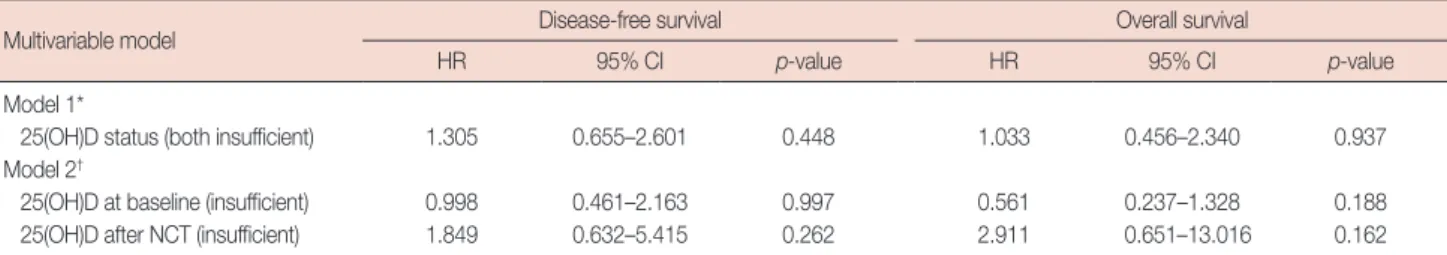
Table 3. Multivariable analysis for survival
Multivariable model Disease-free survival Overall survival
HR 95% CI p-value HR 95% CI p-value
Model 1*
25(OH)D status (both insufficient) 1.305 0.655–2.601 0.448 1.033 0.456–2.340 0.937 Model 2†
25(OH)D at baseline (insufficient) 0.998 0.461–2.163 0.997 0.561 0.237–1.328 0.188 25(OH)D after NCT (insufficient) 1.849 0.632–5.415 0.262 2.911 0.651–13.016 0.162 HR=hazard ratio; CI=confidence interval; 25(OH)D=25-hydroxyvitamin D; NCT=neoadjuvant chemotherapy.
*In model 1, the serum 25(OH)D level before and after NCT was used in the multivariable analyses; †In model 2, each 25(OH)D status prior to or after NCT was
en-tered into the multivariable analyses. Age, clinical stage at diagnosis, histologic grade, molecular phenotype, postsurgical pathology, type of surgery, endocrine therapy, and targeted therapy were adjusted in each multivariable model.
Figure 3. Kaplan-Meier curves accord-ing to the 25-hydroxyvitamin D (25[OH] D) status stratified by molecular phe-notype. Disease-free survival (A, C, E, and G) and overall survival (B, D, F, and H) curves are presented in the luminal A-like (A and B), luminal B-like (C and D), human epidermal growth factor receptor 2 (HER2)-enriched (E and F), and triple-negative breast can-cer (G and H) phenotypes. The green line shows patients with either suffi-cient 25(OH)D level at baseline or after neoadjuvant chemotherapy (NCT) and the blue line exhibits those with defi-cient 25(OH)D at both baseline and af-ter NCT. 1.0 0.8 0.6 0.4 0.2 0 1.0 0.8 0.6 0.4 0.2 0 1.0 0.8 0.6 0.4 0.2 0 1.0 0.8 0.6 0.4 0.2 0 1.0 0.8 0.6 0.4 0.2 0 1.0 0.8 0.6 0.4 0.2 0 1.0 0.8 0.6 0.4 0.2 0 1.0 0.8 0.6 0.4 0.2 0 0 12 24 36 48 60 72 84 0 12 24 36 48 60 72 84 0 12 24 36 48 60 72 84 0 12 24 36 48 60 72 84 0 12 24 36 48 60 72 84 0 12 24 36 48 60 72 84 0 12 24 36 48 60 72 84 0 12 24 36 48 60 72 84 Months after surgery
Months after surgery
Months after surgery
Months after surgery
Months after surgery
Months after surgery
Months after surgery
Months after surgery
Disease-fr ee survival Disease-fr ee survival Disease-fr ee survival Disease-fr ee survival Overall survival Overall survival Overall survival Overall survival
Both deficient 25(OH)D Either sufficient 25(OH)D p=0.090 p=0.191 p=0.513 p=0.810 p=0.060 p=0.203 p=0.614 p=0.560 A C E G B D F H
12 months [11]. However, daily supplements of Vit D3 (400 IU) for 1 year in patients with premenopausal status modestly increased 25(OH)D levels and partially prevented a decrease in serum concentrations by chemotherapy [17].
It has not been clearly determined whether chemotherapy directly affects changes in serum 25(OH)D levels, plays indi-rect roles through gastrointestinal side effects and behavioral changes toward avoiding sunlight exposure, or whether 25(OH)D concentrations are not significantly affected by che-motherapy [7,10,17]. Miyoshi et al. [18] suggested that docetaxel could upregulate the cytochrome P450 3A4 en-zyme, which might convert active forms of Vit D to inactive metabolites. The administration of corticosteroids as anti-emetics during chemotherapy or estrogen deprivation by che-motherapy-induced amenorrhea might partially affect Vit D metabolism and serum concentrations [11].
In addition to dietary intake, supplementation, and sun-light, serum 25(OH)D levels are influenced by many genetic, environmental, and lifestyle factors, including genetic poly-morphisms, race, ethnicity, age, sex, pregnancy, season, body mass index, pregnancy, skin pigmentation, and hereditary and acquired disorders, such as liver failure or chronic renal dis-ease [19,20]. The anticancer actions of Vit D signaling are ex-erted via antiproliferation, anti-inflammation, anti-invasion and metastasis, antiangiogenesis, and induction of apoptosis and differentiation [21,22]. Calcitriol, an active Vit D form, has various functions through endocrine, paracrine, or auto-crine modes. After binding to Vit D or retinoid X receptors, calcitriol plays a role via both genomic and nongenomic ac-tions [23]. The net effects of altered gene expression are bene-ficial antitumor effects [2]. ER pathways may be influenced by calcitriol, like the suppression of aromatase in adipose tissues and the suppression of ER or estrogen-mediated signaling in cancer cells [24]. In patients with breast cancer, Vit D defi-ciency is associated with aggressive prognostic features, in-cluding advanced tumor stage, high grade, high Ki-67 index-es, or negative ER expression [3,25,26]. Consistently, our study demonstrated that the “either sufficient” group was associated with postmenopausal status, rural residence, summer exami-nations, and molecular phenotypes. However, the body mass index, stage, grade, and Ki-67 index were not different be-tween the groups. More comprehensive studies are necessary to determine the associations between serum 25(OH)D levels and clinicopathological characteristics in breast cancer.
Recent studies of the impact of Vit D on the responsiveness to NCT showed no significant association of serum 25(OH)D levels with pCR, similarly to that determined in our study [6,7]. When stratified by molecular phenotype, changes in Vit D status are not a predictive factor for pCR. Although the
baseline patient characteristics differed from those in our study, patients with a favorable response of >90% decrease in tumor cells showed increased serum Vit D levels at the end of NCT [7]. Based on in vitro cell line and animal experiments, calcitriol has been shown to increase chemotherapy-induced cell death; however, in the in vivo milieu, the tumoricidal ef-fects exerted by chemotherapeutic agents might be more complex [6]. As we could not evaluate Vit D receptor expres-sion in the cancer tissues, further studies are required to con-clude the association between serum Vit D concentrations and pCR in neoadjuvant settings.
Additionally, the present study demonstrated no clinical as-sociation between 25(OH)D status and survival in patients with breast cancer treated with NCT. Irrespective of the mo-lecular phenotype, changes in 25(OH)D levels were not a prognostic factor. Although the follow-up duration of our co-hort was relatively sco-hort, subgroup analysis from the I-SPY trial similarly determined no evidence of Vit D levels as a prognostic factor [6]. However, previous meta analyses showed that high 25(OH)D levels were significantly associat-ed with lower risks of breast cancer mortality and overall death in adjuvant settings [27,28]. On the contrary, an adju-vant clinical trial showed no evidence of the association be-tween Vit D blood levels and relapse-free survival, breast can-cer-specific survival, or OS [5]. More research should be con-ducted to determine the implications of 25(OH)D levels on survival outcomes in patients with breast cancer receiving NCT.
A strength of our study is the relatively large sample size with paired evaluation of serum 25(OH)D concentrations be-fore initiation and after completion of NCT. We also analyzed the prognostic power of Vit D levels according to molecular phenotype. However, the retrospective nature and relatively short follow-up period are important limitations. Further, we could not assess several confounding factors for serum 25(OH)D concentrations, including dietary intake, supple-ments, application of sunblock, or physical activity. Finally, for the definition of molecular phenotype, Ki-67 indexes were substituted with histologic grade in 17.6% of cases.
In conclusion, Vit D3 deficiency was highly prevalent at the time of diagnosis in Korean patients with breast cancer and a significant decrease in serum 25(OH)D levels was demon-strated after completion of NCT. Sufficient 25(OH)D status either before or after NCT was not significantly associated with a favorable response to NCT or improved survival out-comes. Therefore, correction or maintenance of appropriate serum Vit D3 levels as comprehensive management of pa-tients receiving NCT should be urgently focused on for skele-tal health, but not for oncological outcomes until more
evi-dence is accumulated. Additionally, the possible oncological aspects of Vit D3 should be further explored and researched comprehensively considering breast cancer subtypes.
CONFLICT OF INTEREST
The authors declare that they have no competing interests.
REFERENCES
1. Wang D, Vélez de-la-Paz OI, Zhai JX, Liu DW. Serum 25-hydroxyvitamin D and breast cancer risk: a meta-analysis of prospective studies. Tumour Biol 2013;34:3509-17.
2. Rose AA, Elser C, Ennis M, Goodwin PJ. Blood levels of vitamin D and early stage breast cancer prognosis: a systematic review and meta-analy-sis. Breast Cancer Res Treat 2013;141:331-9.
3. Yao S, Kwan ML, Ergas IJ, Roh JM, Cheng TD, Hong CC, et al. Associa-tion of serum level of vitamin D at diagnosis with breast cancer survival: a case-cohort analysis in the pathways study. JAMA Oncol 2017;3:351-7.
4. Chlebowski RT, Johnson KC, Kooperberg C, Pettinger M, Wactawski-Wende J, Rohan T, et al. Calcium plus vitamin D supplementation and the risk of breast cancer. J Natl Cancer Inst 2008;100:1581-91. 5. Lohmann AE, Chapman JA, Burnell MJ, Levine MN, Tsvetkova E,
Pritchard KI, et al. Prognostic associations of 25 hydroxy vitamin D in NCIC CTG MA.21, a phase III adjuvant randomized clinical trial of three chemotherapy regimens in high-risk breast cancer. Breast Cancer Res Treat 2015;150:605-11.
6. Clark AS, Chen J, Kapoor S, Friedman C, Mies C, Esserman L, et al. Pretreatment vitamin D level and response to neoadjuvant chemother-apy in women with breast cancer on the I-SPY trial (CALGB 150007/150015/ACRIN6657). Cancer Med 2014;3:693-701. 7. Charehbili A, Hamdy NA, Smit VT, Kessels L, van Bochove A, van
Laarhoven HW, et al. Vitamin D (25-0H D3) status and pathological response to neoadjuvant chemotherapy in stage II/III breast cancer: data from the NEOZOTAC trial (BOOG 10-01). Breast 2016;25:69-74. 8. Taylor CL, Sempos CT, Davis CD, Brannon PM. Vitamin D: moving
forward to address emerging science. Nutrients 2017;9:E1308. 9. van Schoor N, Lips P. Global overview of vitamin D status. Endocrinol
Metab Clin North Am 2017;46:845-70.
10. Jacot W, Pouderoux S, Thezenas S, Chapelle A, Bleuse JP, Romieu G, et al. Increased prevalence of vitamin D insufficiency in patients with breast cancer after neoadjuvant chemotherapy. Breast Cancer Res Treat 2012;134:709-17.
11. Kim HJ, Koh BS, Yu JH, Lee JW, Son BH, Kim SB, et al. Changes in serum hydroxyvitamin D levels of breast cancer patients during tamox-ifen treatment or chemotherapy in premenopausal breast cancer pa-tients. Eur J Cancer 2014;50:1403-11.
12. Moukayed M, Grant WB. The roles of UVB and vitamin D in reducing risk of cancer incidence and mortality: a review of the epidemiology, clinical trials, and mechanisms. Rev Endocr Metab Disord 2017;18: 167-82.
13. Ross AC, Manson JE, Abrams SA, Aloia JF, Brannon PM, Clinton SK, et al. The 2011 report on dietary reference intakes for calcium and vita-min D from the Institute of Medicine: what clinicians need to know. J Clin Endocrinol Metab 2011;96:53-8.
14. Hammond ME, Hayes DF, Dowsett M, Allred DC, Hagerty KL, Badve S, et al. American Society of Clinical Oncology/College of American Pathologists guideline recommendations for immunohistochemical testing of estrogen and progesterone receptors in breast cancer. J Clin Oncol 2010;28:2784-95.
15. Wolff AC, Hammond ME, Hicks DG, Dowsett M, McShane LM, Allison KH, et al. Recommendations for human epidermal growth factor recep-tor 2 testing in breast cancer: American Society of Clinical Oncology/ College of American Pathologists clinical practice guideline update. J Clin Oncol 2013;31:3997-4013.
16. Oh MG, Han MA, Park J, Ryu SY, Choi SW. The prevalence of vitamin D deficiency among cancer survivors in a nationwide survey of the Korean population. PLoS One 2015;10:e0129901.
17. Crew KD, Shane E, Cremers S, McMahon DJ, Irani D, Hershman DL. High prevalence of vitamin D deficiency despite supplementation in premenopausal women with breast cancer undergoing adjuvant che-motherapy. J Clin Oncol 2009;27:2151-6.
18. Miyoshi Y, Ando A, Takamura Y, Taguchi T, Tamaki Y, Noguchi S. Pre-diction of response to docetaxel by CYP3A4 mRNA expression in breast cancer tissues. Int J Cancer 2002;97:129-32.
19. Holick MF. Vitamin D deficiency. N Engl J Med 2007;357:266-81. 20. Fuleihan Gel-H, Bouillon R, Clarke B, Chakhtoura M, Cooper C,
McClung M, et al. Serum 25-hydroxyvitamin D levels: variability, knowledge gaps, and the concept of a desirable range. J Bone Miner Res 2015;30:1119-33.
21. Zhang X, Harbeck N, Jeschke U, Doisneau-Sixou S. Influence of vitamin D signaling on hormone receptor status and HER2 expression in breast cancer. J Cancer Res Clin Oncol 2017;143:1107-22.
22. Welsh J. Function of the vitamin D endocrine system in mammary gland and breast cancer. Mol Cell Endocrinol 2017;453:88-95. 23. Picotto G, Liaudat AC, Bohl L, Tolosa de Talamoni N. Molecular
aspects of vitamin D anticancer activity. Cancer Invest 2012;30:604-14. 24. Hu K, Callen DF, Li J, Zheng H. Circulating vitamin D and overall
survival in breast cancer patients: a dose-response meta-analysis of cohort studies. Integr Cancer Ther 2018;17:217-25.
25. de Sousa Almeida-Filho B, De Luca Vespoli H, Pessoa EC, Machado M, Nahas-Neto J, Nahas EA. Vitamin D deficiency is associated with poor breast cancer prognostic features in postmenopausal women. J Steroid Biochem Mol Biol 2017;174:284-9.
26. Buono G, Giuliano M, De Angelis C, Lauria R, Forestieri V, Pensabene M, et al. Pretreatment serum concentration of vitamin D and breast cancer characteristics: a prospective observational mediterranean study. Clin Breast Cancer 2017;17:559-63.
27. Kim Y, Je Y. Vitamin D intake, blood 25(OH)D levels, and breast cancer risk or mortality: a meta-analysis. Br J Cancer 2014;110:2772-84. 28. Li M, Chen P, Li J, Chu R, Xie D, Wang H. Review: the impacts of
circu-lating 25-hydroxyvitamin D levels on cancer patient outcomes: a sys-tematic review and meta-analysis. J Clin Endocrinol Metab 2014;99: 2327-36.

![Figure 1. Kaplan-Meier curves according to 25-hydroxyvitamin D (25[OH]D) status. Disease-free survival (A) and overall survival (B) curves are presented](https://thumb-ap.123doks.com/thumbv2/123dokinfo/5070207.71338/4.871.448.791.110.776/kaplan-according-hydroxyvitamin-disease-survival-overall-survival-presented.webp)
![Figure 3. Kaplan-Meier curves accord- accord-ing to the 25-hydroxyvitamin D (25[OH] D) status stratified by molecular phe-notype](https://thumb-ap.123doks.com/thumbv2/123dokinfo/5070207.71338/6.871.67.708.110.1103/figure-kaplan-meier-curves-accord-hydroxyvitamin-stratified-molecular.webp)
