LETTER TO THE EDITOR
Copyright © 2017 The Korean Association of Internal Medicine
This is an Open Access article distributed under the terms of the Creative Commons Attribution Non-Commercial License (http://creativecommons.org/licenses/ by-nc/3.0/) which permits unrestricted noncommercial use, distribution, and reproduction in any medium, provided the original work is properly cited.
pISSN 1226-3303 eISSN 2005-6648 http://www.kjim.org
Korean J Intern Med 2017;32:548-551 https://doi.org/10.3904/kjim.2015.269
Department of Internal Medicine, Hanyang University College of Medicine, Seoul, Korea
Lymphoid follicular proctitis resembling rectal
carcinoid tumor, confirmed by endoscopic
resection
Seungjun Gim, Kang Nyeong Lee, Donghoon Lee, Hye Young Lee, Ki Sul Chang, Oh Young Lee, and Ho Soon Choi
Received : August 11, 2015 Revised : October 6, 2015 Accepted : November 4, 2015 Correspondence to Kang Nyeong Lee, M.D.
Department of Internal Medicine, Hanyang University College of Medicine, 222-1 Wangsimni-ro, Seongdong-gu, Seoul 04763, Korea Tel: +82-2-2290-8339
Fax: +82-2-2290-8314
E-mail: [email protected]
To the Editor,
The rectum can be affected by a vari-ety of malignant and benign diseases: malignant diseases are rectal cancer, carcinoid tumor, and lymphoma, and benign diseases are polyps, solitary rectal ulcer syndrome, and other in-flammatory diseases including pseudo-membranous colitis, ulcerative colitis (UC), radiation proctitis, and lymphoid follicular proctitis (LFP). LFP is a very rare benign lymphoproliferative dis-ease involving the rectum [1]. Among lymphoproliferative diseases, LFP is the rarest inflammatory disease, which was previously regarded as a form of UC, or can be misdiagnosed as a muco-sa-associated lymphoid tissue (MALT)
lymphoma [2]. In contrast to UC and lymphoma, its endoscopic and histo-pathologic findings are different from them: the endoscopic findings of LFP are usually multiple nodular, granu-lar or congested mucosa without ul-cerations, and histologic findings are characterized by lymphoid follicular hyperplasia without acute inflammato-ry infiltration. Here, we report the case of LFP accidently found during screen-ing colonoscopy as an unusual form of a subepithelial tumor (SET)-like lesion resembling a carcinoid tumor.
A 66-year-old woman underwent screening colonoscopy in our digestive disease center in Hanyang University Hospital. She was taking statins for
dys-A B
Figure 1. Colonoscopic findings. (A) Colonoscopy shows a nodular polypoid lesion about 5
mm in size in the distal rectum. (B) Narrow band imaging shows mucosal capillary net-work arranged in a honeycomb pattern.
549 www.kjim.org
https://doi.org/10.3904/kjim.2015.269
Gim S, et al. Lymphoid follicular proctitis
lipidemia. Three years ago she had undergone gastrosco-py and colonoscogastrosco-py for screening, which was unremark-able except reflux esophagitis and atrophic gastritis. The present colonoscopy incidentally revealed a somewhat pale and whitish SET-like elevated lesion in the rectum. It was a hard, solitary sessile mass of about 5 mm in size at 10 cm from the anal verge (Fig. 1A). The overlying mu-cosa of the mass appeared normal, but there was neither cushion sign nor rolling sign, resembling a carcinoid tu-mor involving the submucosa being not ruled out. Also, in narrow band imaging (NBI) mucosal capillary network was arranged in a honeycomb pattern and there were no disorganized structure or increased density of mi-cro-vessels, indicating similar NBI findings to carcinoid tumors (Fig. 1B).
This soliltary SET-like lesion in the rectum was con-sidered a carcinoid tumor at first because of its SET-like
appearance with a pale whitish surface, and was decided to be removed by the endoscopist for both diagnosis and treatment. After informed consent, the rectal lesion was completely resected by an endoscopic submucosal dis-section technique without complications.
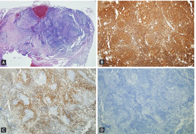
Histopathologic findings of the resected specimen in-cluded dense and diffuse lymphoid infiltration and con-fluent aggregates of large reactive lymphoid follicles in the mucosal and submucosa (Fig. 2A). Together with the lymphoid hyperplasia, reactive proliferation was evident in the germinal center while the mantle zone conserved in the submucosal layer. However, there were no findings of crypt abscess or granuloma. Also, the diffuse follicular lymphoid hyperplasia was immunohistochemically pos-itive for CD20 (Fig. 2B), but negative for Bcl-2, a marker for lymphoma (Fig. 2C) and for synaptophysin, a marker for neuroendocrine tumor (Fig. 2D). Therefore, the final
Figure 2. Histopathologic findings (×40). (A) Dense and diffuse lymphocytes and lymphoid hyperplasia are evident in the
mu-cous and submumu-cous layers (H&E, ×40). (B) Immunohistochemistry for CD20 is diffusely positive, indicating the presence of B-lymphocytes. (C) Immunohistochemistry for Bcl-2, a marker of lymphoma, is negative in the germinal center. (D) Immuno-histochemistry for synaptophysin, a marker of neuroendocrine tumor, is negative in the germinal center.
A
C D
550 www.kjim.org https://doi.org/10.3904/kjim.2015.269
The Korean Journal of Internal Medicine Vol. 32, No. 3, May 2017
diagnosis was LFP. After establishing the diagnosis and completing the treatment, the patient was placed on a regular follow-up schedule without any symptoms.
A solitary elevated lesion in the rectum should be dif-ferentiated from benign lymphoproliferative diseases such as lymphoid polyps of the rectum and also malig-nant diseases such as carcinoid tumor and lymphoma. On the other hand, LFP should also be differentiated from non-neoplastic diseases such as distal UC, and al-lergic proctitis. For these differentiations, histopatholog-ic findings are important: the histopathologhistopatholog-ic findings were prominent lymphoid follicular hyperplasia with reactive germinal center but conserved mantle zone. The histopathologic difference from UC was the absence of any crypt abscess and of neutrophil and eosinophil infil-tration [2]. The difference from benign lymphoid polyp was the absence of well-demarcated proliferative lym-phoid follicles in the surrounding tissue, and the differ-ence from malignant lymphoma of the MALT type was the absence of malignant lymphoid cells and destructive follicular structure [3]. For histopathologic confirmation it is mandatory to obtain intact follicular structure of lymphoid tissues, but forceps biopsy alone may be insuf-ficient because of small, crushed, and distorted tissues obtained [3]. Therefore, to accurately diagnose and differ-entiate LFP from other possible diseases involving the rectum, endoscopic resection is required, as performed in our case. On the other hand, endoscopic ultrasonogra-phy (EUS) must have been helpful in providing import-ant information on this lesion. However, we performed ESD before EUS for pathologic examination and com-plete removal. A previous report described that EUS im-age in LFP showed rectal wall thickening limited to the mucosal and submucosal layers, compared with that of carcinoid tumor showing hypoechoic lesion in the deep mucosal layer.
What causes LFP has not been clearly defined. A pre-vious report suggested a role of vascular or mechani-cal causes in its development showing diversion colitis (nonspecific inflammation of a dysfunctional segment after diversion of the fecal stream) after surgery due to UC as well as after surgery due to severe hemorrhoids and rectal prolapse [4]. However in our patient, as there was no surgery or vascular or mechanical injury, the cause of her LFP is unclear. Concerning symptoms of LFP, our patient did not have any associated symptoms or signs.
According to a study of 20 patients with rectal biopsies showing LFP, its common symptoms are reported to be rectal bleeding and abdominal pain, while tenesmus and mucus discharge were also reported [2]. Most of the symptomatic cases showed multiple lesions with a con-gested granular appearance at endoscopy, whereas our patient had a small solitary lesion producing no symp-toms, probably because there was only one rectal lesion.
Is treatment needed in patients with asymptomatic LFP? In cases with symptoms such as rectal bleeding or abdominal pain, treatment should be directed to allevi-ating or arresting these symptoms. These symptoms as well as endoscopic findings have been reported to re-spond to topical anti-inflammatory agents such as mesa-lamine or sulphasalazine, but systemic mesamesa-lamine and steroids have been found to be ineffective [5]. Although the natural course of LFP is unclear, it is possible that LFP presenting with a solitary lesion, as in our case, might progress to multiple nodular lesions and there-by produce symptoms such as rectal bleeding. In terms of treatment modality, alternative treatment modality include cap-assisted endoscopic mucosal resection in which the lesion is removed using a snare after it is suc-tioned into a cap and ligated by a band, as in rectal car-cinoid tumor. Considering a benign nature of the lesion with small size confined to the mucosa and submucosa, this modality could completely remove the lesion.
In conclusion, this case concerns a very rare benign in-flammatory disease of the rectum, LFP. It was presented as a nodular, SET-like lesion resembling a rectal carci-noid tumor without any symptoms, and was endoscopi-cally removed for both diagnosis and treatment. There-fore, we show that endoscopic resection of the solitary LFP may be an effective and safe treatment as well as a diagnostic tool for confirmation. However, further stud-ies are needed to establish the natural course of LFP as well as its clinical and endoscopic presentations.
Keywords: Rectal; Proctitis; Carcinoid tumor
Conflict of interest
No potential conflict of interest relevant to this article was reported.
551 www.kjim.org
https://doi.org/10.3904/kjim.2015.269
Gim S, et al. Lymphoid follicular proctitis
REFERENCES
1. Kim MH, Jung JT, Kim EJ, et al. A case of mucosa-asso-ciated lymphoid tissue lymphoma of the sigmoid colon presenting as a semipedunculated polyp. Clin Endosc 2014;47:192-196.
2. Flejou JF, Potet F, Bogomoletz WV, et al. Lymphoid fol-licular proctitis: a condition different from ulcerative proctitis? Dig Dis Sci 1988;33:314-320.
3. Shami VM, Waxman I. Lymphoid follicular proctitis mimicking rectal lymphoma: diagnosis by EMR. Gas-trointest Endosc 2004;60:648-652.
4. Geraghty JM, Talbot IC. Diversion colitis: histological features in the colon and rectum after defunctioning co-lostomy. Gut 1991;32:1020-1023.
5. Seo GS, Choi SC, Jo EY, et al. The usefulness of mesala-zine suppositories for the treatment of lymphoid follic-ular proctitis. Korean J Gastroenterol 2006;47:420-424.