The role of ASK1 in selective striatal
lesion formation induced by neuronal
injury
Kyoung Joo Cho
Department of Medical Science
The Graduate School, Yonsei University
The role of ASK1 in selective striatal
lesion formation induced by neuronal
injury
Directed by Professor Jong Eun Lee
The Doctoral Dissertation
submitted to the Department of Medical Science,
the Graduate School of Yonsei University
in partial fulfillment of the requirements for the degree
of Doctor of Philosophy
Kyoung Joo Cho
This certifies that the Doctoral
Dissertation of Kyoung Joo Cho is
approved.
---
Thesis Supervisor : Jong Eun Lee
---
Thesis Committee Member#1 : Won Taek Lee
---
Thesis Committee Member#2 : Kyung Ah Park
---
Thesis Committee Member#3 : Se Hoon Kim
---
Thesis Committee Member#4 : Chul Hoon Kim
The Graduate School
Yonsei University
TABLE OF CONTENTS
ABSTRACT
···
1
PART I
Inhibition of ASK1 reduces ER stress and nuclear huntingtin fragments
in a mouse model of Huntington Disease
I. INTRODUCTION
···
4
II. MATERIALS AND METHODS
···
6
1. Animal model
···
6
2. Immunohistochemistry
···
6
3.Western blot analysis and co-immunoprecipitation
···
7
4. RT-PCR Analysis
···
8
5. Neutralization of Ask1 using an anti-Ask1-antibody
···
9
6. Rotarod test
···
9
7. Striatal lesion analysis
···
10
8. Statistical analysis
···
10
III. RESULTS
···
11
1. Levels of ASK1 protein and ER stress are increased in HD mice
···
11
2. ASK1 interacts with htt fragments and aids in the translocation of htt
···
14
3. Inhibition of ASK1 facilitates BDNF transport to the striatum and improves motor dysfunction
···
17
IV. DISCUSSION
···
20
PART II
Apoptosis signal-regulating kinase-1 aggravates ROS-mediated striatal
degeneration in 3-nitropropionic acid-infused mice
I. INTRODUCTION
···
25
II. MATERIALS AND METHODS
···
27
1. Animal model
···
27
2. ASK1gene silencing with siRNA and Administration of ASK1-peptide
···
27
3.Immunohistochemistry
···
28
4.Western blot analysis
···
29
5.Apoptotic cell-death assay
···
29
6. ROS detection by staining
···
30
7. ROS detection by flow cytometry analysis
···
30
8. Detection of DNA oxidation in mouse striatum
···
31
9.Rotarod test
···
31
10. Statistical analysis
···
31
III. RESULTS
···
33
1. Systemic infusion of 3-NP led to the formation of selective striatal lesions in wt, but SOD-tg mice
···
33
2. Greater ROS production, oxidative damage and ASK1 levels and activity were detected in striatal lesions
···
36
3. ASK1 amounts mediated the striatal cell death without change of ROS level
···
41
IV. DISCUSSION
···
45
PART III
Apoptosis signal-regulating kinase 1 mediates 3-nitropropionic acid
toxicity and regulates C1q level via astrocyte TGF-beta
I. INTRODUCTION
···
50
II. MATERIALS AND METHODS
···
54
1. Primary neuronal cell culture
···
54
2. Animal model and lesion analysis
···
54
3. Behavior and motor function test
···
55
4. PCR and realtime PCR
···
55
5.ATP assay
···
57
6. Immunocytochemistry / immunohistochemistry and confocal microscopy
···
57
7. Cell death assay
···
58
8. ROS detection by in situ HEt and AP sites counting
···
59
9.Detection of DNA oxidation in mouse striatum
···
60
10. RNA interference
···
60
11. Astrocyte conditioned medium
···
61
12.ELISA
···
62
III. RESULTS
···
63
1. Selective striatal lesions were formed by systemic infusion of 3-NP in mice
···
63
2. Mitochondrial dysfunction was triggered and ROS level was raised by 3-NP infusion
···
66
3. Brain-damage and apoptotic cell death were induced in the lesion of 3-NP infusion
···
68
4. Downregulation of ASK1 prevented 3-NP induced cell death, BDNF depletion, and subsequently improved the neurologic impairment
···
73
5. Infusion of 3-NP leads to striatum-selective astrocyte reactivation and changes the distribution of secreted BDNF, TGF-β1, and C1q protein in the damaged subregions
···
76
6. TGF-β1 is differentially expressed in the damaged brain subregions and astrocyte derived ASK 1 by 3-NP involves in TGF-β1
···
79
7. TGF-β1 secreted by astrocyte triggers neuronal C1q degenerating
neuronal dendrites and ASK1 mediates the event
···
82
IV. DISCUSSION
···
87
V. CONCLUSION
···
91
REFERENCES
···
92
ABSTRACT(IN KOREAN)
···
104
LIST OF FIGURES
PART I
Inhibition of ASK1 reduces ER stress and nuclear huntingtin
fragments in a mouse model of Huntington Disease
Figure 1. Expression of Ask1 in HD transgenic mice (tg) and
HD littermates (wt)···12
Figure 2. ER stress and expression pattern of Ask1 in HD
transgenic mice (tg) and HD littermates (wt)···13
Figure 3. The effect of inhibition of Ask1 on the levels of ER
stress and htt fragments in HD···15
Figure 4. The effect of neurtralizing Ask1 on htt fragments
localiziation in HD···16
Figure 5. The effect of inhibition of Ask1 on expression of
BDNF ···18
Figure 6. The effect of inhibition of Ask1 on expression of
PART II
Apoptosis signal-regulating kinase-1 aggravates
ROS-mediated striatal degeneration in 3-nitropropionic
acid-infused mice
Figure 1. Pathological examination in the striatum after
systemic infusion of 3-NP···34
Figure 2. Apoptotic cell death in wt and SOD-tg mice ···35
Figure 3. Environmental changes and ROS level changes in
the striatum after 3-NP infusion···37
Figure 4. The alteration of ROS scavenging enzyme protein
level in the striatum of wt and SOD tg mice after
3-NP infusion ···38
Figure 5. Oxidative DNA damage in the striatum of wt and
SOD tg mice after 3-NP infusion···39
Figure 6. The changes of ASK1 protein and activity···40
Figure 7. Down regulation and induction of ASK1···42
Figure 8. The effect of ASK1-peptide on ROS production level
···43
Figure 9. The role of ASK1 in cell death and behavioral
PART III
Apoptosis signal-regulating kinase 1 mediates 3-nitropropionic
acid toxicity and regulates C1q level via astrocyte TGF- β1
Figure 1. Chronic infusion of 3-NP induces selective striatal
lesion ···64
Figure 2. Chronic infusion of 3-NP induces behavioral and
motor dysfunction ···65
Figure 3. Infusion of 3-NP increases mitochondrial
dysfunction and DNA damage ···67
Figure 4. Apoptotic cell death was induced in the lesion of
3-NP infusion ···69
Figure 5. ASK1 protein level and activation were induced in
the lesion of 3-NP infusion ···70
Figure 6. Protein level of p53 increased in the lesion of 3-NP
infusion ···71
Figure 7. Infusion of 3-NP decreases BDNF expression ···72
Figure 8. Down-regulating ASK1 prevents 3-NP induced
BDNF depletion ···74
Figure 9. Down-regulation of ASK1 reduced striatal lesion
Figure 10. Astrocytes were strongly reactivated by infusion of
3-NP ···77
Figure 11. TGF-β1and C1q secretion was triggered by 3-NP
infusion ···78
Figure 12. TGF-β1 is differentially expressed in brain
subregions and astrocyte derived ASK 1 by 3-NP
involves in TGF-β1···80
Figure 13. Infusion of 3-NP alters TGF- β1 and ASK1 in
astrocyte ···81
Figure 14. Astrocyte conditioned media (ACM) affect on
neuron to trigger neuronal C1q degenerating
neuronal dendrites and ASK1 mediates the event
···83
Figure 15. Infusion of 3-NP increases C1qR mediated by
1
<ABSTRACT>
The role of ASK1 in selective striatal lesion formation induced by
neuronal injury
Kyoung Joo Cho
Department of Medical Science
The Graduate School, Yonsei University
(Directed by Professor Jong Eun Lee)
Apoptosis signal-regulating kinase-1 (ASK1), an early signaling element in the cell death pathway, has been suggested to participate in the pathology of neurodegenerative diseases, which may be associated with environmental factors that impact the diseases. The systemic administration of 3-nitropropionic acid (3-NP) facilitates the development of selected striatal lesions and it remains unclear whether specific neurons are selectively targeted in 3-NP infused striatal degeneration. Although not entirely elucidated, the mechanisms of neurotoxicity induced by 3-NP have been shown to include the exhaustion of adenosine triphosphate, mitochondrial membrane depolarization, dysregulation of intracellular calcium homeostasis, calpain activation, and the release of pro-apoptotic proteins from mitochondria. The present study is to characterize the regulation of BDNF in each cortical and striatal subregion. This study investigates that mild and chronic exposure of mitochondrial toxin can modulate the C1q level both in cortex and striatum via regulation of TGF-beta
2
from astrocyte. Consequently we investigate how the BDNF is dominantly depleted in striatum, and eventually whether striatal lesion is established in involving in ASK1 pathway.
The results of the present work show an alteration of ASK1 pathway molecules, TGF-beta, C1q level, and BDNF level as a final standard to striatal degeneration. By ASK1 down-regulation, improvement in each molecules containing behavioral impairment was evaluated in 3NP- infused mice and 3-NP treated primary neuronal cells. We propose the hypothesis that (1) ASK1 overexpression by systemic infusion of 3-NP promotes the formation of selective striatal lesions, and this occurs apart from just ROS generation. (2) ASK1 may differentially regulate C1q secretion level via active TGF-beta in each brain subregion of cortex and striatum, consequently involved in axon degeneration of corticostriatal projection neuron. When brain is mildly and chronically exposed to mitochondrial toxin, presynaptic neuron (in cortical neuron) degrades first, and then postsynaptic neuron of striatal MSN neuron withers as a consequence of it.
Consolidating these results, we suggest that the increased ASK1 is linked to regulation of TGF-beta secreted in astrocytes, and differential C1q expression in neurons triggered by TGF-beta leads degradation of cortical projection and depletion of BDNF in striatal neuron in mice brains systemically infused with 3-NP.
--- Key words : apoptosis signal-regulating kinase 1, 3-nitropropionic acid, Huntington’s disease, brain derived neurotrophic factor, siRNA, superoxide dismutase
3
PART I
Inhibition of ASK1 reduces ER stress and nuclear huntingtin
fragments in a mouse model of Huntington Disease
4 I. INTRODUCTION
Accumulation of misfolded proteins within the endoplasmic reticulum (ER) lumen induces ER stress, which has been implicated in human diseases such as Alzheimer’s disease 1
, Parkinson’s disease 2, and Huntington’s disease (HD) 3. HD is characterized clinically by chorea, psychiatric disturbances, and dementia, while it is characterized pathologically by neuronal inclusions as well as striatal and cortical neurodegeneration. The neurodegeneration is arisen from the loss of long projection neurons in the cortex and striatum 4. HD is inherited in an autosomal dominant manner, and is caused by the presence of an elongated polyglutamine (polyQ) tract (> 40) in the huntingtin (htt) gene 5.
Htt fragments with pathogenic repeat lengths have been reported to co-localize with various molecular chaperones 6,7 and proteasome components and to impair the function of the ubiquitin-proteasome system (UPS) 8, resulting in ER stress 9. Apoptosis signal-regulating kinase 1 (Ask1) is reported to be involved in ER stress triggered by htt fragments, which results in neuronal cell death. ER stress activates Ask1 through the formation of the IRE1-TRAF2-Ask1 complex. Furthermore, the expanded polyQ portion of htt activates Ask1, and it has been shown conclusively that Ask1 is required for polyQ-induced neuronal cell death 3,4,10,11. Nevertheless, despite the numerous in
vitro studies supporting a correlation between Ask1 and ER stress, it is not
known whether the level of ER stress and Ask1 protein levels or activity are increased in vivo in HD transgenic (tg) animals. In HD, Ask1 is thought to be a modifier of htt at the gene level 12, a signal transducer at the protein level 10, and a cell death modulator at the post-translational level 13. It has also been suggested that the manifestation, progression, and outcome of HD may be affected by modifier genes and environmental factors that impact the disease’s
5 presentation 12.
In this study, we investigated the role of Ask1 in the pathogenesis of HD transgenic (tg) mice. Specifically, we analyzed the expression of Ask1 and htt within the striatum and cortex along with ER stress in each region, and determined whether Ask1 interacts with htt fragments in vivo. Additionally, we assessed the levels of brain-derived neurotrophic factor (BDNF) in a region-specific manner, and examined the effects of inactivation of Ask1 on striatal atrophy and motor dysfunction.
6 II. MATERIALS AND METHODS
1. Animal model
In this study, 8-week and 12-week R6/2 HD male tg (The Jackson Laboratory, Bar Harbor, ME, U.S.A.), littermate wt mice were used. All procedures were performed in accordancewith the guidelines for the care and use of laboratory animals (Yonsei University, Seoul, Korea), which are approved by the Association for Assessment and Accreditation of Laboratory Animal Care (AAALAC). Animals were anesthetized with 2.0% isoflurane under 30% oxygen and 70% nitrous oxide using a vaporizer (VMC Anesthesia Machine, MDS matrix, Orchard Park, NY, U.S.A.). Rectal temperature was controlled at 370.5°Cwith a homeothermic blanket.
2. Immunohistochemistry
To determine the pattern of localization of htt (Epitomics, Burlingame, CA, U.S.A; 1C2, Chemicon, Bedford, MA, U.S.A.) and Ask1 (Santa Cruz Biotechnology, Santa Cruz, CA, U.S.A.), we performed immunofluorescent staining. After sacrificing the animals, their brains were removed, postfixed o vernight in 3.7% formaldehyde, and stored in 30% sucrose. After fixation, the brains were cut into coronal sections of 20 m thickness on a cryostat section and processed for immunohistochemistry.Fixed sections were incubated with the blocking solution as described previously 14 and incubated with a prim ary antibody, rabbit polyclonal anti-htt antibody (1:100, Epitomics). After washing, the sections were incubated withFITC-conjugated donkey anti-rabbit IgG (1:200, Jackson ImmunoResearch, West Grove, PA, USA). For counter-staining, the sections were incubated with propidum iodine (PI, 1:5000, Sigma, St. Louis, MO, U.S.A.). The stained tissue samples were washed and
7
mounted using Vectashield mounting medium (Vector Lab, Burlingame, CA, U.S.A.). These sections were observed under a LSM510 confocal laser scanning microscope (Carl Zeiss, Thornwood, NY, U.S.A.).
For immunofluorescent double-labeling with htt and Ask1, htt (1:100, Epitomics) immunohistochemistry was performed as described above, followed by incubation with CyTM 3-conjugated donkey anti-rabbit IgG (1:200, Jackson ImmunoResearch). After washing, the sections were incubated with the blocking solution and reacted with goat anti-Ask1 antibody (1:100, Santa Cruz Biotechnology), followed by FITC-conjugated donkey anti-goat IgG (1:200, Jackson ImmunoResearch). The sections were then covered with a cover-slip and examined under a confocal microscope (Carl Zeiss).
3. Western blot analysis and co-immunoprecipitation
The expression levels of proteins were analyzed by western blotting. The cortex or striatum of dissected animal brains were lysed in lysis buffer (20 mM Hepes–KOH, pH 7.5, 250 mM sucrose, 10 mM KCl, 1.5 mM MgCl2, 1 mM
EDTA, 1 mM EGTA, 0.5 mM PMSF, 0.1 mM sodium vanadate, and 0.1 mM proteinase inhibitor cocktail). The samples were homogenized by douncing with a Teflon homogenizer (Wheaton, Millville, NJ, U.S.A.) and centrifuged at 8,000
g for 20 min at 4℃. The lysates were loadedonto SDS-polyacrylamide gels. After migration, the proteins were electrotransfered onto a polyvinylidine difluoride membrane (PVDF) (Millipore, Bedford, MA, U.S.A), which was blocked in5% skim milk, incubated with primary antibodies; Ask1 (1:1000, Cell Signaling, Danvers, MA, U.S.A.), pAsk1 (1:1000, Cell Signaling), 1C2 (1:5000, Chemicon, Bedford, MA, U.S.A.), htt (1:1000, Epitomics), or BDNF (1:500, Abchem, Cambridge, UK). After washing, blots were incubated with HRP-conjugated secondary antibodies(1:5000, Roche Diagnostics, Indianapolis,
8
IN, U.S.A.) and the bands were visualized withan enhanced chemiluminescence reagent (Amersham Biosciences, Piscataway, NJ, U.S.A.). To separate the cytosolic and nuclear fractions of the proteins, each sample was homogenized briefly in lysis buffer and then spun down at 750 g for 10 minutes at 4ºC. The pellet containing the nuclear fraction was resuspended with lysis buffer. The resuspened solution was centrifuged at 16,000 g for 20 min at 4℃and the supernatant was used as the nuclear fraction. The supernatant obtained after centrifugation at 750 g for 10 minutes was then centrifuged at 10,000 g for 20 min at 4℃, and the supernatant containing the cytosolic fraction was transferred to a new tube. This tube was centrifuged at 100,000 g for 1 hr at 4℃ and the supernatant obtained was designated the cytosol.
The procedure for co-immunoprecipitation was performed as described previously with some modifications 15. The cortex and striatum were removed from mice brains, and preparedin the same manner as for western blotting. The protein samples were incubated in a 50% slurry of protein A-sepharose (Amersham Biosciences), and this mixed sample was centrifuged at 12,000 g for 1 min at 4℃. The supernatant was incubated with 20 µl of protein A-sepharose for 2 hr at 4°C and then incubated with 2 µg of goat anti-Ask1 antibody (1:1000; Santa Cruz Biotechnology) overnight at 4°C. The pellets were washed by centrifugation at 14,000 g for 3 min at 4℃. After centrifugation, the supernatant was immunoblotted with polyclonal rabbit anti-htt antibody (1:1000; Epitomics) and with mouse anti-Ask1 antibody as a control.
4. RT-PCR Analysis
Total RNA was isolated from the striatum and cortex of the mice. Total RNA (1 g) was reverse-transcribed and cDNA was synthesized using PCR. The primers used are shown in Table 1. PCR was performed as follows: initial
9
denaturation at 95°C for 10 min, amplification (30 cycles) for 30 sec at 95°C, 30 sec at 60°C, and 30 sec at 72°C, and a final extension at 70°C for 10 min. To semi-quantify BDNF mRNA levels without performing real-time PCR, we performed the PCR reaction for different numbers of cycles (15, 20, 25, and 30 cycles). RT-PCR products were visualized with a UV illuminator after electrophoresis on an agarose gel.
5. Neutralization of Ask1 using an anti-Ask1-antibody
To inactivate Ask1 for an extended period of time (several weeks), we used an Ask1 antibody raised against an epitope corresponding to amino acids at the C-terminus of the Ask1 protein. The antibody was added to the dried BioPORTER (Quiktease protein delivery kit, Sigma Aldrich) reagent and allowed to incubate at room temperature for 10 min according to the manufacturer’s protocol 16
. The mixture of Ask1 antibody and BioPORTER was loaded into an osmotic reservoir. Mice were anesthetized as described above and placed in a stereotaxic apparatus. A micro-osmotic pump reservoir (Alzet, Cupertino, CA, U.S.A.) containing anti-Ask1 antibody (0.2 mg/ml in saline, Santa Cruz) was placed subcutaneously on the backs of the animals 17. A brain infusion cannula connected to the pump was positioned at the intra-striatum (anterial, 0.7 mm; lateral, 1.2 mm; and depth, -3.3 mm). As a control, rabbit IgG or pre-immune Ask1 antibody was infused in the same way. The infusion rate was 1.0 l/h and the pump infused the designated solution for 4 weeks into the brains of HD tg mice. After implantation of the osmotic pump at the age of 8-weeks, the mice were sacrificed 4 weeks later 18.
6. Rotarod test
10
apparatus (Ugo Basile North America Inc., Schwenksville, PA, U.S.A.), where the time a mouse was able to remain on the rod was measured at an accelerating speed from 4 to 40 rpm 19. Each mouse was trained for 5 min and the training session was followed by a 30 min rest period. Mice were then placed back on the rotarod for five trials of a maximum of 5 min at accelerating speed. Mice were evaluated for 3 consecutive days.
7. Striatal lesion analysis
Four weeks after Ask1 inhibition with antibody, animals were anesthetized and decapitated. Brains were frozen and cut on a cryostat into 20
m sections from the anterior to the posterior at 500 m intervals. The sections were stained with cresyl violet and then scanned with an LAS1000 imaging densitometer (Fujifilm, Tokyo, Japan). Striatal atrophy was identified as a striatal region size with pallor or loss of cresyl violet staining. The area of the striata was measured using Sigmascan Pro (ver 6.0).
8. Statistical analysis
Data are expressed as mean ± SD.The significance of differences among multiple groups was evaluated by ANOVA followed by Fisher's post hoc protected least-significantdifference test, while two groups were compared using unpaired t tests (StatView, version 5.01; SAS Institute Inc, Cary, NC).
11 III. RESULTS
1. Levels of ASK1 protein and ER stress are increased in HD mice
In vitro studies in neuronal cell cultures have shown that Ask1 is involved
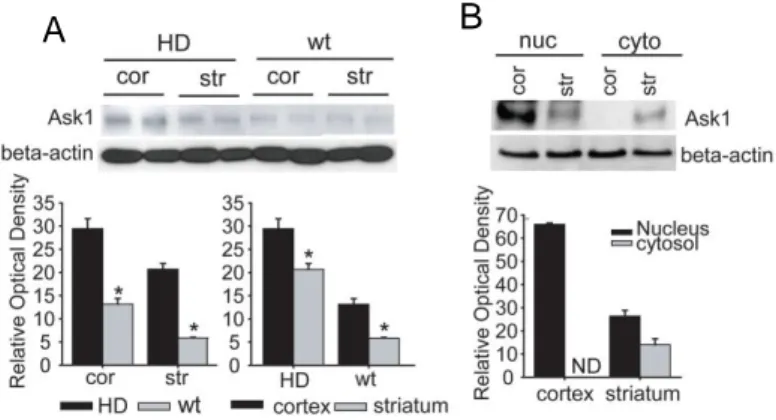
in the pathogenesis of HD 3. In our western blot assay (Figure 1; n = 6 each), the amount of Ask1 protein was increased in HD mice compared to wt mice; furthermore, higher levels of Ask1 were detected in the cortex than in the striatum of both HD tg and wt mice. Ask1 protein in the cortex was mostly present in the nuclear fraction, and barely detectable in the cytosol; similarly, in the striatum, more Ask1 protein was present in the nuclear fraction than the cytosolic fraction (Figure 1; n = 5).
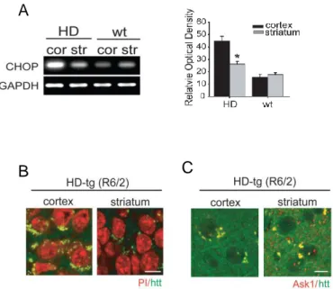
ER stress was evaluated by assessing the level of CHOP expression using RT-PCR (Figure 2; n = 5 each). Levels of CHOP, a member of the C/EBP family of bZIP transcription factors and an indicator of ER stress, were higher in HD than wt mice. Specifically, CHOP expression was higher in the cortex than in the striatum, which correlates with protein expression level of Ask1.
We confirmed the pattern and cellular localization of fragmented htt in HD mice by immunohistochemistry (Figure 2; n = 5 each). High-magnification imaging of htt immunoreactive neurons in the cortex and striatum revealed a perinuclear pattern of fragmented htt staining. The striatal neurons of HD mice stained relatively weakly for htt, which may reflect the lower level of htt expression in the striatum compared with cortical neurons. When we double-stained for both Ask1 and htt, we found that Ask1 and htt fragments colocalized in the cytsol in both the cortex and striatum (Figure 2; n = 3 each).
12
Figure 1. Expression of Ask1 in HD transgenic mice (tg) and HD littermates (wt). (A) Western blot analysis of Ask1 protein in HD and wt mice demonstrates that the Ask1 protein is expressed in cortical and striatal regions, respectively. (B) Ask1 protein is expressed at higher levels in the nuclear than in the cytosolic fraction; this is especially noticeable in the cortex.
13
Figure 2. ER stress and expression pattern of Ask1 in HD transgenic mice (tg) and HD littermates (wt). (A) CHOP mRNA expression, a marker of ER stress, was evaluated in the cortex and striatum of HD and wt mice using RT-PCR. CHOP mRNA was highly expressed in the cortex of HD mice. (B) immunohistochemical staining of htt. Htt-positive cells show a perinuclear staining pattern in the cytosol of both the cortex and striatum of HD mice. (C) Double immunohistochemistry of Ask1 and htt fragments demonstrates that the two molecules are co-localized.
Scale bar = 10 m; cor, cortex; str, striatum; *p < 0.01
A
14
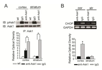
2. ASK1 interacts with htt fragments and aids in the translocation of htt We inhibited Ask1 activity for 4 weeks using an anti-Ask1 antibody, and confirmed that HD mice brains exhibited diminished Ask1 activity under these conditions (Figure 3A; n = 5 each). Infusion of the Ask1 antibody into the striatum of HD mice resulted in a reduction of CHOP expression in both of cortex and striatum (Figure 3B; n = 5 each).
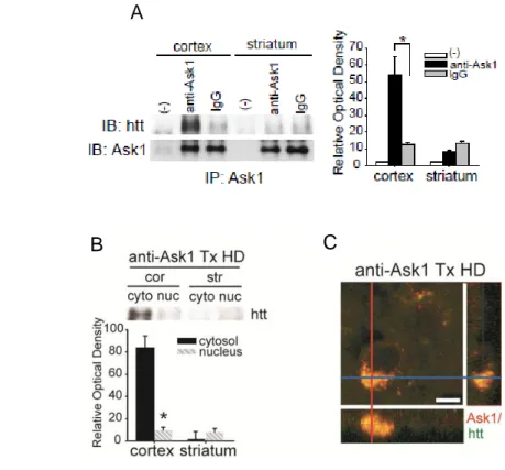
Co-immunoprecipitation and double-immunohistochemistry experiments revealed that inactivated Ask1 bound to htt fragments, and this occurred to a greater extent in the cortex than in the striatum (Figure 4; n = 4 each). The complex of two molecules was mostly shown in the cortex moreover the combined complex was detected in the cytosol than in the nucleus of the cortex. (Figure 4; n = 3 each). Double immunohistochemistry of Ask1 and htt confirmed that inactivated Ask1 and htt fragments co-localized in the cytosol of cortical neurons (Figure 4; n = 4).
15
Figure 3. The effect of inhibition of Ask1 on the levels of ER stress and htt fragments in HD. (A) Co-immunoprecipitation of Ask1 and pAsk1 (Thr 845) determined whether Ask1 was inhibited effectively in Ask1 antibody-treated mice. The amount of pAsk1 is significantly reduced in Ask1 antibody-treated HD mice. (B) RT-PCR results for CHOP show that ER stress is decreased in both the cortex and striatum of Ask1 antibody-treated HD mice. *p < 0.01
16
Figure 4. The effect of neurtralizing Ask1 on htt fragments localiziation in HD. (A) Inactivated Ask1 interacts with htt fragments. Co-immunoprecipitation of Ask1 and htt fragments in the cortex and striatum of Ask1 antibody-treated HD and IgG/preimmune antibody-treated HD mice. (B) Co-immunoprecipitation of Ask1 and htt fragments was performed in the subcellular fraction in the both of cortex and striatum. More htt immunoreactive products are present in the cytosol fraction than in the nuclear fraction of the cortex in Ask1 antibody-treated HD mice. (C) Cells double positive for Ask1 and htt are detected in the cytosol of the cortical region of Ask1 antibody-treated HD mice.
Scale bar = 5 m. (-), without antibody; cor, cortex; str, striatum; *p < 0.01
A
17
3. Inhibition of ASK1 facilitates BDNF transport to the striatum and improves motor dysfunction
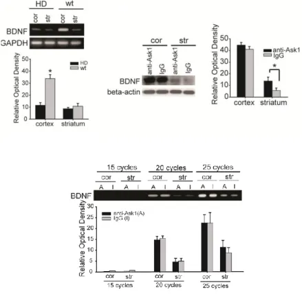
BDNF mRNA levels were shown to differ between different brain regions of HD mice, including the cortex and striatum (Figure 5; n = 5 each). There were more BDNF transcripts present in the cortex than in the striatum of wt mice, whereas BDNF mRNA levels in the cortex of HD mice were significantly reduced compared to the level of striatal BDNF mRNA in wt mice. Western blotting of BDNF revealed that inhibition of Ask1 significantly prohibit the reduction of BDNF protein levels in the striatum of HD mice (Figure 5; n = 7 each). Although levels of BDNF protein increased as a result of Ask1 inhibition, there were no significant changes in BDNF mRNA levels between Ask1-inhibited HD (anti-Ask1 treated HD, A) and vehicle-treated HD mice (rabbit IgG treated HD, I) (Figure 5; n = 5 each). Thus, the increase in BDNF protein as a result of the inhibition of Ask1 was not due to an increase in the amount of BDNF transcript.
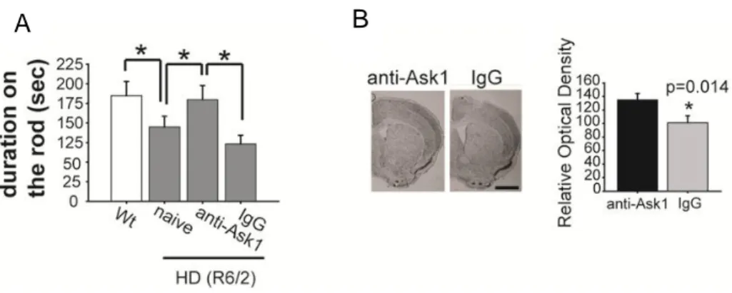
We next evaluated motor function in HD mice at 12 weeks of age, at which stage brain Ask1 had been inhibited for 4 weeks. There was significant performance impairment in HD mice at 12 weeks of age, indicated by the reduced times these mice were able to stay on the rotarod compared to wt mice (Figure 6; n = 4 each). HD mice treated with Ask1 antibody, however, showed improved motor performance compared to preimmune antibody-treated HD mice. We also assessed the changes in striatal atrophy in Ask1 antibody-treated HD mice by measuring the size of the striatal region (Figure 6, n = 3 each). The striatum was larger in the Ask1 antibody-treated HD mice than naïve HD mice or preimmune antibody-treated HD mice (p = 0.014).
18
Figure 5. The effect of inhibition of Ask1 on expression of BDNF. BDNF mRNA expression was reduced in both the cortex and striatum of HD mice, whereas BDNF mRNA is expressed at a high level in the cortex of wt mice. Western blot analysis of BDNF protein demonstrates that the level of BDNF expression in the HD mice treated with Ask1 antibody (anti-Ask1) or IgG/preimmune antibody (IgG) increased in the striatum of Ask1
antibody-treated HD mice; BDNF mRNA levels are not significantly different between Ask1 antibody-treated HD (anti-Ask1, A) and IgG/preimmune antibody-treated HD mice (IgG, I)
19
Figure 6. The effect of inhibition of Ask1 on expression of motor dysfunction. (A) Motor function was evaluated as the amount of time spent on the rotarod. HD mice were not able to remain on the rod for long periods of time, but Ask1-inhibited HD mice remained on the rotarod longer than vehicle-treated mice. There was no significant difference in motor performance between naive HD mice and vehicle-treated HD mice. cor, cortex; str, striatum;
*p < 0.01. (B) Inhibition of Ask1 prevents striatal atrophy. Representative
photographs of brain sections from Ask1 antibody-treated or IgG/preimmune antibody-treated HD mice ; Sizes of the striatal area and ventricle in Ask1 antibody-treated HD mice (anti-Ask1) and IgG/preimmune antibody-treated HD mice (IgG). Scale bar = 500 m; *p = 0.014.
20 IV. DISCUSSION
In this study, we presented two novel findings suggesting that increased Ask1 may play a critical role in HD progression in mice. First, we showed for the first time in vivo that the level of Ask1 protein is increased in HD (R6/2) mice, as is ER stress. Second, we demonstrated that inhibition of Ask1 prevents the translocation of htt fragments and improves motor dysfunction in mice, suggesting that Ask1 may be closely correlated with disease development in HD mice.
Our findings indicate that Ask1 is an element in ER stress-triggered neuronal cell death in a HD animal model (Figure 1). This finding is consistent with a study performed in a cell culture system that showed that Ask1 mediates proteasome dysfunction and ER stress-induced neuronal cell death by neuropathological alterations in polyQ disease 3. Moreover, the levels of Ask1 protein and CHOP transcripts were greater in the cortex than the striatum of HD mice (Figure 1). Our study showed that the level of Ask1 protein was increased in the cortex and striatum of HD mice (Figure 1). Based on our previous study of Ask1 14, phosphorylation of Ask1 is required for its activation, but if insufficient Ask1 protein is present, cell death may not be initiated. Normal htt is balanced with the pro-apoptotic role of the N-terminal mutant htt fragments 20. However, expansion of the htt fragments could disrupt this balance because of the sustained presence of htt fragments. In HD, loss of intact htt along with accumulation of htt fragments may move the cell towards a toxic state 21, which is known to damage corticostriatal projection neurons thereby reducing growth factors such as BDNF in the cortex. Finally, reduced BDNF transport from the cortex to the striatum may result in the death of striatal cells and behavioral abnormalities 5. In the present work, we demonstrated that intact htt was reduced to a greater extent in HD than in wt mice, and that levels of htt are
21
lower in the striatum than in the cortex of HD mice (Figure 2), suggesting that htt fragmentation may occur to a greater extent in the striatum of HD mice. Furthermore, the vast majority of htt fragments was detected in the cytoplasm (Figure 2), consistent with previous reports that cytoplasmic htt cleavage precedes nuclear uptake 20,21.
Ask1 activity, as assessed by the amount of pAsk1 (Thr845) present, was effectively inhibited in both the cortex and striatum of the Ask-1 antibody-treated group compared to the control group (Figure 3). Because Ask1 antibody was infused in the striatum, Ask1 activity was scarcely detected in the striatal fraction in contrast to the cortical fraction. When Ask1 activity was inhibited (Figure 3), CHOP induction decreased significantly. Inactivated Ask1 binding to htt fragments was mainly detected in the cytosolic fraction of cortical cells, as shown by the immunoprecipitation and double-immunohistochemistry experiments (Figure 4).Larger htt fragment do not readily enter the nucleus 22, and the translocation of htt fragments to the nucleus is associated with increased htt toxicity in vitro and neuropathology in vivo 20. Because caspases interact with htt and produce cytotoxic htt fragments, the interaction of caspases and htt amplifies caspase activation and initiates the caspase cascade in the brains of patients with HD 20,22. We speculate that Ask1 may recruit caspases thereby facilitating htt fragmentation. In future studies, we plan to investigate this hypothesis. Alternatively, Ask1 in the normal state may act as a type of chaperone and help the smaller htt fragments enter the nucleus, but when inhibited, might bind to htt fragments in the cytosol and hinder the translocation of htt fragments into the nucleus. Based on our results, we suggest that Ask1 is a key modulator of cellular signal transduction in R6/2 HD mice. Ask1 has a dual role: it is activated by various harmful stimuli and simultaneously activates or binds to other cellular molecules. We suggest that Ask1 is activated by ER stress
22
and that active Ask1 interacts with htt fragments, allowing them to translocate into the nucleus more efficiently. This interaction may amplify ER stress and may contribute to an htt toxicity feedback loop. This interaction may be one of the explanations why ER stress was reduced in Ask1 antibody-treated HD mice. We speculate that Ask1 positively regulates ER stress rather than simply respond to the ER stress signal. Under the condition of continuous ER stress, ER stress-induced neuronal cell death could be suppressed by BDNF-mediated neuroprotection 23,24. Prevention of BDNF protein reduction by inhibition of Ask1 could be inferred to suppress CHOP expression, which may supply another explanation for positive regulating role of Ask1 in CHOP.
In HD pathogenesis, the hypothesis that dysfunction of the corticostriatal pathway may contribute to the onset of HD has been supported by the observation that BDNF is expressed in cortical neurons and delivered to striatal neurons 25. BDNF co-localizes with htt in cortical neurons that project to the striatum 5. In this study, when Ask1 was inactivated in the striatum, the levels of BDNF protein in the cortex increased compared to preimmune antibody-treated HD mice, although there was no difference in the expression level of BDNF transcripts. These findings imply that inactivated Ask1 modifies the levels of BDNF protein at the post-translational level. Because cortex-specific deletion of BDNF is known to elicit progressive striatal degeneration in mice 25, BDNF reduction is expected to contribute to striatal pathogenesis in HD. Although striatal toxicity caused by htt fragments alone can not explain progressive locomotor deficits 26, our rotarod test results demonstrate that inhibition of Ask1 activity in the striatum can alleviate motor dysfunction symptoms in HD mice (Figure 6). Along with the improvement in motor dysfunction, striatal atrophy was decreased in Ask1 antibody-treated HD mice compared to that in naïve HD and IgG/preimmune antibody-treated HD mice (Figure 6).
23 V. CONCLUSION
In this study, we demonstrated that ASK1 was involved in the translocation of small htt fragments into the nucleus rather than htt cleavage. Increased levels of Ask1 may interact with htt fragments and subsequently induce ER stress. ER stress can be diminished by inhibiting Ask1, resulting in prevention of BDNF depletion in the striatum and an improvement in behavioral and anatomical abnormalities. Therefore, regulating the amount and activity of Ask1 protein are promising novel treatment strategies for HD.
24
PART II
Apoptosis signal-regulating kinase-1 aggravates ROS-mediated
striatal degeneration in 3-nitropropionic acid-infused mice
25 I. INTRODUCTION
The compound3-nitropropionic acid (3-NP) produces selective striatal lesions in animal models 27. Although not entirely elucidated, the mechanisms of neurotoxicity induced by 3-NP have been shown to include the exhaustion of adenosine triphosphate, mitochondrial membrane depolarization, dysregulation of intracellular calcium homeostasis, calpain activation, and the release of pro-apoptotic proteins from mitochondria 28-30. The neurotoxic mechanism and the reason for the selective vulnerability of the striatum are not yet well understood.
Superoxide dismutase (SOD) functions to protect cells from the effects of superoxide radicals by eliminating reactive oxygen species (ROS). Several reports have shown that SOD overexpression reduced the size of striatal lesions after 3-NP injection 31. Increased cytosolic Cu/ZnSOD is a compensatory response to the generation of ROS to reduce the toxic effects of the superoxide anion. In addition, the cumulative effects of ROS exposure cause the activation of various harmful cellular pathways 32-34.
Apoptosis signal-regulating kinase-1 (ASK1), an early signaling element in the cell death pathway 35, is activated by ROS 36 and is required for ROS-induced apoptosis 37. Previous studies have indicated that increased activated ASK1impacts the presentation of neurodegenerative diseases 10,12.The rationale for targeting ASK1 was based on findings that oxidative stress is causally related to apoptotic neuronal cell death in neurodegenerative lesions commonly associated with abnormal protein fragments and aggregates 34,38. Although many investigations have shown that ASK1 is essentially involved in neuronal cell death triggered by various external injuries and expanded polyglutamine, the relationship of ASK1 and ROS in the selective striatal cell
26
death by 3-NP chronic and systemic infusion in mice remains is to be elucidated. In addition, ASK1 was suggested to play a role in mediating the cell death signal in striatal cells of aged mice that received 3-NP injections directly into the striatum.
Here, we examined whether increased levels of ASK1 are reciprocal to the amount of ROS and also investigated the relationship between ASK1 expression and striatal degeneration in 3-NP systemic infusion mice. We suggest that overexpressed ASK1 amplifies ROS damage, which can lead to cell death, and is a deleterious primary responder to oxidative stress in the striatum of 3-NP systemically infused mice.
27 II. MATERIALS AND METHODS
1. Animal model
Male SOD1-tg mice (C57BL/6-TgN; Jackson Laboratory, USA) and wild type (wt) male littermates were used in this study. All procedures were performed in accordance with the guidelines for the care and use of laboratory animals (Yonsei University), which have been approved by the Association for Assessment and Accreditation of Laboratory Animal Care (AAALAC). The animals were anesthetized with 2.0% isoflurane under 30% oxygen and 70% nitrous oxide using a vaporizer (VMC Anesthesia Machine, MDS Matrix, USA). The rectal temperatures were maintained at 37 ± 0.5°C. For the sustained chronic administration of 3-NP infusion, 3-NP (Sigma, USA) was dissolved in saline at a concentration of 0.5 mg per µl (pH 7.4). The prepared 3-NP solution or vehicle control was delivered by sustained infusion (180~220 mg/Kg/day) using osmotic mini-pump at an infusion rate of 0.5 µl/h for 7 days (Alzet, USA).
2. ASK1gene silencing with siRNA and Administration of ASK1-peptide The ASK1 gene was silenced using siRNA against ASK1 (sense,
GCUCGUAAUUUAUACACUGtt; antisense,
CAGUGUAUAAAUUACGAGCtt; concentration 5µM; Ambion, Austin, TX, USA). After confirmation of efficiency in vitro, the transfecting reagent SiPORTNeoFX (Ambion, USA) and ASK1-siRNA or nonfunctional mutantRNA (control siRNA; 5'-AAG AGA AAA AGC GAA GAG CCA-3'; Ambion) were combined, mixed gently, and allowed to form siRNA liposomes for an additional 10 min at room temperature. An alzet micro-osmotic pump (Durect, USA) containing 100 µl of the transfection reagent or ASK1-siRNA
28
was then placed subcutaneously on the backs of the animals, and a brain infusion cannula connected to the pump was positioned in the striatum (A, 0.7 mm; L, 1.2 mm, and D, 3.3 mm) for 7 days during 3-NP infusion. The mice were sacrificed 7 days after surgery, and the brains were processed for experiments.
To restore ASK1, we synthesized an ASK1 peptide containing active sites (Thr845) from amino acids 836 to 875. The ASK1 protein was transported with the dried BioPORTER Quiktease (Sigma Aldrich) reagent to the striatum of SOD tg mice concomitant with 3-NP infusion for 7 days by a micro-osmotic pump containing 100 µl of ASK1 protein (0.1 mg/ml in saline). As a control, control-peptides were infused in the 3-NP-infused SOD-tg mouse striatum. To confirm the delivery, western blot analysis was performed with antibodies recognizing the Thr845 region.
3. Immunohistochemistry
To determine thepatterns of DARPP32 (Epitomics, USA) and ASK1 (SantaCruz Biotechnology, Cell signaling) expression, we performed immunofluorescent staining. After sacrificing the animals, brains were removed, fixed, and cut into 20µm thickness coronal sections on a cryostat. The fixed sect ions were incubated with a blocking solution, as described previously 39, a nd incubated with the appropriate primary antibodies. After washing, the sections were incubated with fluorescein isothiocyanate (FITC)-conjugated secondary antibodies (Jackson ImmunoResearch, USA). For counter-staining, the sections were incubated with propidium iodide (PI, Sigma). After washing, stained tissue samples were mounted using Vectashield mounting medium (Vector Lab, USA) and coverslipped. The sectionswere then observed under a LSM510 confocal laser scanning microscope (Carl Zeiss, USA).
29 4. Western blot analysis
The protein expression levels were analyzed by western blotting. Tissues were lysed in lysis buffer (20 mM Hepes–KOH, pH 7.5, 250 mM sucrose, 10 mM KCl, 1.5 mM MgCl2, 1 mM EDTA, 1 mM EGTA, 0.5mM PMSF,
0.1mMsodium vanadate, 0.1mM proteinase inhibitor cocktail), the samples were homogenized with a Teflon homogenizer (Wheaton, USA), and then centrifuged at 8,000 g for 20 min. The lysates were loaded onto SDS-polyacrylamide gels and after migration, the proteins were electro-transferred onto a polyvinylidenedifluoride membrane (PVDF) (Millipore, USA), which was then blocked in 5% skim milk and incubated with one of the following primary antibodies: Cu/ZnSOD, MnSOD, ASK1, pASK1. After washing, the blots were incubated with horseradish peroxidase-conjugated secondary antibodies (Roche Diagnostics), and the bands were visualized with an enhanced chemiluminescence reagent (Amersham Biosciences).
5. Apoptotic cell-death assay
Apoptosis-related DNA fragmentation assays were quantified using a commercial enzyme immunoassay kit (Chemicon) and cytoplasmic histone-associated DNA fragment kit (Roche Diagnostics). According to the manufacturers’ protocols, cytosolic samples were used in each assay. In addition, we performed a TUNEL assay to detect cell death following the manufacturer’s protocol (Roche Diagnostics). Tissue were prepared as described above, and immune-fluorescent labeling was performed as previously described 40,41. The sections
sections were counterstained with Hoechst (Molecular Probe, USA), mounted, and observed under a confocal laser scanning microscope (Carl Zeiss). The number of TUNEL-positive cells was counted in the striatum in three sections
30 from each mouse at different levels.
6. ROS detection by staining
ROS were investigated in the brains of 3-NP-infused mice using green-fluorescence (CM-H2DCFDA) in situ detection (Molecular Probes). Each
mouse was placed in a stereotaxic frame (Stoelting) under general anesthesia, the CM-H2DCFDA (2 ul, 50ug/50ul dimethylsulfoxide) was infused for 10min
into the striatum with a Hamilton syringe. The animals were sacrificed and trans-cardially perfused with heparin and formaldehyde 1h after infusion of CM-H2DCFDA. After preparing a frozen block of brain tissue, ROS was
detected to appear as green dots by CM-H2DCFDA when examined under
confocal laser scanning microscope (Carl Zeiss).The nucleus was stained with PI (Sigma).
7. ROS detection by flow cytometry analysis
CMXRosamine (MitoTracker-Red™) and CMH2-DCFDA (Molecular
Probe) were obtained from Molecular Probes, dissolved in DMSO, and stored at 20°C in the dark. Both MitoTracker (50 M) and CM-H2DCFDA (50 M) were
administered into the cortex and striatum at the position described above 30 min before sacrifice. As a control, DMSO was injected in the same locations at the same time before sacrifice in some mice. Following this procedure, the mouse brain was removed, dissected out striatum, and minced finely with a scalpel. Minced striatum tissue was placed in separate tubes containing 2 ml digestion solution, and cells were dissociated with Accumax (Millipore) for 1 h at 37°C with agitation in the dark. The cell suspension was filtered through a 100-µ m mesh (BD Biosciences) and diluted with PBS after fixing the single cells with 3.7% formaldehyde in the dark, the fluorescence of the bound dyes within each
31
cell was analyzed using a FACSCalibur (BD Biosciences; over 100,000 total events; 5,000 events / gate), and the data were analyzed using Cell Quest software (version 3.3; BD Biosciences). Negative controls were prepared by reacting the tissues with PBS, FITC-conjugated IgG, or PE-conjugated IgG (BD Biosciences).
8. Detection of DNA oxidation in mouse striatum
We evaluated DNA oxidation usinga monoclonal antibody against 8-OHdG according to a previous report 32. The brain tissue was fixed, and treated with DNase-free RNase (Roche) at 37°C for 1 h. Next, the sections were denatured in 4N HCl and neutralized with 50 mM Tris-base. After washing, the samples were incubated with an anti-mouse 8-OHdG antibody and also incubated with secondary antibody (MOM kit, Vector Laboratory, USA). The slides were mounted with Vectashield and coverslipped.
9. Rotarod test
The motor function of the animals was assessed with a rotarod apparatus (UgoBasile, USA), where the time that the animals were able to remain on the rod was measured at an accelerating speed from 4 to 40 rpm. Each mouse was pre-trained before implanting the osmotic pump filled with 3-NP or saline. After 30min rest period, and mice were then placed back on the rotarod for five trials of a maximum of 5 min at an accelerating speed.
10. Statistical analysis
Data are expressed as the mean ± SD. The statistical comparisons among multiple groups were performed using analysis of variance followed by Fisher's
32
two groups were performed using the unpaired t test (StatView, version 5.01; SAS Institute Inc., Cary, NC, USA).
33 III. RESULTS
1. Systemic infusion of 3-NP led to the formation of selective striatal lesions in wt, but SOD-tg mice
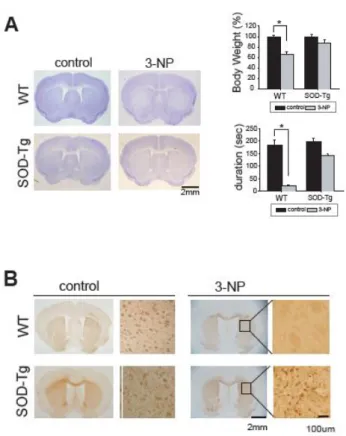
Mice infused with 3-NP were examined for symptoms, and the degenerated brain region was validated with cresyl violet staining. Primarily, we confirmed an obvious decrease in body weight by 7 days after systemic3-NP reservoir implantation. The mouse body weight reduced an average of 70% of the initial body weight. Seven days after 3-NP administration, most mice presented with severe motor dysfunction in addition to weight loss. Histological evaluation of the 3-NP-infused mice at day 7 demonstrated striatal abnormalities and cell loss (Figure 1A).
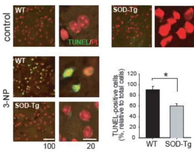
DARPP32 (dopamine- and cyclic adenosine monophosphate-regulated phosphoprotein), an established of striatal neuron marker, showed weak immunoreactivity in 3-NP-infused wt mice; however, the immunoreactivity in SOD- tg mice was significantly more dense (Figure 1B). The induction of DNA damage in striatal neurons was evaluated by terminal TUNEL staining (Figure 2). The average number of TUNEL-positive nuclei was determined using multiple images in the four different mice of each group. Figure 2 reveals a statistically significant increase in TUNEL-positive nuclei in the 3-NP-infused wt mice, which was scarcely demonstrated by the DARPP32-positive cells.
34
Figure 1. Pathological examination in the striatum after systemic infusion of 3-NP. (A) Histological analysis was performed by cresyl violet staining (left). Body weight which measured on day 7 after infusion of 3-NP is expressed as a percentage of the weight before 3-NP infusion (right upper). Motor function is shown the duration sustained on the rotarod (right lower). (B) DARPP32-postive cells shown by immunohistochemistry was sparsely detected in the wt mice, while positive cells were more plentiful in the SOD-tg.
35
Figure 2. Apoptotic cell death in wt and SOD-tg mice. TUNEL-positive cells were more densely detected in the 3-NP-infused wt mice compared to the SOD-tg mice (left). A quantification graph is presented showing relative values of positive cells to counterstained cells in the unit area (right). *p<0.05.
36
2. Greater ROS production, oxidative damage and ASK1 levels and activity were detected in striatal lesions
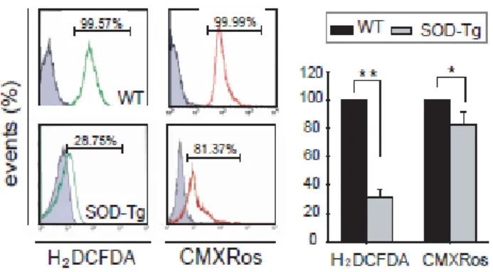
Using FACS analysis to quantify ROS, the total amount of ROS in the striatum significantly decreased from 9.57% in wt to 28.75% of SOD-tg mice (Figure 3). In contrast, there was a slight difference in the ROS quantity in mitochondria that were directly injured after 3-NP infusion between wt and SOD-tg mice (Figure 3), as revealed by FACS (wt, 99.99%; SOD-tg, 81.37%).
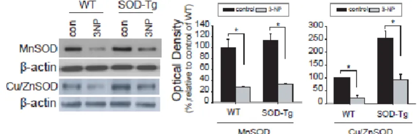
As an alternative way to compare the changes in the amount of ROS, levels of MnSOD, and Cu/ZnSOD proteins, were determined by western blot analysis in the lesioned striatum from each brain. MnSOD protein amounts were not different in the damaged striatum between wt and SOD-tg, whereas relatively higher levels of Cu/ZnSOD were detected in SOD-tg than wt (Figure 4). Unaltered pattern of MnSOD likely dues the direct attack of the mitochondrial dysfunction reagent, 3-NP. Cytosolic SOD levels, however, were greater in the SOD-tg than in the wt mice because of the restoring effect of SOD overexpression in the SOD tg mice.
Representing DNA oxidation, 8-OHdG-positive cells were scarcely detected in the 3-NP-infused SOD-tg striatum; however, the immune-reactivity was strong in striatum of wt mice (Figure 5), suggesting that the ROS was produced following mitochondrial dysfunction and subsequent DNA damage (Figure 6). Under oxidative conditions, increased ASK1 protein levels, one of the first ROS-responsive molecules, were measured, and also measured increased pASK1 activated by phosphorylation at Thr845. The 3-NP infusion elevated total ASK1 protein amounts the SOD-tg and wt mice however, this increase was meager in the SOD-tg group. Corresponding to total ASK1 amounts, the levels of pASK1 were also significantly lower in SOD-tg mice (Figure 2D).
37
Figure 3. Environmental changes and ROS level changes in the striatum after 3-NP infusion. After 3-NP infusion, CM-H2DCFDA-for evaluating total
ROS levels, and MitoTracker-Red for mitochondrial ROS levels were assayed by flow cytometry (left), and the events are presented in a quantified graph (right).
38
Figure 4. The alteration of ROS scavenging enzyme protein level in the striatum of wt and SOD tg mice after 3-NP infusion. The MnSOD and Cu/ZnSOD protein levels in the striatum are shown in western blot analysis (left) and the quantitative values are presented in the graphs (right).
39
Figure 5. Oxidative DNA damage in the striatum of wt and SOD tg mice after 3-NP infusion. The 8-OHdG illustrates that the 8-OHdG-positive cells were more densely detected in the wt mice compared to the SOD-tg mice. A quantified graph is presented showing relative values of positive cells to counterstained cells in the unit area.
40
Figure 6. The changes of ASK1 protein and activity. The changes of ASK1 and pASK1expression levels were assayed by western blot (left) and presented the quantified graph (right). *p<0.05.
41
3. ASK1 amounts mediated the striatal cell death without change of ROS level ASK1 gene silencing by siRNA sufficiently regulated ASK1 protein down (Figure 7). However, ASK1 silencing did not alter the amounts of ROS by 3-NP infusion. Indeed, ROS levels were similar or abundant both in the si-ASK1-treated and the si-control-treated wt mice. ROS detected with CM-H2DCFDA were scant in 3-NP-infused SOD-tg mice, while the
CM-H2DCFDA in normal and ASK1-silenced 3NP infusion mice abundantly
stained the striatal region with a punctuate shape (Figure 7). The results showed that the down-regulation of ASK1 did not affect ROS generation. Although the down-regulation of ASK1 did not affect ROS scavenging, it was sufficient to reduce cell death and improve the motor function (Figure 9). On the other hand, ASK1-peptide administration triggered cell death without increasing ROS levels.
To confirm whether overexpression of ASK1 plays a role in mediating cell death, synthesized ASK-peptide was infused for 7 days in the SOD-tg mice striatum starting with 3-NP systemic infusion (Figure 8). In 3-NP-infused SOD-tg mice, the ROS levels were assessed in the control-peptide and ASK1-peptide treated groups by FACS (Figure 8). ASK1-peptide did not alter the amount of ROS generated. A DNA fragmentation assay showed that striatal apoptotic cell death occurred in the 3-NP-infused wt mice and decreased significantly in both SOD-tg mice and siASK1-treated wt mice with 3-NP infusion. In contrast, the DNA fragmentation recurred in the ASK1-peptide-treated SOD tg mice and also aggravate movement ability (Figure 9).
42
Figure 7. Down regulation and induction of ASK1. In the striatum, ASK1 proteins were declined by siRNA-ASK1 (left), and quantified graph (right). CM-H2DCFDA in the striatum was detected in abundance in the si-control- or
si-ASK1-treated wt mice, but was decreased in the 3-NP-infused SOD-tg mice. H2DCFDA, green; PI, red.
43
Figure 8. The effect of ASK1-peptide on ROS production level. Western blot analysis shows the expression levels of ASK1 in the striatum of wt or ASK1-peptide-added SOD-tg mice with 3-NP infusion. After 3-NP infusion, total ROS levels was evaluated with CM-H2DCFDA in each group (con-pep vs
44
Figure 9. The role of ASK1 in cell death and behavioral impairment. Apoptotic cell death using DNA fragmentation assay was found in each treated mouse striatum. Motor function is shown as the duration sustained on the rotarod in each treated group. *p<0.05.
45 IV. DISCUSSION
The results of the present work show an improvement in behavioral impairment by ASK1 down-regulation, despite 3-NP infusion. We propose the hypothesis that ASK1 overexpression by systemic infusion of 3-NP promotes the formation of selective striatal lesions, and this occurs apart from ROS generation. The results of our study showed that increased ASK1andpASK1expression by harmful ROS signals augmented neuronal cell death in the striatum.
Mitochondria are key players in the production of ROS and have been reported to have an important role in 3-NP injury-induced pathology 42. We evaluated amounts of total ROS produced in mitochondria and cytosol MitoTracker-Red CMH2XRos (MT Red CM-H2XRos) is a molecule that has
been used to measure mitochondrial free radicals (MFRs) in various cell cultures. Because this dye has been reported to be sequestered into the nucleus and vesicles 43,44, we administered it to detect the generation of MFRs in 3-NP-infused mice and to measure MFR generation in situ. Besides of mitochondrial dysfunction, 3-NP also raises cellular oxidative stress. The alteration of Cu/Zn-SOD is a compensatory mechanism that protects cells from free radical-induced damage, and systemic infusion of 3-NP altered Cu/Zn-SOD enzyme levels in this study. It has been previously reported that 3-NP induced major changes in SOD activity in the striatum of 3-NP-injected rats 45. Our study displayed that 3-NP caused a reduction in Cu/Zn-SOD, but had no effect on MnSOD protein levels. It can be inferred that the overexpression of Cu/ZnSOD is primarily responsible for cellular anti-oxidant protection in 3-NP-infused SOD-tg mice, possibly by compensating for the overexpression of ROS. In wt mice, however, the endogenous antioxidant system could not enough to eliminate the increased ROS, as Cu/ZnSOD amounts were
46
diminished. The lesions of 3-NP indicate that striatal oxidative damage has occurred, and that this damage is associated with changes in the cellular antioxidant system 46. The Cu/ZnSOD levels were much greater in the SOD-tg mice compared to the wt mice, likely to overcome the oxidative damage. However, the changes in SOD protein levels during oxidative stress do not determine whether protect or not, although it is certain that overexpressed SOD amounts protect against harmful oxidative environments 33.
Oxidative DNA damage results from direct ROS attacks. There are some types of oxidative DNA damage detective method. Among them, 8-OHdG results in oxidative DNA damage. The results presented here show that the oxidative DNA lesions are markedly augmented in the 3-NP-infused wt mouse brain.
Once ROS are generated, they can damage mitochondria causing additional free-radical generation and loss of antioxidant capacity, leading to a deleterious cycle 4. Therapeutic prevention of oxidative stress has been proposed to “break the cycle” of cell death 4, and studies have been carried out in the neurodegenerative field attempting to modulate key enzymatic components that regulate oxidative stress 33. Our study was performed on the assumption that mitochondrial complex II inhibitors generate ROS, and contribute to cell death. Furthermore, because ASK1is known to be involved in ROS-induced cell death, we propose that ASK1plays an instrumental role and even amplifies this process. The ASK1 signaling-mediated cell fate decision appears to depend in part on the extent and duration of ASK1 amounts and its activation. A previous report found that a mutated SOD induced ER stress and activated the ASK1-mediated cell death pathway 13. Also, the deletion of ASK1 mitigated neuronal loss and extended the life span of SOD1-mutated mice 13.
47
causative factor in the initiation and progression of Alzheimer’s disease (AD) and Parkinson’s disease, where antioxidants have the capacity to attenuate the phenotypes associated with these neurodegenerative disorders. Although antioxidants can prevent the oxidative stress-mediated progression of neurodegenerative diseases 47, antioxidant treatment is not sufficient to halt disease progression. We implied that inhibition of ASK1, the major downstream activator, may disrupt the positive feedback Therefore we propose that mitochondrial dysfunction by 3-NP induces an increase in ROS level; ROS-activated ASK1 mediates harmful oxidative signals; and cells eventually undergo apoptosis. The precise mechanisms have not yet been elucidated and have to be investigated.
48 V. CONCLUSION
This study is summarized as followings: (1) systemic infusion of 3-NP increased ROS and led striatal cell loss; (2) ASK1 down-regulation decreased striatal cell death without scavenging ROS; (3) adding ASK1protein to SOD tg aggravated striatal degeneration; and (4) ASK1 activation induced by ROS is an important step of the 3-NP pathophysiology. Conclusively, this study indicates that ROS-induced ASK1 is an important step in the pathogenesis of 3-NP-mediated striatal lesion, and suggests that ASK1 acts as an amplifier of the ROS signal cascade. Taken together, we suggest a combination of ASK1 inhibition and ROS elimination for more effective therapy.
49
PART III
Apoptosis signal-regulating kinase 1 mediates 3-nitropropionic acid
toxicity and regulates C1q level via astrocyte TGF-beta
50 I. INTRODUCTION
The systemic administration of 3-nitropropionic acid (3-NP) facilitates the development of selected striatal lesions and it remains unclear whether specific neurons are selectively targeted in 3-NP infused striatal degeneration. It is well known that 3-NP inhibits succinate dehydrogenase (SDH), mitochondrial complex II, to the same extent in both the cerebrum and the striatum 4,25, suggesting that 3-NP neurotoxicity occurs to the same extent in the cerebrum and the striatum. Although not entirely elucidated, the mechanisms of neurotoxicity induced by 3-NP have been shown to include the exhaustion of adenosine triphosphate, mitochondrial membrane depolarization, dysregulation of intracellular calcium homeostasis, calpain activation, and the release of pro-apoptotic proteins from mitochondria 28,30,48. The neurotoxic mechanism and the reason for the selective vulnerability of the striatum are not yet well understood. 3-NP has been used a pharmacologic model to study mitochondrial dysfunction in relation to Huntington’s disease (HD). However, speculation of selective regional neurotoxicity induced by 3-NP in different areas may not be accurate, and the molecular mechanisms underlining 3-NP-induced striatal lesion remain to be fully defined 49.
Mitochondrial dysfunction can bring energy metabolism defects, oxidative stress, and defects in mitochondrial calcium usage. A number of studies have examined whether alterations in mitochondrial respiration contribute to the observed bioenergetic defects. Biochemical studies of brain and peripheral tissues from HD patients, as well as studies on HD cells and animal models, revealed decreased activity of several enzymes involved in oxidative phosphorylation such as complex I, II, III, and IV 23,27,50-52. Mitochondria are both a target and an important source of reactive oxygen