Notes Bull. Korean Chem. Soc. 2014, Vol. 35, No. 12 3647 http://dx.doi.org/10.5012/bkcs.2014.35.12.3647
Synthesis and Biological Evaluation of Kojic acid Derivatives as Tyrosinase Inhibitors
Yeong Jin Choi, Ho Sik Rho,†,* Heung Soo Baek,† Yong Deog Hong,† Yung Hyup Joo,† Song Seok Shin,† and Ji Man Kim*
Functional Material Laboratory, Department of Chemistry, Sungkyunkwan University, Gyeonggi-do 440-746, Korea *E-mail: jimankim@skku.edu
†R & D Center, AmorePacific Corporation, Yongin 446-729, Korea. *E-mail: thiocarbon@hanmail.net Received June 25, 2014, Accepted August 12, 2014
Key Words : Kojic acid, Ester, Thioether, Tyrosinase, Depigmenting activity
Tyrosinase1 is a copper-containing enzyme which is wide-ly distributed in nature. This enzyme is associated with the production of melanin for the protection of the skin from solar irradiation. However, overproduction of melanin in the skin is of particular concern to woman, for example, in the conditions melasma and lentigo.2 Tyrosinase catalyzes the first two steps of melanin biosynthesis, namely the hydrox-ylation of L-tyrosine to L-dopa and the subsequent oxidation of L-dopa to dopaquinone. Tyrosinase is contained in veget-ables and fruits and is responsible for the undesirable enzy-matic browning that occurs upon long term storage of these.3 Therefore, tyrosinase inhibitors have attracted attention as important depigmenting agents for the treatment of hyper-pigmentation and as anti-browning agents in vegetables and fruits. Kojic acid4 (1) is a well known tyrosinase inhibitor. However, its inhibitory activity is not potent enough for the above purposes. To overcome this drawback, many semi-synthetic kojic acid derivatives have been synthesized, usually by modification of the C-2 hydroxyl group.5 Recent-ly, we synthesized kojic acid derivatives containing thioether6 and ester7 linkers and evaluated their tyrosinase inhibitory activities. Among them, 2-((cyclohexylthio)methyl)-5-hydr-oxy-4H-pyran-4-one (2b), showed the most potent inhibitory activity. Compound 2b is composed of three parts, including the kojic acid moiety, a thioether linker, and a hydrophobic cyclohexane group. We synthesized and evaluated additional compounds to get more structural insight into the tyrosinase inhibitory activity of the kojic acid derivatives. The crystallo-graphic structure of tyrosinase has recently been established, enabling closer examination of its three-dimensional structure, which revealed the presence of a hydrophobic protein pocket adjoining the binuclear copper active site.8 We also conduct-ed a molecular docking study to elucidate the structural importance of the kojic acid derivatives in binding with the
active site of tyrosinase.
The synthetic pathways of the kojic acid derivatives are shown in Scheme 1. Thioether derivatives (2a-2e) were syn-thesized by the condensation of kojyl chloride4 with the potassium salts of thiols. Ester derivatives (3a-3e) were syn-thesized by the condensation of kojyl chloride4 with the potassium salts of acids.
The inhibitory activities of the kojic acid derivatives on mushroom tyrosinase were investigated using kojic acid as a positive control. The results are summarized in Table 1.
Figure 1. Structures of the kojic acid derivatives.
Scheme 1. Reaction conditions: (a) thionyl chloride (1.5 equiv.), dimethylformamide, room temperature, 0.5 h, 72% yield; (b) the potassium salts of thiols (1.0 equiv.), dimethylformamide, 110 o C-120 oC, 1 h, 65%-81% yields; (c) the potassium salts of acids (1.0 equiv.), dimethylformamide, 110 oC-120 oC, 1 h, 70%-84% yields.
3648 Bull. Korean Chem. Soc. 2014, Vol. 35, No. 12 Notes
Kojic acid (1) inhibited mushroom tyrosinase with an IC50 value of 50.07 μM. Dramatically enhanced activities were observed with the thioether compounds (2a-2e). Compound 2a, which contains a hexyl group, showed a potent inhibitory activity (IC50= 0.87μM). Compound 2b, 2-((cyclohexyl-thio)methyl)-5-hydroxy-4H-pyran-4-one, showed the most potent inhibitory activity (IC50 = 0.25 μM). However, com-pounds with a greater hydrophobic character, for example, those with the adamantine or planar aromatic groups such as phenyl and naphthyl, had a negative influence on inhibitory activity (2c, 2d, and 2e). The activities of compounds con-taining an ester linker were lower than those of the thioether compounds. Compound 3b, (5-hydroxy-4-oxo-4H-pyran-2-yl)methyl cyclohexanecarboxylate, exhibited decreased activity (IC50 = 2.74 μM). The ester derivatives (3a-3e) were found to show a similar activity trend to the thioether derivatives. The adamantanyl compound (3c) and phenyl compound (3d) showed decreased inhibitory activities (IC50 = 3.26 μM and 5.90 μM, respectively). In the naththyl compound 3e, a greater decrease in activity was observed (IC50 = 21.01 μM). In the tyrosinase assay, the thioether linkage, and flexible normal alkyl and cycloalkyl groups were critical factors for the inhibitory activity.
To understand the binding mode of the kojic acid deriva-tives in the active site of tyrosinase, we conducted a model-ing study. The results are summarized in Table 2.
The docking results agreed with the observed in vitro data, which showed that compound 2b had the most potent tyrosinase inhibitory activity (IC50 = 0.25 μM), exhibiting a
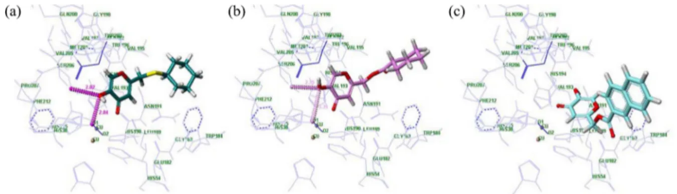
higher docked score (5.51); whereas compound 3e, showed decreased tyrosinase inhibitory activity (IC50 = 21.01 μM) and exhibited a lower docked score (3.98). The selected docked conformations of compounds 2b, 3b, and 3e in the tyrosinase binding site are shown in Figure 2.
The major factors for a tight binding mode are an H-bonding interaction between the oxygen atoms of the ligands and a π-π interaction with His 194 in the active site. Com-pound 2b had a stronger interaction with peroxide, with a hydrogen bond distance of 2.80 Å, than that of compound 3b (3.34 Å). Moreover, compound 2b also formed H-bond-ing interactions with residue Ser 206 in the bindH-bond-ing pocket. In addition, a π-π interaction between compound 2b and His 194 was found, and the position of the pyranone ring of kojic acid was nearly identical to that of superimposed L -tyrosin-ase (Fig. 2(a)). However, compound 3e (3.98) exhibited a different binding pattern due to the naphthyl moiety. The planer and bulky naphthyl group caused a steric hindrance that changed its binding to the active site of tyrosinase (Fig. 2(c)). For this reason, ligand 3e also lost important actions, such as the H-bond with peroxide and π-π inter-action with His 194 in the active site.
After evaluating the tyrosinase inhibitory activity, we selected compound 2b as a candidate for an effective depig-menting agent and evaluated its inhibitory potency against melanin formation. The results are summarized in Table 3.
In this study, B16/F1 melanoma cells were used without α-melanocyte stimulating hormone treatment as a stimulant of melanogenesis. We treated B16/F1 melanoma cells with compound 2b at various concentrations (2.5 μM-20 μM). A
Table 1. Inhibitory activities of the kojic acid derivatives (2a-2e) and (3a-3e) on mushroom tyrosinase
Compounds IC50a Compounds IC50a 2a 0.87 µM 3a 2.55 µM 2b 0.25 µM 3b 2.74 µM 2c 1.39 µM 3c 3.26 µM 2d 1.59 µM 3d 5.90 µM 2e 1.10 µM 3e 21.01 µM Kojic acid 50.07 µM
aValues were determined from the logarithmic concentration-inhibition
curves and are given as the mean values of the results of three experi-ments.
Table 2. Docked scores of the kojic acid derivatives (2a-2e) and (3a-3e)
Compounds Docked scorea Compounds Docked scorea
2a 5.43 3a 4.45 2b 5.51 3b 4.50 2c 5.18 3c 4.39 2d 4.89 3d 3.84 2e 5.22 3e 3.98 Kojic acid 3.69
aMolecular modeling study was carried out using SYBYL-X version 1.2
Figure 2. Docked conformation of compounds 2b, 3b, and 3e in the binding site of tyrosinase: (a) compound 2b, (b) compound 3b, (c) compound 3e.
Notes Bull. Korean Chem. Soc. 2014, Vol. 35, No. 12 3649
slight decrease in cell viability was observed at 20 μM concentration. After confirming cell viability, we evaluated the depigmenting activity of compound 2b at the same con-centrations (2.5 μM-20 μM). Lower melanin content (%) equated to a higher inhibition of melanin synthesis. Compound 2b significantly inhibited melanogenesis in a dose-depen-dent manner. At concentrations of 5μM and 10 μM, the melanin content of compound 2b was 74.41% and 48.38%, respectively. However, kojic acid (1) inhibited melanin production at higher concentrations (0.25 mM-2 mM). Thus, compound 2b, containing a thioether linkage and hydro-phobic cyclohexane group, is more effective than kojic acid in inhibiting melanin production.
In conclusion, we synthesized a series of kojic acid thio-ether derivatives (2a-2e) and kojic acid ester derivatives (3a-3e). The tyrosinase inhibitory activities for the ten synthesiz-ed compounds were evaluatsynthesiz-ed to investigate the structure-activity relationships (SAR). The molecular docking results closely agreed with the observed in vitro data and clearly explained the SAR pattern. Among them, 5-hydroxy-2-((cyclo-hexylthio)methyl)-4H-pyran-4-one (2b) exhibited the most potent tyrosinase inhibitory activity (IC50= 0.25 μM). In the cell-based assay, compound 2b decreased the melanin con-tent in a dose-dependent manner without cytotoxicity. Overall, these results suggest that compound 2b could be used as an effective depigmenting agent.
Experimental Section
5-Hydroxy-2-((phenylthio)methyl)-4H-pyran-4-one (2d). To a stirred solution of kojyl chloride 4 (4.80 g, 30.0 mmol) in DMF (100 mL) under N2 was added potassium benzenethiolate (4.43 g, 30.0 mmol). The reaction mixture was stirred for 1 h at 110oC-120oC, after which DMF was evaporated in vacuo. The residue was extracted with ethyl acetate (500 mL), washed with water. The organic layer was dried with anhydrous MgSO4 and concentrated to give a crude product. The resultant was purified by crystallization from ethyl acetate-hexane to give 2d (5.18 g) in 74% yields.
1H NMR (300 MHz, DMSO-d 6) δ 9.07 (s,1H), 7.97 (s, 1H), 7.26-7.35 (m, 5H), 6.21 (s, 1H), 4.10 (s, 2H). 13C-NMR (125 MHz, DMSO-d6) δ 174.0, 164.2, 146.1, 140.26, 140.21, 134.3, 130.2, 129.6, 127.4, 113.1, 112.9, 34.8. FABMS: (m/e) 235 [M+H]+.
(5-Hydroxy-4-oxo-4H-pyran-2-yl)methyl benzoate (3d). To a stirred solution of kojyl chloride 4 (4.80 g, 30.0 mmol) in DMF (100 mL) under N2 was added potassium benzoate (4.79 g, 30.0 mmol) with benzoic acid (2.74 g, 22.5 mmol). The reaction mixture was stirred for 1 h at 110oC-120oC, after which DMF was evaporated in vacuo. The residue was extracted with ethyl acetate (500 mL), washed with water. The organic layer was dried with anhydrous MgSO4 and concentrated to give a crude product. The resultant was purified by crystallization from ethyl acetate-hexane to give 3d (5.00 g) in 68 % yields. 1H NMR (300 MHz, DMSO-d 6) δ 9.29 (s, 1H), 8.11 (s, 1H), 8.02 (m, 2H), 7.71 (m, 1H), 7.58 (m, 2H), 6.55 (s, 1H), 5.22 (s, 2H). 13C-NMR (125 MHz, DMSO-d6) δ 173.5, 164.9, 161.4, 146.0, 139.9, 133.7, 129.3, 128.8, 128.7, 112.6, 61.9. FABMS, m/e 245.1 [M-H]+.
Mushroom Tyrosinase Assay. Mushroom tyrosinase and
L-tyrosine were purchased from Sigma Chemical (Saint Louis, Missouri, USA). The reaction mixture for mushroom tyrosinase activity consisted of 150 μL of 0.1 M phosphate buffer (pH 6.5), 3 μL of sample solution, 8 μL of mushroom tyrosinase (2,100 unit/mL, 0.05 M phosphate buffer at pH 6.5), and 36 μL of 1.5 mM L-tyrosine. Tyrosinase activity was determined by reading the optical density at 490 nm using a microplate reader (Bio-Rad 3550, Richmond, CA, USA) after incubation for 20 min at 37 oC. The inhibitory activity of each sample is expressed as the concentration that inhibits enzyme activity by 50% (IC50).
Molecular Modeling. Molecular modeling study was carried out on linux system using SYBYL-X version 1.2. To prepare the tyrosinase structure, the crystal structure of the oxy form of S. castaneoglobisporus tyrosinase was taken from the Protein Data Bank (PDB cod 1 wx2) because there is no crystal structure of mushroom tyrosinase published yet. The caddie protein and water molecules were removed. Hydro-gen atoms were added to the enzyme using the SYBYL. For the molecular docking method, Surflex-Dock version 2.5 was used using standard parameters and allowing the hydro-gen of protein movement.
Cell Culture. The B16/F1 melanoma cells line was obtain-ed from the Korean cell Line Bank (Seoul, Korea). Cells were cultured in Dulbecco’s Modified Eagle medium (DMEM) containing Fetal Bovine Serum (FBS, 10%), penicillin (100 U/mL), and streptomycin (0.1 mg/mL) at 37 oC in a humidi-fied atmosphere of 5% CO2.
Measurements of Cell Viability. Cell viability was mea-sured using the (3-(4,5-dimethylthiazol-2-yl)-2,5-diphenyl-tetrazolium bromide (MTT) method. Cells were plated in 96-well plates and cultured for 24 h. After treatment with kojic acid and kojic acid derivatives (2a-2e) and (3a-3e), 100 μL MTT (5 mg/mL in PBS) was added to each well. Cells were incubated at 37oC for 30 min, dimethyl sulfoxide (DMSO) was added to dissolve the formazan crystals, and the absorbance was measured at 560 nm using a microplate
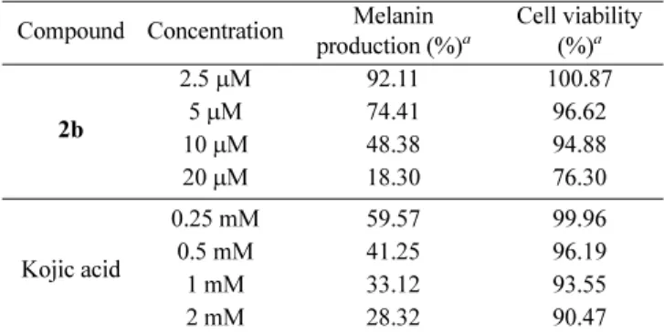
Table 3. Depigmenting activities of compound 2b and kojic acid Compound Concentration Melanin
production (%)a Cell viability (%)a 2b 2.5 µM 92.11 100.87 5 µM 74.41 96.62 10 µM 48.38 94.88 20 µM 18.30 76.30 Kojic acid 0.25 mM 59.57 99.96 0.5 mM 41.25 96.19 1 mM 33.12 93.55 2 mM 28.32 90.47
aValues were determined from the logarithmic concentration-inhibition
curves and are given as the mean values of the results of three experi-ments.
3650 Bull. Korean Chem. Soc. 2014, Vol. 35, No. 12 Notes
reader a (Molecular Devices Co., Sunnyvale, CA, USA). Measurements of Melanin Content. Cells (2 × 10⁴ cells/ mL) were seeded into 24-well plates and dicinnamoylamide derivatives were added in triplicate. The medium was changed daily and after 4 d of culture, the cells were lysed with 0.1 mL of 1 N NaOH. Then 100 μL of each crude cell extract was transferred to a 96-well plate. Relative melanin content was measured at 400 nm with a microplate reader (Molecular Devices).
References
1. (a) Sanchez-Ferrer, A.; Rodriguez-Lopez, J. N.; Garcia-Canavas, F.; Garcia-Carmona, F. Biochim. Biophys. Acta 1995, 1247, 1. (b) Khatib, S.; Nerya, O.; Musa, R.; Shmuel, M.; Tamir, S.; Vaya, J. Bioorg. Med. Chem. 2005, 13, 433.
2. (a) Ando, H.; Kondoh, H.; Ichihashi, M.; Hearing, V. J. J. Invest. Dermatol. 2007, 127, 751. (b) Wang, H. M.; Chen, C. Y.; Wen, Z. H. Exp. Dermatol. 2010, 20, 242.
3. (a) Chen, J. S.; Wei, C. I.; Rolle, R. S.; Otwell, W. S.; Balaban, M. O.; Marshall, M. R. J. Agric. Food. Chem. 1991, 39, 1396. (b) Martinez, M. V.; Whitaker, J. R. Trends Food Sci. Tech. 1995, 6, 195.
4. (a) Ohyama, Y.; Mishima, Y. Fragrance J. 1990, 6, 53. (b) Battaini, G.; Monzani, E.; Casella, L.; Santagostini. L.; Pagliarin, R. J. Biol. Inorg. Chem. 2000, 5, 262.
5. (a) Manosroi, A.; Wongtrakul, P.; Manosroi, J.; Midorikawa, U.;
Hanyu, Y.; Yuasa, M.; Sugawara, F.; Sakai, H.; Abe, M. Int. J. Pharm. 2005, 298, 13. (b) Kodokawa, J.; Nishikura, T.; Muraoka, R.; Tagaya, H.; Fukuoka, N. Synth. Commun. 2003, 33, 1081. (c) Kwak, S.-Y.; Noh, J.-M.; Park, S.-H.; Byun, J.-W.; Choi, H.-R.; Park, K.-C.; Lee, Y.-S. Bioorg. Med. Chem. Lett. 2010, 20, 738. (d) Rho, H. S.; Baek, H. S.; You, J. W.; Kim, S.; Lee, J. Y.; Kim, D. H.; Chang, I. S. Bull. Korean Chem. Soc. 2007, 28, 471. (e) Rho, H. S.; Baek, H. S.; Ahn, S. M.; Kim, D. H.; Chang, I. S. Bull. Korean Chem. Soc. 2008, 29, 1569.
6. (a) Rho, H. S.; Ahn, S. M.; Yoo, D. S.; Kim, M. K.; Cho, D. H.; Cho, J. Y. Bioorg. Med. Chem. Lett. 2010, 20, 6569. (b) Rho, H. S.; Baek, H. S.; Ahn, S. M.; Kim, M. K.; Ghimeray, A. K.; Cho, D. H.; Hwang, J. S. Bull. Korean Chem. Soc. 2010, 31, 2375. (c) Rho, H. S.; Yoo, D. S.; Ahn, S. M.; Kim, M. K.; Cho, D. H.; Cho, J. Y. Bull. Korean Chem. Soc. 2010, 31, 3463.
7. (a) Ahn, S. M.; Rho, H. S.; Beak, H. S.; Joo, Y. H.; Hong, Y. D.; Shin, S. S.; Park, Y.-H.; Park, S. N. Bioorg. Med. Chem. Lett. 2011, 21, 7466. (b) Rho, H. S.; Lee, C. S.; Ahn, S. M.; Hong, Y. D.; Shin, S. S.; Park, Y.-H.; Park, S. N. Bull. Korean Chem. Soc. 2011, 32, 4411. (c) Rho, H. S.; Goh, M.; Lee, J.; Ahn, S. M.; Yeon, J.; Yoo, D. S.; Kim, D. H.; Kim, H. G.; Cho, J. Y. Bull. Korean Chem. Soc. 2011, 32, 1411. (d) Cho, C.-C., Rho, H. S.; Baek, H. S.; Ahn, S. M.; Woo, B. Y.; Hong, Y. D.; Cheon, J. W.; Heo, J. M.; Shin, S. S.; Park, Y.-H.; Suh, K. D. Bioorg. Med. Chem. Lett. 2012, 22, 2004. (e) Cho, C.-C.; Rho, H. S.; Joo, Y. H.; Lee, C. S.; Lee, J.; Ahn, S. M.; Kim, J. E.; Shin, S. S.; Park, Y.-H.; Suh, K. D.; Park, S. N. Bioorg. Med. Chem. Lett. 2012, 22, 4159. 8. Nithitanakool, S.; Pithayanukul, P.; Bavovada, R.; Saparpakron, P.