40
-Vol. 13, No. 1(June), 2016 The Journal of Medicine and Life Science
Recent studies demonstrate that FDG PET/CT can find active infectious or inflammatory lesions. Acute sialadenitis is a usual complication due to radioiodine therapy, but late-onset sialadenitis is a rare condition. We report an uncommon case of FDG PET/CT detecting late-onset sialadenitis after radioiodine therapy.
A 45 year-old woman underwent total thyroidectomy due to papillary thyroid cancer in 2011. High-dose radioiodine ablation of 3.7 GBq (100 mCi) was performed 2 months after the operation. During hospitalization the patient had not complained of symptoms, suggesting an acute complication due to the therapy. For 3 years after discharge,
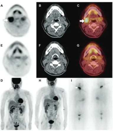
she had routine follow-up tests that included physical examination, serum thyroid function tests, ultrasonography and F-18-fluoro-2-deoxyglucose positron emission tomography/computed tomography (18F-FDGPET/CT), and the tests results were found to be normal. Thereafter she complained of a mass-like lesion that grew around the right submandibular area. Sialadenitis was suspected upon physical examination, and a planned 18F-FDGPET/CT, as routine follow-up, was carried out after 6 days. The images revealed a diffuse and intense 18F-FDG uptake in right submandibular area on the transaxial and on the maximum intensity projection views(Fig. 1A-D, empty arrows; maximum standardized uptake value, 6.8). The present authors retrospectively reviewed imagery that had been captured 19 months prior, and found that those images contained a faint physiologic 18F-FDG uptake in both submandibular glands(Fig. 1E-H). The 131I scintigraphy taken on the second day after radioiodine therapy of 3.7 GBq was reviewed additionally. The images showed a focal 131I uptake in the thyroid bed area, suggesting the presence of a remnant of thyroid tissue. The other focal 131I uptake in the
방사성요오드 치료 후 지연 발생된 침샘염의
18F-FDG PET/CT 소견
이진호
1, 고관표
2, 송희성
31제주대학교 의학전문대학원, 2제주대학교 의학전문대학원 내과학교실
3제주대학교 의학전문대학원 핵의학교실
(Received May 16, 2016; Revised May 23, 2016; Accepted May 30, 2016)
18
F-FDG PET/CT Detects Late-onset Sialadenitis After Radioiodine Ablation Therapy
Jin Ho Lee
1, Gwanpyo Koh
2, Heesung Song
31Jeju National University School of Medicine, 2Department of Internal Medicine and 3Nuclear Medicine, Jeju National University School of Medicine, Jeju, Republic of Korea
Sialadenitis is a common complication arising from radioactive iodine therapy. F-18-fluoro-2-deoxyglucose positron emission tomography/computed tomography (18F-FDGPET/CT) has been proposed to be useful in detecting not only a malignant lesion but also inflammatory changes. An 18F-FDG uptake pattern can vary according to inflammatory changes, and the present study reports on a case of late-onset sialadenitis 3 years after the treatment, showing a diffuse and intense 18F-FDGuptakeon 18F-FDGPET/CT. To the best of our knowledge, this is the first report on such an 18F-FDG pattern in late-onset sialadenitis after radioiodine therapy. (J Med Life Sci 2016;6(1):40-42)
Key Words
: 18F-FDG,PETScan,Radioiodinetherapy,SialadenitisIntroduction
Correspondence to : Heesung Song
Department of Nuclear Medicine, Jeju National University School of Medicine, 15, Aran 13gil, Jeju-si, Jeju Special self–governing province, 63241, Republic of Korea
E-mail : [email protected]
Abstract
Case Report
41
-18F-FDG PET/CT Detects Late-onset Sialadenitis After Radioiodine Ablation Therapy
right submandibular area was seen with an asymmetric pattern(Fig. 1I, empty arrows).
The present study showed that diffuse and intense 18 F-FDG uptake in right submandibular gland on 18F-FDG PET/CT. Other salivary disease such as Warthin’s tumor can also have the same pattern on 18F-FDG PET/CT1). The present authors could exclude the presence of a salivary tumor because after conservative management, the
complaint was resolve. And therefore they presume that this case of sialadenitis occurred as a result of previous radioiodine treatment, considering there was a significant 131I uptake on the salivary area during post-therapy scintigraphy even though a late-onset condition after 3 years was rare2,3). Furthermore, previous studies have shown that 131I uptake in salivary glands is more likely to cause sialadenitis after radioiodine therapy4,5). Recent research revealed that 18F-FDG PET/CT was useful for detecting not only malignant lesions but also infectious or inflammatory lesions6-9). This study showed the pattern of 18F-FDG PET/CT of late-onset sialadenitis after radioiodine ablation therapy and demonstrated that 18F-FDG PET/CT helps to diagnose inflammatory diseases, such as sialadenitis.
1) Basu S, Houseni M, Alavi A. Significance of incidental fluorodeoxyglucose uptake in the parotid glands and its impact on patient management. Nucl Med Commun. 2008;29:367-73.
2) Alexander C, Bader JB, Schaefer A, Finke C, Kirsch CM. Intermediate and long-term side effects of high-dose radioiodine therapy for thyroid carcinoma. J Nucl Med. 1998;39:155-4.
3) An YS, Yoon JK, Lee SJ, Song HS, Yoon SH, Jo KS. Symptomatic late-onset sialadenitis after radioiodine therapy in thyroid cancer. Ann Nucl Med 2013;27:386-91. 4) Jo KS, An YS, Lee SJ, Soh EY, Lee J, Chung YS, et al.
Significance of salivary gland radioiodine retention on post-ablation 131I scintigraphy as a predictor of salivary gland dysfunction in patients with differentiated thyroid carcinoma. Nucl Med Mol Imaging. 2014;48:203-11. 5) Mandel SJ, Mandel L. Radioactive iodine and the
salivary glands. Thyroid. 2003;13:265-71
6) Jamar F, Buscombe J, Chiti A, Christian PE, Delbeke D, Donohoe KJ, et al. EANM/SNMMI guideline for 18F-FDG use in inflammation and infection. J Nucl Med. 2013;54:647-58.
7) Oh JR, Song HC, Kang SR, Yoo SW, Kim J, Chong A, et al. The clinical usefulness of (18)F-FDG PET/CT in patients with systemic autoimmune disease. Nucl Med Mol Imaging. 2011;45:177-84.
8) Glaudemans AW, de Vries EF, Galli F, Dierckx RA, Slart RH, Signore A. The use of (18)F-FDG-PET/CT for diagnosis and treatment monitoring of inflammatory and infectious diseases. Clin Dev Immunol. 2013; doi:10. 1155/2013/623036
Discussion
Figure 1. 18F-FDG PET/CT as a routine follow-up
showed a diffuse and intense 18F-FDG uptake in right
submandibular area on the transaxial and on the maximum intensity projection views(A-D, empty arrows;
maximum standardized uptake value, 6.8). 18F-FDG
PET/CT which had been conducted 19 months prior demonstrated that a faint physiologic 18F-FDG uptake in
both submandibular glands(E-H). The 131I scintigraphy
taken on the second day after radioiodine therapy of 3.7 GBq revealed a focal 131I uptake in the thyroid bed area,
suggesting the presence of a remnant of thyroid tissue. The other focal 131I uptake in the right submandibular
area was seen with an asymmetric pattern(I, empty arrows).
References
9) Basu S, Kumar R, Alavi A. PET and PET-CT imaging in infection and inflammation: its critical role in assessing
complications related to therapeutic interventions in patients with cancer. Indian J Cancer. 2010;47:371-9.
42
-Jin Ho Lee, Gwanpyo Koh, Heesung Song