R E S E A R C H Open Access
Clinical changes of TMD and condyle stability
after two jaw surgery with and without preceding TMD treatments in class III patients
Sang-Yong Yoon, Jae-Min Song, Yong-Deok Kim, In-Kyo Chung, Sang-Hun Shin
*and Pusan Korea Pusan National University
Abstract
Background: This study are to identify the symptomatic changes and condylar stability after 2 jaw surgery without preceding treatments for Temporomandibular joints(TMJ) in class III patients with the TMJ symptoms; and to assess therapeutic effect of 2 jaw surgery and the necessity of preceding treatment for alleviation of TMJ symptoms.
Methods: 30 prognathic patients with preexisting TMJ symptoms were divided into 2 groups according to presence or absence of preceding treatments before the surgery. We evaluated symptomatic changes on both TMJ by questionnaires and clinical examinations. And we reconstructed 3D cone beam computed tomography images before 2 jaw surgery, immediately after the surgery, and 6 months or more after the surgery with SimPlant software, and analyzed the stability of condylar position on 3D reconstruction model. Significances were assessed by the Wilcoxon signed rank test on SPSS ver. 20.0.
Results: Both groups had favorable changes of TMJ symptoms after orthognathic surgery. And postoperative position of condyle had good stability during follow-up period.
Conclusion: 2 jaw surgery without preceding treatments for TMD can have therapeutic effect for TMD patients with class III malocclusion.
Keywords: Orthognathic surgery; 2 jaw surgery; TMJ; Condylar stability; TMD
Background
Many patients with dentofacial deformity often have various symptoms and signs on temporomandibular joints (TMJ) and its related structures. The symptoms and signs of temporomandibular disorders (TMD) typic- ally include: (1) Pain during resting, palpation or joint movement, (2) TMJ noise such as clicking, popping, and crepitus, (3) Joint dysfunction such as limitation of mouth opening (LOM), jaw locking, and jaw deviations.
TMD can be the manifestation of multifactorial dysfunc- tion in oral and maxillofacial area [1], and the occlusion accounts for only a small portion. McNamara et al. [2]
expected that the contribution of occlusal factors to TMJ symptoms is only 10 ~ 20%.
Orthognathic surgery, especially 2-jaw surgery is a treat- ment to resolve severe skeletal discrepancies through sur- gical corrections of maxillomandibular complex, and can change the congenital interocclusal relationship and con- dylar position. Many studies have been reported various degree of improvement, deterioration or no effect in symptoms and signs of TMD after orthognathic surgery [3-13]. Recently, it is generalized that there is no contra- indication related with TMD in orthognathic surgery ex- cept for acute symptoms or inflammatory diseases on TMJ. But there is still controversial about the necessity of preceding treatment for stabilization of preoperative TMJ condition.
The purpose of this retrospective study are (1) to identify changes of clinical symptoms and signs and postoperative stability of condyle according to pres- ence or absence of preceding treatments in 2-jaw surgery patients with prior TMD, and (2) to evaluate
* Correspondence:[email protected]
Department of Oral and Maxillofacial Surgery, School of Dentistry, Pusan National University, 49 Busandaehak-ro, Mulgeum-eup, Yangsan 626-870, Korea
© 2015 Yoon et al.; licensee Springer. This is an open access article distributed under the terms of the Creative Commons Attribution License (http://creativecommons.org/licenses/by/4.0), which permits unrestricted use, distribution, and reproduction in any medium, provided the original work is properly credited.
the necessity of preceding treatment for alleviation of TMD.
Methods
SubjectsThe initial subjects consisted of 54 patients (18 males and 36 females) who complained of TMD before 2-jaw surgery. Inclusion criteria were: 2-jaw surgery by Le Fort I osteotomy and bilateral sagittal split ramus osteotomy (BSSRO), no history of orofacial trauma, check-up with cone beam computed tomography (CBCT) imaging at 3 periods (T0: preoperative period, T1: postoperative 1 week or less, T2: postoperative 6 months or more). Ex- clusion criteria were: congenital developmental disorders such as cranio-facial syndromes and clefting, inflamma- tory TMJ disease such as acute capsulitis and osteoarth- ritis. On the basis of the criteria, 32 patients were recruited finally. The patients were divided into two groups according to presence or absence of preceding treatments for the purposes of alleviation of TMD and stabilization of condyles before the surgery: The study group consisted of 15 patients that had no preoperative TMD treatments (sex: 4 males and 11 females, mean age: 24.8 ± 2.76 years, range: 21 - 31 years). And the con- trol group consisted of 15 patients had been treated until the symptoms and signs of TMD alleviated (sex: 7 males and 8 females, mean age: 24.4 ± 4.29 years, range: 18 - 31 years). The treatments for stabilization of pre- operative TMJ condition included medication therapy, physical therapy, splint therapy, and self-regulation ther- apy. This study was exempted by the Institutional Review Board at Pusan National University Dental Hospital, and we followed the guidelines of Helsinki Declaration in this study.
Surgical procedure
All patients underwent 2-jaw surgery by 1 experienced surgeon from January, 2007 to June, 2012 in the clinic of Oral and Maxillofacial surgery, Pusan National University Dental Hospital. During the BSSRO, the mandibular prox- imal segments were manually repositioned and fixated with single miniplate (4 holes) & four monocortical screws (2.0 mm diameter) through intraoral approach. Intermaxil- lary fixation with the occlusion guided wafer was applied for 1 week after the surgery. Since the 1 week, the postop- erative physical trainings for mandibular function were progressed gradually.
Clinical examination of TMD
In this study, we collected the data by self-reported questionnaires and clinical and functional examinations:
(1) TMJ pain during function(mouth opening or masti- cation), (2) TMJ noise on jaw movement, (3) LOM under 35 mm. The study group were examined in three
times: before the preceding treatments for TMD, before surgery and 6 months after surgery. The control group were examined in two times: before surgery and 6 months after surgery. Self-reported questionnaire con- sisted of several questions regarding the subjective changes of TMJ symptoms. Clinical and functional ex- aminations were performed for the diagnosis of TMD according to the Research Diagnostic Criteria for TMD (RDC/TMD) Axis I [14]. The severity of TMJ pain and noise was rated on Numerical Analogue Scales (NAS) composed of 11 rating points. The NAS ranged from 0 to 10 with 0 indicating ‘no symptom and sign’, 10 repre- senting ‘worst possible symptom and sign’, and 5 docu- menting an intermediate level at ‘moderate symptom and sign’. The change of LOM was evaluated as three grades: ‘improvement (+)’, ‘deterioration (-)’, and ‘no change (0)’.
Analysis of condylar position with 3D CBCT
The patients underwent 3D CBCT imaging with the closed mouth (Pax-Zenith 3D, VATECH, Korea). For as- certainment of positional changes in both condyles, den- tal CBCT (DCT) images were reconstructed with 3D dental image software (SimPlant Pro Crystal for Intel X86 Platform V13. 0. 1. 4, Belgium). On the basis of three reference planes and twenty-five reference points set up on 3D reconstruction model, fifteen measure- ments were obtained (Figures 1 and 2, Tables 1 and 2).
Statistical methods
The data were analyzed with a commercial statistical software package (SPSS for windows ver. 20.0). Signifi- cances of differences between the times were assessed by the Wilcoxon signed rank test. The significant level is set at P < 0.05.
Results
Changes of clinical TMJ symptoms
Changes between preoperative and postoperative TMJ symptoms in 30 orthognathic patients are summarized (Tables 3, 4, 5 and 6). The significances of symptomatic changes after orthognathic surgery are analyzed statisti- cally, but the statistical process of LOM was excluded because LOM was rare in patients (Table 7).
Study group (patients without preoperative treatments for TMJ symptoms)
Ten patients (66.7%) experienced TMJ noises without
TMJ pain before orthognathic surgery, and there were
decrease to 6 patients (40%) after the surgery. 5 patients
(33.3%) reported both TMJ pain and noise before the
surgery were reduced to 2 patients (13.3%) after the sur-
gery and those with no symptom increased from none to
7 (46.7%). The average scores of TMJ pain and noise
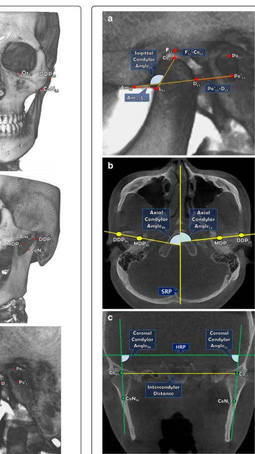
Figure 1 Reference points on 3D reconstruction model.
a. Coronal view; b. Oblique view (Left); c. Sagittal view (Left).
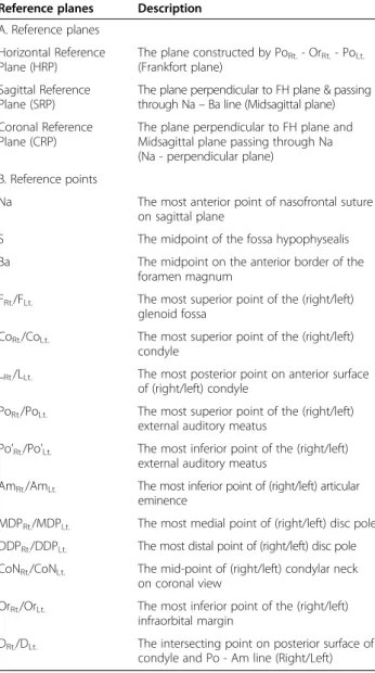
Figure 2 Measurements on 3D CBCT images. a. Sagittal view (Left);
b. Axial view; c. Coronal view.
were 1.5 (range : 0-7) and 4.0 (range : 2-8) on T0. But the scores were decreased to 0.4 (range : 0-4) and 1.2 (range : 0-4) on T2. All TMJs in study group had varying degrees of symptomatic alleviations and no deteriora- tions in comparison between the T0 and T2. The symp- tomatic decreases between preoperative and postoperative pain and noise (T0-T1, T0-T2) were significant statistically (P < .05).
Control group (patients with preoperative treatments for TMJ symptoms)
The number of patients with TMJ symptoms was de- creased slightly and the number of patients with no TMJ symptoms was increased from none to 5 (33.3%) in con- trol group. The average pain score was 2.3 (range : 0-7) on T0, and 1.1 (range : 0-4) on T2. The average noise score was reduced from 3.1 (range : 0-5) to 0.9 (range : 0-5) after 2 jaw surgery (T2). Although very few of the patients (2 of 11 patients with TMJ pain and 1 of 12 patients with TMJ noise) reported slight worsening comparing T0 versus T2, the majority of the patients were relieved or unchanged.
Moreover two patients with preoperative LOM showed improvement unexceptionally. In the results of statistical analysis, changes of TMJ noise between T0 and T2 were statistically significant (P < .05) even though TMJ pain had no significant changes (p > .05).
Changes of condylar position with analysis of 3d cbct Surgical change (T0– T1)
Between T0 and T1, study group had significant changes of F
Rt.- Co
Rt., F
Lt.- Co
Lt., Am
Rt.- L
Rt., Axial Axis angle (Rt.), Intercondylar Distance, and control group
Table 1 Definitions of reference points & planesReference planes Description A. Reference planes
Horizontal Reference Plane (HRP)
The plane constructed by PoRt.- OrRt.- PoLt.
(Frankfort plane) Sagittal Reference
Plane (SRP)
The plane perpendicular to FH plane & passing through Na– Ba line (Midsagittal plane) Coronal Reference
Plane (CRP)
The plane perpendicular to FH plane and Midsagittal plane passing through Na (Na - perpendicular plane)
B. Reference points
Na The most anterior point of nasofrontal suture on sagittal plane
S The midpoint of the fossa hypophysealis
Ba The midpoint on the anterior border of the foramen magnum
FRt./FLt. The most superior point of the (right/left) glenoid fossa
CoRt./CoLt. The most superior point of the (right/left) condyle
LRt./LLt. The most posterior point on anterior surface of (right/left) condyle
PoRt./PoLt. The most superior point of the (right/left) external auditory meatus
Po’Rt./Po’Lt. The most inferior point of the (right/left) external auditory meatus
AmRt./AmLt. The most inferior point of (right/left) articular eminence
MDPRt./MDPLt. The most medial point of (right/left) disc pole DDPRt./DDPLt. The most distal point of (right/left) disc pole CoNRt./CoNLt. The mid-point of (right/left) condylar neck
on coronal view
OrRt./OrLt. The most inferior point of the (right/left) infraorbital margin
DRt./DLt. The intersecting point on posterior surface of condyle and Po - Am line (Right/Left)
Table 2 Definitions of measurements
Plane Measurement Description
Sagittal Po’Rt.- DRt./Po’Lt.- DLt. Distance between Po’ & D FRt.- CoRt./FLt.- CoLt. Distance between F & Co AmRt.- LRt./AmLt.- LLt. Distance between Am & L Po’Rt.- AmRt./Po’Lt.- AmLt. Distance between Po’ & Am Sagittal condylar angleRt./Lt. Angle composed of Co - L line &
Po’ - Am line
Axial Axial condylar angleRt./Lt. Angle composed of MDP - DDP line & SRP
Intercondylar Distance Distance between CoRt.& CoLt.
Coronal Coronal condylar angleRt./Lt. Angle composed of CoN - Co line & HRP
Table 3 Clinical data in both TMJs, A. Distributions Study group Control group
Symptoms T0 T2 T0 T2
TMJ pain only 0 (0) 0 (0) 3 (20) 3 (20)
TMJ noise only 10 (66.7) 6 (40) 4 (26.7) 2 (13.3)
LOM only 0 (0) 0 (0) 0 (0) 0 (0)
TMJ pain and noise only 5 (33.3) 2 (13.3) 6 (40) 5 (33.3) TMJ pain, noise and LOM 0 (0) 0 (0) 2 (13.3) 0 (0)
No symptoms 0 (0) 7 (46.7) 0 (0) 5 (33.3)
Numbers of patients (%).
Table 4 Clinical data in both TMJs, B. TMJ pain and noise B-1. Change of NAS values
Study group Control group
Symptoms T0 T2 T0 T2
TMJ pain 1.5 (0-7) 0.4 (0-4) 2.3 (0-7) 1.1 (0-4) TMJ noise 4.0 (2-8) 1.2 (0-4) 3.1 (0-5) 0.9 (0-5) Average (range).
had significant changes of F
Rt.- Co
Rt.,F
Lt.- Co
Lt.,. But, other measurements were not changed significantly during 2 jaw surgery.
Postoperative stability (T1– T2)
Between T1 and T2, most measurements were not chan- ged significantly after the surgery. Changes of only three measurements (F
Rt.- Co
Rt., F
Lt.- Co
Lt., Intercondylar Distance) were significant in both groups. Intercondylar distance significantly increased immediately after the surgery, but returned near the existing position during follow-up interval.
Definitive change of condylar position (T0– T2)
The final changes of condylar positions were evaluated from identification of positional changes between T0 and T2. Any measurements of both groups did not have significant changes in this period (Table 8).
Discussions
Many studies have suggested that surgical corrections of dento-facial deformities can improve the symptoms re- lating to TMJ pain and dysfunction [3-11]. However, Henrikson et al. [15] suggested that short-term decrease of the painful tenderness may be due to altered activity of the muscles, and Onizawa et al. [16] also speculated that alteration of TMJ sounds after orthognathic surgery were associated with postoperative reduction of man- dibular mobility. Unlike these studies, we think that improvements of the TMJ symptoms are not solely due to postoperative reduction of muscular function or jaw mobility and may be relevant to the improvements of
occlusal, skeletal and neuromuscular balance after the surgery. The aim of this study is to identify postoperative changes of TMD symptoms and condylar stability and to evaluate additional therapeutic effect of the surgery and necessity of preoperative TMD treatments on the orthognathic patients with TMD definitively.
The symptomatic results of this study are almost con- sistent with previous studies in which TMJ symptoms improved. Ueki et al. [17] found that the incidence of TMJ symptoms decreased after SSRO although SSRO did not change the disk position. According to Togashi et al. [18], the incidence of TMJ signs and symptoms sig- nificantly decreased from 29.5% before orthognathic sur- gery to 12.1% at one year after the surgery. Also, TMJ signs and symptoms decreased in 82.4% of symptomatic patients before the surgery. In this study, both symp- tomatic patients with and without preoperative TMD treatment had favorable changes of TMJ symptoms.
Especially, TMJ noise decreased significantly in both groups. Although deteriorations of the symptoms were unusually shown, TMJ pain generally improved after the surgery. There were significant improvements and no de- teriorations in study group, but a few deteriorations and
Table 5 Clinical data in both TMJs, B-2: Symptomaticchange of both TMJs
Symptoms Change Study group Control group
Improved 6 (20) 12 (40)
TMJ Pain Deteriorated 0 (0) 4 (13)
No change 24 (80) 14 (47)
Improved 19 (63.5) 17 (56.5)
TMJ Noise Deteriorated 0 (0) 1 (3.5)
No change 11 (36.5) 12 (40)
Numbers of TMJs (%).
Table 6 Clinical data in both TMJs, C LOM
Symptom Change Study group Control group
LOM Improved 0 (0) 2 (13)
Deteriorated 0 (0) 0 (0)
No change 15 (100) 13 (87)
Numbers of patients (%).
Table 7 Significance in changes of TMJ symptoms
Symptoms Study group Control group
T1-T0 T2-T0 T2-T1 T1-T0 T2-T0 T2-T1
Pain .034* .034* 1.000 .241 .183 .842
Noise .000* .000* .705 .000* .001* .886
*Significant difference by Wilcoxon signed rank test (P < 0.05).
Table 8 Significances in changes of condylar positions
Measurement Study group Control group
T1-T0 T2-T1 T2-T0 T1-T0 T2-T1 T2-T0 Po’Lt.- DLt. .334 .594 .233 .057 .094 .100 Po’Rt.- DRt. .589 .589 .865 .140 .211 .776 FLt.- CoLt. .008* .036* .307 .043* .011* .363 FRt.- CoRt. .001* .009* .058 .002* .036* .147
AmLt.- LLt. .670 .156 .156 .820 .495 .394
AmRt.- LRt. .001* .233 .055 .281 .427 .053
Po’Lt.- AmLt. .733 .256 .100 .551 .394 .460 Po’Rt.- AmRt. .112 .910 .191 .910 .589 .820 Sagittal condylar angle (Lt.) .256 .331 .798 .067 .256 .609 Sagittal condylar angle (Rt.) .078 .910 .057 .733 .307 .211 Axial axis angle (Lt.) .191 .865 .100 .191 .609 .100 Axial axis angle (Rt.) .035* .910 .052 .233 .650 .233 Intercondylar distance .011* .032* .334 .061 .140 .100 Coronal axis angle (Lt.) .712 .363 .427 .609 .281 .334 Coronal axis angle (Rt.) .100 .650 .425 .363 .443 .156
*Significant difference by Wilcoxon signed rank test (P < 0.05).
unsignificant changes in control group. We speculated that it is the reason that the control group included some TMD patients with facial asymmetry. The comparison of LOM was little meaningful because of insufficiency of subjects with preexisting LOM.
Condylar repositioning during BSSRO can influence the changes of TMJ symptoms and positional stability of condyle during postoperative follow-up. Individual physiological adaptation also can affect positional changes of condyles during follow-up period. However, according to Nakata et al. [19], the physiological adaptation to the surgically corrected structures needs long time over two years. Because the follow-up time of this study was less than a year, we eliminated a factor of the physiologic adap- tation. In this study, the condylar position of both groups during postoperative follow-up interval had good stability (P > .05) except for the distances between glenoid fossa and condyle (F – Co) in both sides. It seems that whether or not TMD treatments precede 2 jaw surgery in patients with prior TMD did not have any significant influences on postoperative stability of condylar position. We estimated that the significant changes of both F - Co measurements were because all patients in both groups underwent CBCT wearing the occlusal splint at T1 although showed signifi- cant changes (P < .05). In other words, we considered them as temporary increases due to wearing of the occlu- sal splint for preventing skeletal relapse during 1 month or less after the surgery. Intercondylar distance in study group significantly increased immediately after the sur- gery, but returned near the existing position during follow-up interval. Kim et al. [20] reported that condyle in the glenoid fossa had tendency to return to normal pos- ition during postoperative period in orthognathic patients.
We used a miniplate and 4 monocortical screws for semirigid fixation so that functional stability and slight flexibility for enhanced long term TMJ function could be achieved [21].
2 jaw surgery can have favorable effects on TMJ symp- toms in patients with dentofacial deformities. The results in this study showed that even if TMJ symptoms were not treated before the surgery, 2 jaw surgery could have therapeutic effects for TMJ symptoms while also provide good stability of condyles. This study has some differ- ences from other similar studies. Firstly, we divided orthognathic patients into 2 groups contingent upon presence or absence of preoperative treatment for TMJ symptoms, unlike other studies in which TMJ symptoms concerned as the standard of classification. Secondly, we simultaneously analyzed postoperative stability of con- dylar position with 3D CBCT while examining changes of TMJ symptoms. On the other hand, there are also some limitations: (1) The number of the orthognathic patients without any preceding treatments for TMJ symptoms before the surgery were not enough. (2) We
had only mandibular prognathic patients with class III malocclusion among various dentofacial deformities.
(3) We did not use validated scales such as the modified Helkimo index, craniomandibular index, and the Research Diagnostic Criteria [14,22,23]. We collected only the symp- tomatic data by simple self-report form.
Conclusions
This study showed improvements of preexisting TMJ symptoms and good condylar stability during the post- operative follow-up even though the patients were not treated for the TMJ symptoms before the surgery. Thus, We think that 2 jaw surgery patients with preexisting TMJ symptoms can have therapeutic effect exclusively attributed to the surgery unless the existence of acute and severe TMD before the surgery. However, the symp- toms cannot be always improved and there would be some risk of symptomatic deterioration though the risk is very quite low.
Abbreviations
TMJ:temporomandibular joints; TMD: temporomandibular disorders;
LOM: limitation of mouth opening; BSSRO: bilateral sagittal split ramus osteotomy; CBCT: cone beam computed tomography; NAS: Numerical Analogue Scales; DCT: dental CBCT.
Competing interest
The author declared that they have no competing interest.
Authors' contributions
SYY carried out all processes with other authors. JMS been involved in drafting the many parts of manuscript. YDK involved in revising it critically for all content. IKC made substantial contributions to analysis and interpretation of data. SHS made contributions to conception and design, acquisition of data, analysis and interpretation of data. All author read and approved the final manuscript.
Acknowledgments
All authors have viewed and agreed to the submission.
This work was supported by a 2-Year Research Grant of Pusan National University, Korea.
Received: 9 January 2015 Accepted: 20 January 2015
References
1. Luther F (1998) Orthodontics and the temporomandibular joint: where are we now? Part 1. Orthodontic treatment and temporomandibular disorders.
Angle Orthod 68:295–304
2. McNamara JA, Seligman DA, Okeson JP (1995) Occlusion, Orthodontic treatment, and temporomandibular disorders: a review. J Orofac Pain 9:73–90
3. White CS, Dolwick MF (1992) Prevalence and variance of temporomandibular dysfunction in orthognathic surgery patients. Int J Adult Orthodon Orthognath Surg 7:7–14
4. Westermark A, Shayeghi F, Thor A (2001) Temporomandibular dysfunction in 1,516 patients before and after orthognathic. Int J Adult Orthodon Orthognath Surg 16:145–151
5. Dervis E, Tuncer E (2002) Long-term evaluations of temporomandibular disorders in patients undergoing orthognathic surgery compared with a control group In: United States. 554-560
6. Dujoncquoy JP, Ferri J, Raoul G, Kleinheinz J (2010) Pmc2998459;
Temporomandibular joint dysfunction and orthognathic surgery: a retrospective. Head Face Med 6:27
7. Panula K, Somppi M, Finne K, Oikarinen K (2000) Effects of orthognathic surgery on temporomandibular joint dysfunction. A Int J Oral Maxillofac Surg 29:183–187
8. Karabouta I, Martis C (1985) The TMJ dysfunctionsyndrome before and after sagittal split osteotomy of the rami. J Maxillofac Surg 13:185–188 9. Al-Riyami S, Moles DR, Cunningham SJ (2009) Orthognathic treatment and
temporomandibular disorders: a systematic review. In: Part 1. A new quality- assessment technique and analysis of study characteristics and classifications In: . United States., 624 e1,15; discussion 624-5
10. De Clercq CA, Neyt LF, Mommaerts MY, Abeloos JS (1998) Orthognathic surgery: patients' subjective findings with focus on the. J Craniomaxillofac Surg 26:29–34
11. Moenning JE, Bussard DA, Montefalco PM, Lapp TH, Garrison BT (1997) Medical necessity of orthognathic surgery for the treatment of dentofacial deformities associated with temporomandibular disorders. Int J Adult Orthodon Orthognath Surg 12:153–161
12. Aghabeigi B, Hiranaka D, Keith DA, Kelly JP, Crean SJ (2001) Effect of orthognathic surgery on the temporomandibular joint in patients with anterior open bite. Int J Adult Orthodon Orthognath Surg 16:153–160 13. Wolford LM, Reiche-Fischel O, Mehra P (2003) Changes in temporomandibular
joint dysfunction after orthognathic surgery In: . United States, 2003 American Association of Oral and Maxillofacial Surgeons. J Oral Maxillofac Surg 61:655–660, 2003: 655,60; discussion 661
14. Dworkin SF, LeResche L (1992) Research diagnostic criteria for temporomandibular disorders: review, criteria. J Craniomandib Disord 6:301–355
15. Henrikson T, Nilner M, Kurol J (1999) Symptoms and signs of temporomandibular disorders before, during and after orthodontic treatment. Swed Dent J 23:193–207
16. Onizawa K, Schmelzeisen R, Vogt S (1995) Alteration of temporomandibular joint symptoms after orthognathic surgery: comparison with healthy volunteers In: United States. 117,21; discussion 122-123
17. Ueki K, Marukawa K, Nakagawa K, Yamamoto E (2002) Condylar and temporomandibular joint disc positions after mandibular osteotomy for prognathism. J Oral Maxillofac Surg 60:1424–1432, discussion 1432-4 18. Togashi M, Kobayashi T, Hasebe D, Funayama A, Mikami T, Saito I, Hayashi T,
Saito C (2012) Effects of surgical orthodontic treatment for dentofacial deformities on signs and symptoms of temporomandibular joint
19. Nakata Y, Ueda HM, Kato M, Tabe H, Shikata-Wakisaka N, Matsumoto E, Koh M, Tanaka E, Tanne K (2007) Changes in stomatognathic function induced by orthognathic surgery in patients with mandibular prognathism. J Oral Maxillofac Surg 65:444–451
20. Kim YI, Cho BH, Jung YH, Son WS, Park SB (2011) Cone-beam computerized tomography evaluation of condylar changes and stability following two-jaw surgery: Le Fort I osteotomy and mandibular setback surgery with rigid fixation In: United States, Inc. 681-687
21. Mavili ME, Canter HI, Saglam-Aydinatay B (2009) Semirigid fixation of mandible and maxilla in orthognathic surgery: stability and advantages.
Ann Plast Surg 63(4):396–403
22. Helkimo M (1974) Studies on function and dysfunction of the masticatory system. II. Index for. Sven Tandlak Tidskr 67:101–121
23. Fricton JR, Schiffman EL (1986) Reliability of a craniomandibular index.
J Dent Res 65:1359–1364
Submit your manuscript to a journal and benefi t from:
7 Convenient online submission 7 Rigorous peer review
7 Immediate publication on acceptance 7 Open access: articles freely available online 7 High visibility within the fi eld
7 Retaining the copyright to your article
Submit your next manuscript at 7 springeropen.com