Stability of bimaxillary surgery involving intraoral
vertical ramus osteotomy with or without presurgical
miniscrew-assisted rapid palatal expansion in adult
patients with skeletal Class III malocclusion
Objective: The aim of this study was to evaluate the stability of bimaxillary surgery involving bilateral intraoral vertical ramus osteotomy performed with or without presurgical miniscrew-assisted rapid palatal expansion (MARPE) in adult patients with skeletal Class III malocclusion. Methods: A total of 40 adult patients with skeletal Class III malocclusion were retrospectively divided into two groups (n = 20 each) according to the use of MARPE for the correction of transverse maxillomandibular discrepancy during presurgical orthodontic treatment. Serial lateral cephalograms and dental casts were analyzed until 6 months after surgery. Results: Before presurgical orthodontic treatment, there was no significant differences in terms of sex and age between groups. However, the difference of approximately 3.1 mm in the maxillomandibular intermolar width was statistically significant (p < 0.001). Two days after surgery, the mandible had moved backward and upward without any significant intergroup difference. Six months after surgery, the maxillary intercanine (2.7 ± 2.1 mm), interpremolar (3.6 ± 2.4 mm), and intermolar (2.0 ± 1.3 mm) arch widths were significantly increased (p < 0.001) relative to the values before presurgical orthodontic treatment in the MARPE group; these widths were maintained or decreased in the control group. However, there was no significant difference in surgical changes and the postsurgical stability between the two groups. No significant correlations existed between the amount of maxillary expansion and postsurgical mandibular movement. Conclusions: MARPE is useful for stable and nonsurgical expansion of the maxilla in adult patients with skeletal Class III malocclusion who are scheduled for bimaxillary surgery.
[Korean J Orthod 2020;50(5):304-313]
Key words: Miniscrew-assisted rapid palatal expansion, Transverse maxillo-mandibular discrepancy, Skeletal Class III malocclusion, Stability
Yoon-Soo Ahna
Sung-Hwan Choia,b
Kee-Joon Leea
Young-Soo Jungc
Hyoung-Seon Baika
Hyung-Seog Yua
a
Department of Orthodontics, Institute of Craniofacial Deformity, Yonsei University College of Dentistry, Seoul, Korea
bBK21 PLUS Project, Yonsei University
College of Dentistry, Seoul, Korea
cDepartment of Oral and Maxillofacial
Surgery, Oral Science Research Center, Yonsei University College of Dentistry, Seoul, Korea
Received March 5, 2020; Revised April 3, 2020; Accepted April 25, 2020.
Corresponding author: Hyung-Seog Yu.
Professor, Department of Orthodontics, Institute of Craniofacial Deformity, Yonsei University College of Dentistry, 50-1 Yonsei-ro, Seodaemun-gu, Seoul 03722, Korea.
Tel +82-2-2228-3104 e-mail [email protected]
Yoon-Soo Ahn and Sung-Hwan Choi contributed equally to this study.
How to cite this article: Ahn YS, Choi SH, Lee KJ, Jung YS, Baik HS, Yu HS. Stability of bimaxillary surgery involving intraoral vertical ramus osteotomy with or without presurgical miniscrew-assisted rapid palatal expansion in adult patients with skeletal Class III malocclusion. Korean J Orthod 2020;50:304-313.
© 2020 The Korean Association of Orthodontists.
This is an Open Access article distributed under the terms of the Creative Commons Attribution Non-Commercial License (http://creativecommons.org/licenses/by-nc/4.0) which permits unrestricted non-commercial use, distribution, and reproduction in any medium, provided the original work is properly cited.
pISSN 2234-7518 • eISSN 2005-372X https://doi.org/10.4041/kjod.2020.50.5.304
INTRODUCTION
Some adult patients with skeletal Class III malocclu-sion exhibit severe maxillomandibular anteroposterior discrepancy with transverse maxillary deficiency, which renders the treatment more challenging.1 In such cases,
the clinician can address the transverse discrepancy us-ing both surgical and nonsurgical methods.
Surgical expansion can be achieved with segmental maxillary osteotomy, which can be performed simulta-neously with bimaxillary surgery in the operating room. However, it produces inaccurate and unstable out-comes.2,3 Consequently, surgically-assisted rapid palatal
expansion (SARPE) has been widely performed, although clinicians and patients might be concerned about addi-tional issues such as hospitalization, attendant morbid-ity, increased cost, and surgical complications.4,5
For nonsurgical expansion, clinicians can decide to extract the maxillary premolars for relative expansion of the maxillary arch during presurgical orthodontic treatment.6 However, premolar extraction loses its
ap-plicability if the maxillary arch shows no or mild crowd-ing. In addition, this modality lengthens the presurgical orthodontic treatment period, thus deteriorating the pa-tient’s quality of life. A transpalatal arch or conventional rapid palatal expansion (RPE) is generally not feasible in adults because of possible adverse effects such as buccal tipping, root resorption, and gingival recession around the anchor teeth.7
Recently, Lee et al.8 reported a successful clinical
out-come for a patient who underwent orthognathic surgery and tooth–bone-borne RPE, which was assisted by four palatally placed orthodontic titanium miniscrews (i.e., miniscrew-assisted RPE [MARPE]). The miniscrews can reduce stress on the anchor teeth when the appliance expands the maxillary arch, thus reducing the side ef-fects on the anchor teeth.9
Anterior and inferior movement of the maxilla and subsequent clockwise rotation of the mandible have been reported to occur with use of the aforementioned surgical and nonsurgical procedures for maxillary expan-sion.10-12 If transverse maxillary expansion can lead to
such vertical and anteroposterior changes in the maxil-lomandibular complex, any relapse can have consider-able effects on the skeletal changes after orthognathic surgery. In particular, when bimaxillary surgery such as bilateral intraoral vertical ramus osteotomy (IVRO) is performed, the mandible reportedly tends to undergo clockwise rotation due to muscular pull during the post-operative retention period.13 Even if slight maxillary
con-traction occurs, which is possible after MARPE, it may increase the probability of an anterior open bite after surgery. Moreover, maxillary constriction can lead to pal-atal inclination of the maxillary posterior teeth and an
edge-to-edge bite between the posterior teeth on one or both sides, which could result in premature contact of these teeth. This is because the downward-backward rotation of the mandible increases the possibility of an anterior open bite.14,15
To our knowledge, no study has evaluated the stabil-ity of bimaxillary surgery involving IVRO performed with or without presurgical MARPE in adult patients with skeletal Class III malocclusion. Therefore, the purpose of this study was to evaluate differences in postsurgical stability between bimaxillary surgery with MARPE and bimaxillary surgery without MARPE. The null hypothesis was that the stability of bimaxillary surgery is not influ-enced by expansion of the maxillary arch using MARPE during presurgical orthodontic treatment.
MATERIALS AND METHODS
Study design and patients
This retrospective study received institutional review board approval based on the Declaration of Helsinki (2-2019-0051). Written informed consent was obtained from all patients prior to the initiation of presurgical orthodontic treatment at the Department of Orthodon-tics, Yonsei University Dental Hospital, Seoul, Republic of Korea.
The subjects were patients who underwent bimaxillary surgery involving IVRO between 2013 and 2017 at the Department of Oral and Maxillofacial Surgery, Yonsei University Dental Hospital, Seoul, Republic of Korea. The patients were divided into control and MARPE groups according to the use of MARPE for nonsurgical maxillary expansion during presurgical orthodontic treatment.
Regardless of the group, the inclusion criteria were as follows: (1) age > 18 years; (2) presence of skeletal Class III malocclusion before surgery, with an angle of < 0° formed by point A, the nasion, and point B (ANB); (3) treatment with presurgical orthodontics and con-ventional bimaxillary surgery involving one-piece Le Fort I osteotomy and IVRO; (4) use of nonextraction presurgical orthodontic treatment (except extraction of third molars); and (5) availability of a complete series of identifiable lateral cephalograms and dental casts. The exclusion criteria were as follows: (1) history of congeni-tal malformations such as cleft lip and palate and (2) past history of orthognathic surgery and/or orthodontic treatment.
The inclusion criteria for the MARPE group were as follows: (1) transverse maxillary deficiency with unilater-al or bilaterunilater-al posterior crossbite when the maxillary and mandibular posterior teeth were simulated to a Class I molar key; (2) a maxillomandibular difference of more than 5 mm according to Korean standards,16 measured
or-thodontics; and (3) midpalatal suture opening confirmed on maxillary anterior periapical radiographs within 4 weeks of activation.
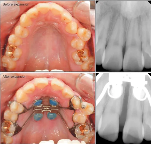
In the MARPE group, rapid maxillary expansion using the MARPE appliance was initiated before orthodontic brackets were bonded (Figure 1).17 After being informed
about other treatment options such as segmental maxil-lary osteotomy and SARPE, patients who refused these options due to avoid extended or additional surgery were included. As previously described,8,18 a Hyrax screw
and four orthodontic miniscrews with a diameter of 1.8 mm and lengths of 7.0 mm and 9.0 mm (self-drilled type, ORLUS; Ortholution, Seoul, Korea) were used for the MARPE appliance. The 9.0-mm miniscrews were placed in the rugae area while the 7.0-mm screws were placed in the paramidline area of the palate. The heads of the miniscrews were covered by light-cured resin to connect the miniscrews with the helical hooks. The ap-pliance was activated by approximately 0.2 mm daily until the desired amount of expansion was achieved.19
The mean amount of expansion in the maxillary in-termolar region was approximately 6.1 ± 7.2 mm (mean 30.6 turns; range, 17–40 turns). After a 3-month con-solidation period required for bone formation in the
median palatal suture,18-20 the MARPE appliance was
removed and orthodontic brackets were bonded. Presur-gical orthodontic treatment was performed for at least 6 months (control group, 11.6 ± 6.4 months; MARPE group, 13.0 ± 4.7 months), with no statistically sig-nificant differences in the treatment duration between the two groups (p = 0.437). The patients in the control group received the same presurgical orthodontic treat-ment, only without MARPE.
Orthognathic treatment
After presurgical orthodontic treatment, all patients underwent maxillary Le Fort I osteotomy and bilateral IVRO performed by a surgeon. Intermaxillary fixation was implemented for 10–14 days after surgery, and physical therapy for rehabilitation was performed for an-other 2 weeks.
Dental cast analysis
Dental casts were analyzed using digital calipers to evaluate the changes in tooth positions before presurgi-cal orthodontic treatment (T0) and at 6 months after surgery (T3). The intercanine width (ICW), interpremolar width (IPMW), and intermolar width (IMW) were
mea-Before expansion
After expansion
Figure 1. Miniscrew-assisted
rapid palatal expansion be-fore bimaxillary surgery. The images show the appliance and periapical radiographs before and after expansion.
sured. The normal values for differences in the maxil-lomandibular ICW, IPMW, and IMW in patients with skeletal Class I malocclusion are 8.18 ± 1.57, 9.01 ± 1.66, and 8.43 ± 2.22 mm, respectively; this means that the maxillary arch width should exceed the mandibular arch width for ideal occlusion.21
Lateral cephalometric analysis
Skeletal changes were evaluated using lateral cepha-lograms obtained at 1 month before surgery (T1), 2 days after surgery (T2), and T3. One observer who was blinded to the patient's clinical condition traced the radiographs using V-ceph 5.5 (Osstem, Seoul, Korea). Seven measurements were recorded to analyze the an-teroposterior and vertical skeletal changes (Figure 2). Reliability
Two weeks after the initial cephalometric and dental cast measurements, all measurements were repeated by the same observer. The method error, calculated using Dahlberg’s formula, ranged from 0.20 to 0.30 mm for linear measurements and from 0.15° to 0.30° for angu-lar measurements.
Statistical analysis
All statistical analyses were performed using IBM SPSS Statistics for Windows, version 22.0 (IBM Corp., Armonk, NY, USA). According to a preliminary study with an ef-fect size of 0.25 for evaluating changes in cephalometric measurements over time in each group, at least 18 sub-jects were required in each group.
Normality was determined using the Shapiro–Wilk test. The independent t-test was used to evaluate the groups at the same time, with the exclusion of data that
did not show normality, such as age and sex. Repeated measures analysis of variance was performed to com-pare skeletal and dental changes over time within and between the two groups. Between-group differences in
Figure 2. Lateral cephalometric analysis performed to
evaluate skeletal changes after bimaxillary surgery with or without presurgical miniscrew-assisted rapid palatal expansion. The x-axis is the horizontal plane that shifts the SN plane up 7° relative to N. The y-axis is the plane through S and perpendicular to the x-axis.
A, Point A; A (x), horizontal position of point A; A (y), vertical position of point A; B, point B; B (x), horizontal position of point B; B (y), vertical position of point B; N, nasion; OP, occlusal plane; S, sella.
x-axis y-axis N S A B A (x) B (x) A (y) B (y) OP
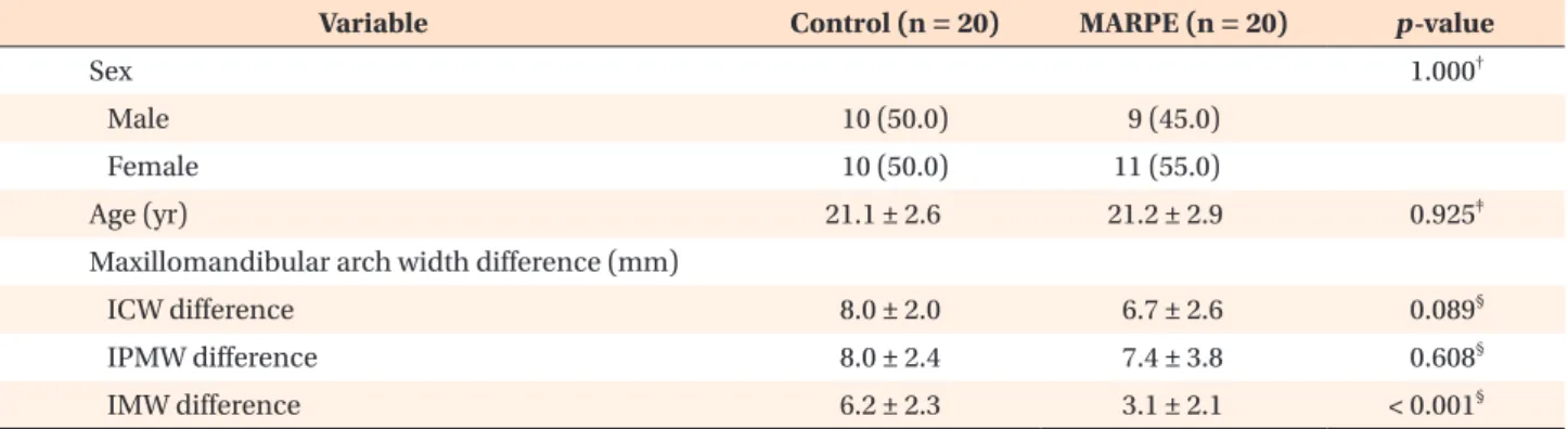
Table 1. Baseline characteristics of patients scheduled for bimaxillary surgery with (MARPE group) or without (control
group) presurgical MARPE
Variable Control (n = 20) MARPE (n = 20) p-value
Sex 1.000†
Male 10 (50.0) 9 (45.0)
Female 10 (50.0) 11 (55.0)
Age (yr) 21.1 ± 2.6 21.2 ± 2.9 0.925‡
Maxillomandibular arch width difference (mm)
ICW difference 8.0 ± 2.0 6.7 ± 2.6 0.089§
IPMW difference 8.0 ± 2.4 7.4 ± 3.8 0.608§
IMW difference 6.2 ± 2.3 3.1 ± 2.1 < 0.001§
Values are presented as number (%) or mean ± standard deviation.
MARPE, Miniscrew-assisted rapid palatal expansion; ICW, intercanine width; IPMW, interpremolar width; IMW, intermolar width.
†The p-value was calculated using the chi-squared test. ‡The p-value was calculated using the Mann–Whitney U test. §The p-value was calculated using the independent t-test.
changes over time were analyzed using the independent t-test with Bonferroni correction (α = 0.05/3). The cor-relations of maxillary arch width changes (T3–T0) and surgical changes (T2–T1) with postoperative changes (T3–T2) in all patients were evaluated using the Pearson correlation analysis.
RESULTS
From 2013 to 2017, 109 patients underwent orthog-nathic surgery with MARPE. Among these, 32 patients had undergone presurgical orthodontic treatment for < 6 months, 38 patients exhibited menton deviation > 4 mm, 16 patients exhibited skeletal Class I or II malocclu-sion; and three patients did not have their digital
cepha-Table 2. Changes in maxillary arch widths (T0 to T3) in
patients who underwent bimaxillary surgery with (MARPE group) or without (control group) presurgical MARPE
Variable Control group MARPE group p-value MxICW (mm) 0.4 ± 1.5 2.7 ± 2.1 < 0.001 MxIPMW (mm) 0.2 ± 1.2 3.6 ± 2.4 < 0.001 MxIMW (mm) −0.6 ± 1.2 2.0 ± 1.3 < 0.001 Values are presented as mean ± standard deviation. The p-value was calculated using the independent t-test. T0, Before presurgical orthodontic treatment; T3, 6 months after bimaxillary surgery; MARPE, miniscrew-assisted rapid palatal expansion; MxICW, maxillary intercanine width; MxIPMW, maxillary interpremolar width; MxIMW, maxillary intermolar width.
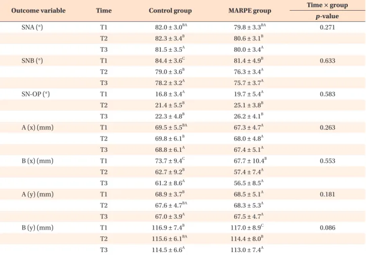
Table 3. Cephalometric measurements before and after bimaxillary surgery with (MARPE group) or without (control
group) presurgical MARPE
Outcome variable Time Control group MARPE group Time × group p-value SNA (°) T1 82.0 ± 3.0BA 79.8 ± 3.3BA 0.271 T2 82.3 ± 3.4B 80.6 ± 3.1B T3 81.5 ± 3.5A 80.0 ± 3.4A SNB (°) T1 84.4 ± 3.6C 81.4 ± 4.9B 0.633 T2 79.0 ± 3.6B 76.3 ± 3.4A T3 78.2 ± 3.2A 75.7 ± 3.7A SN-OP (°) T1 16.8 ± 3.4A 19.7 ± 5.4A 0.583 T2 21.4 ± 5.5B 25.1 ± 3.8B T3 22.3 ± 4.8B 26.2 ± 4.1B A (x) (mm) T1 69.5 ± 5.5BA 67.3 ± 4.7A 0.263 T2 69.8 ± 6.1B 68.0 ± 4.8A T3 68.8 ± 6.1A 67.4 ± 5.1A B (x) (mm) T1 73.7 ± 9.4C 67.7 ± 10.4B 0.553 T2 62.7 ± 9.2B 57.4 ± 7.4A T3 61.2 ± 8.6A 56.5 ± 8.5A A (y) (mm) T1 68.9 ± 3.7B 68.5 ± 5.1A 0.181 T2 67.6 ± 4.7BA 68.3 ± 5.3A T3 67.0 ± 3.9A 67.5 ± 4.7A B (y) (mm) T1 116.9 ± 7.4B 117.0 ± 8.9C 0.086 T2 115.6 ± 6.1BA 114.4 ± 8.0B T3 114.5 ± 6.6A 113.0 ± 7.4A
Values are presented as mean ± standard deviation.
Within each column, significant differences are represented by uppercase letters.
The p-value was calculated using repeated measures analysis of variance with Bonferroni correction.
MARPE, Miniscrew-assisted rapid palatal expansion; SNA, angle formed by the lines connecting sella, nasion, and point A; SNB, angle formed by the lines connecting sella, nasion, and point B; SN-OP, angle between the sella–nasion plane and the occlusal plane; A (x), horizontal position of point A; B (x), horizontal position of point B; A (y), vertical position of point A; B (y), vertical position of point B; T1, 1 month before surgery; T2, 2 days after surgery; T3, 6 months after surgery.
lograms or dental casts. Twenty patients were eventually included in the MARPE group.
The control group comprised 10 males and 10 fe-males with an average age of 21.1 ± 2.6 years, while the MARPE group comprised nine males and 11 females with an average age of 21.2 ± 2.9 years (Table 1). The demographic characteristics before presurgical orthodon-tic treatment were not significantly different between the two groups. The mean IMW discrepancy was 6.2 ± 2.3 mm in the control group and 3.1 ± 2.1 mm in the MARPE group, with a statistically significant between-group difference (p < 0.001). This indicated that the maxillary IMW in patients in the MARPE group was ap-proximately 3.1 mm lesser than that in patients in the control group.
Table 2 shows that all maxillary arch width changes (i.e., T3–T0), measured on the maxillary dental casts, were significantly different between the two groups (p < 0.001). In the MARPE group, the maxillary ICW (2.7 ± 2.1 mm), IPMW (3.6 ± 2.4 mm), and IMW (2.0 ± 1.3 mm) were significantly increased (p < 0.001) relative to the values before presurgical orthodontic treatment. In the control group, however, the widths were maintained or even decreased.
Table 3 shows that all cephalometric measurements at the different time points were not significantly differ-ent between the two groups. At T1, T2, and T3 in the MARPE group, point A was located 68.5 ± 5.1 mm, 68.3 ± 5.3 mm, and 67.5 ± 4.7 mm vertically on the x-axis, respectively. However, the values were not significantly different.
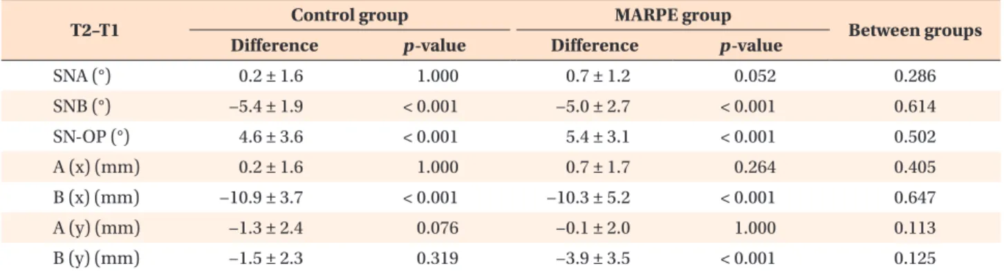
From T1 to T2, point B moved 10.9 ± 3.7 mm poste-rior (p < 0.001) and 1.5 ± 2.3 mm supeposte-rior in the con-trol group and 10.3 ± 5.2 mm posterior (p < 0.001) and 3.9 ± 3.5 mm superior (p < 0.001) in the MARPE group (Table 4). However, none of the surgical changes from T1 to T2 showed significant differences between the two groups.
During the postsurgical period (T2 to T3), the angle between the sella–nasion plane and the occlusal plane increased by 0.8 ± 2.3° in the control group and 1.0 ± 2.0° in the MARPE group; however, this angular change was not significant within each group or between the two groups (Table 5). Point B moved 1.5 ± 1.7 mm backward (p = 0.003) and 1.0 ± 2.1 mm upward in the control group and 0.8 ± 2.0 mm backward and 1.3 ± 1.6 mm upward (p = 0.004) in the MARPE group. However, the two groups showed no significant differences in the postoperative changes in all measurements over time.
In both groups, the amount of upward mandibular movement at 6 months after surgery (T2 to T3) de-creased as the amount of upward mandibular movement during surgery (T1 to T2) increased (r = −0.330; p = 0.038; Table 6). However, the amounts of horizontal man-dibular movements during surgery (T1 to T2) and the amount of expansion in the maxillary intermolar region during the overall treatment period (T0 to T3) showed no significant correlation with the postsurgical changes in the mandible (T2 to T3).
Table 4. Surgical changes (T1 to T2) in cephalometric measurements for patients who underwent bimaxillary surgery
with (MARPE group) or without (control group) presurgical MARPE
T2–T1 Control group MARPE group Between groups
Difference p-value Difference p-value
SNA (°) 0.2 ± 1.6 1.000 0.7 ± 1.2 0.052 0.286 SNB (°) −5.4 ± 1.9 < 0.001 −5.0 ± 2.7 < 0.001 0.614 SN-OP (°) 4.6 ± 3.6 < 0.001 5.4 ± 3.1 < 0.001 0.502 A (x) (mm) 0.2 ± 1.6 1.000 0.7 ± 1.7 0.264 0.405 B (x) (mm) −10.9 ± 3.7 < 0.001 −10.3 ± 5.2 < 0.001 0.647 A (y) (mm) −1.3 ± 2.4 0.076 −0.1 ± 2.0 1.000 0.113 B (y) (mm) −1.5 ± 2.3 0.319 −3.9 ± 3.5 < 0.001 0.125
Values are presented as mean ± standard deviation.
Group comparisons were tested using the independent t-test with Bonferroni correction (p < 0.05/3). Positive and negative values indicate, respectively, the anterior and posterior horizontal changes, inferior and superior vertical changes, and increased and decreased dimensional changes.
The p-value was calculated using repeated measures analysis of variance with Bonferroni correction.
T1, 1 month before surgery; T2, 2 days after surgery; MARPE, miniscrew-assisted rapid palatal expansion; SNA, angle formed by the lines connecting the sella, nasion, and point A; SNB, angle formed by the lines connecting the sella, nasion, and point B; SN-OP, angle between the sella–nasion plane and the occlusal plane; A (x), horizontal position of point A; B (x), horizontal position of point B; A (y), vertical position of point A; B (y), vertical position of point B.
DISCUSSION
Nonsurgical maxillary expansion using MARPE leads to a pyramidal pattern of changes in the circummaxil-lary structures, the center of rotation of which is located near the frontonasal suture.22 Lim et al.20 investigated
the stability of the treatment outcome at 1 year after MARPE expansion and reported that more than half of the expansion occurred in the dentoalveolar portion. Hong23 reported that the axial angulation of the
maxil-lary first molar in adults increased by 2.29 ± 8.09° upon
removal of the fixed orthodontic appliance after comple-tion of nonsurgical orthodontic treatment with MARPE; this indicated that the maxillary molar is slightly buc-cally inclined after orthodontic treatment. In view of this finding, relapse of the tilted molars and alveolar bone after MARPE could cause occlusal interferences in the postsurgical period. Previous studies13,24 have reported
posterior and inferior relapse with clockwise rotation of the mandible after setback surgery using IVRO. Because IVRO does not involve rigid fixation between the proxi-mal and distal segments immediately after surgery,
con-Table 5. Postoperative relapse (T2 to T3) in cephalometric measurements for patients who underwent bimaxillary surgery
with (MARPE group) or without (control group) presurgical MARPE
T3–T2 Control group MARPE group Between groups
Difference p-value Difference p-value
SNA (°) −0.7 ± 0.7 0.001 −0.5 ± 0.9 0.029 0.475 SNB (°) −0.7 ± 0.8 0.002 −0.5 ± 1.1 0.133 0.485 SN-OP (°) 0.8 ± 2.3 0.342 1.0 ± 2.0 0.103 0.780 A (x) (mm) −0.9 ± 1.0 0.002 −0.5 ± 1.0 0.075 0.256 B (x) (mm) −1.5 ± 1.7 0.003 −0.8 ± 2.0 0.215 0.268 A (y) (mm) −0.5 ± 1.8 0.698 −0.8 ± 1.5 0.096 0.584 B (y) (mm) −1.0 ± 2.1 0.116 −1.3 ± 1.6 0.004 0.622
Values are presented as mean ± standard deviation.
Group comparisons were tested using the independent t-test with Bonferroni correction (p < 0.05/3). The positive and negative values indicate, respectively, the anterior and posterior horizontal changes, inferior and superior vertical changes, and increased and decreased dimensional changes.
The p-value was calculated using repeated measures analysis of variance with Bonferroni correction.
T2, 2 days after surgery; T3, 6 months after surgery; MARPE, miniscrew-assisted rapid palatal expansion; SNA, angle formed by the lines connecting the sella, nasion, and point A; SNB, angle formed by the lines connecting the sella, nasion, and point B; SN-OP, angle between the sella-nasion plane and the occlusal plane; A (x), horizontal position of point A; B (x), horizontal position of point B; A (y), vertical position of point A; B (y), vertical position of point B.
Table 6. Correlations of maxillary arch width changes (T3–T0) and surgical changes (T2–T1) with postoperative changes
(T3–T2) in patients who underwent bimaxillary surgery with (MARPE group) or without (control group) presurgical MARPE
Variable
Postsurgical change 6 months after surgery (T3–T2)
B (x) (mm) B (y) (mm) r p-value r p-value MxICW (T3–T0) (mm) 0.185 0.254 −0.155 0.339 MxIPMW (T3–T0) (mm) 0.120 0.460 0.032 0.845 MxIMW (T3–T0) (mm) −0.068 0.677 0.208 0.198 B (x) (T2–T1) (mm) −0.221 0.170 0.193 0.233 B (y) (T2–T1) (mm) −0.027 0.868 −0.330 0.038
The p-value was calculated using Pearson correlation analysis.
T0, Before presurgical orthodontics; T1, 1 month before surgery; T2, 2 days after surgery; T3, 6 months after surgery; MARPE, miniscrew-assisted rapid palatal expansion; B (x), horizontal position of point B; B (y), vertical position of point B; r, Pearson’s correlation coefficient; MxICW, maxillary intercanine width; MxIPMW, maxillary interpremolar width; MxIMW, maxillary intermolar width.
siderable caution should be exercised to prevent anterior open bite due to temporary condylar sagging or muscu-lar pull of the masticatory muscles up to 3 months after surgery. Considering these two factors (i.e., relapse of maxillary constriction and mandibular vertical instabil-ity with IVRO), we were skeptical about the results of mandibular setback by IVRO combined with nonsurgi-cal maxillary expansion using MARPE in patients with skeletal Class III malocclusion and transverse maxillary deficiency.
Before presurgical orthodontic treatment, the MARPE group exhibited a greater arch width discrepancy in the molar region than did the control group. From T0 to T3, the maxillary IMW was significantly increased in the MARPE group (2.0 ± 1.3 mm), but not in the control group (−0.6 ± 1.2 mm). The mean amount of expansion with the MARPE appliance was approximately 6 mm (30 turns × 0.2 mm per turn) in this study, although the ac-tual remaining amount of expansion at T3 was approxi-mately 2 mm in the molar region. These phenomena may be attributed to lingual repositioning of the molars during the alignment phase, depending on the arch form during the pre- and postsurgical orthodontic treat-ments.18 This factor was considered before surgery, and
the maxilla was over-expanded using MARPE until the palatal cusps of the maxillary posterior teeth contacted the buccal cusps of the mandibular posterior teeth, simi-lar to the outcome after conventional RPE.
Handelman et al.25 stated that SARPE should be used
when the required increase in the maxillary IMW exceeds 8 mm. Choi et al.18 stated that MARPE caused skeletal
expansion that is approximately 43% of the maxillary IMW expansion, unlike appliances that induce buc-cal tilting of the molars, such as the transpalatal arch. Therefore, according to the results of this study, we recommend the use of MARPE in cases where the maxil-lomandibular IMW difference is approximately 3–4 mm smaller than the normal value than wire expansion with a transpalatal arch or arch width control by maxillary premolar extraction. This is because MARPE opens the midpalatal suture, inducing bone formation between the separated sutures that ensures maintenance of the maxillary arch width even after the molars are lingually uprighted due to the intrinsic buccolingual inclination of the brackets during presurgical orthodontics.18-20,26
In several previous studies,27-29 skeletal tissues have
been reported to be stable at 6 months after orthogna-thic surgery. Normally, debonding in cases undergoing presurgical orthodontic treatment is performed within 6 months of surgery. Accordingly, we also followed our cases for 6 months after surgery. Point A did not show significant anteroposterior or vertical movement at T1, T2, and T3 in the MARPE group. Hong23 reported that
maxillary expansion using MARPE in adults induced
forward and downward movement of the maxilla, with no change in the tilt of the palatal plane relative to the cranial base. This displaced maxillary position is report-edly maintained after debonding. In the present study, forward and downward movements of the maxilla may have occurred during presurgical orthodontic treat-ment with MARPE; however, the position of point A was relatively stable between 2 days and 6 months after orthognathic surgery. This indicates that the maxilla was anteroposteriorly and vertically stable after the surgery.
Two days after surgery, there were no significant inter-group differences in the amount of surgical movement of point B in any direction. At 6 months after surgery, the stability was not different between the two groups. Both groups showed backward and upward mandibular movement during surgery which was also seen after sur-gery. However, some of the latter changes were not in-significant. The postsurgical backward movement of the mandible was consistent with the findings of some pre-vious studies performed using the IVRO technique.13,30
In addition, no significant correlation existed between the amount of maxillary expansion and the postsurgical movement of the mandible.
This study has many limitations. First, it was a ret-rospective study. Second, only two-dimensional radio-graphs were analyzed. Third, the sample size was small, although it did not affect the statistical analyses. There-fore, it is difficult to make a generalization based on the results of this study. However, the outcomes at 6 months after bimaxillary surgery, including mandibular setback by IVRO, did not differ significantly, regardless of the use of MARPE before surgery. These findings legitimize the use of nonsurgical maxillary expansion using MARPE and the IVRO technique in patients with skeletal Class III malocclusion and transverse maxillomandibular discrep-ancy.
CONCLUSION
The findings of this study suggest that MARPE is use-ful for stable and nonsurgical expansion of the maxilla in adult patients with skeletal Class III malocclusion and transverse maxillary deficiency who are scheduled for bi-maxillary surgery.
CONFLICTS OF INTEREST
No potential conflict of interest relevant to this article was reported.
ACKNOWLEDGEMENTS
This research was partially supported by the Basic Science Research Program through the National
Re-search Foundation of Korea (NRF) funded by the Mi-nistry of Science, ICT and Future Planning (NRF-2018R1C1B6000989).
REFERENCES
1. Ahn J, Kim SJ, Lee JY, Chung CJ, Kim KH. Trans-verse dental compensation in relation to sagittal and transverse skeletal discrepancies in skeletal Class III patients. Am J Orthod Dentofacial Orthop 2017;151:148-56.
2. Proffit WR, Turvey TA, Phillips C. The hierarchy of stability and predictability in orthognathic surgery with rigid fixation: an update and extension. Head Face Med 2007;3:21.
3. Kim H, Cha KS. Evaluation of the stability of maxil-lary expansion using cone-beam computed tomog-raphy after segmental Le Fort I osteotomy in adult patients with skeletal Class III malocclusion. Korean J Orthod 2018;48:63-70.
4. Dergin G, Aktop S, Varol A, Ugurlu F, Garip H. Com-plications related to surgically assisted rapid palatal expansion. Oral Surg Oral Med Oral Pathol Oral Ra-diol 2015;119:601-7.
5. Haas Junior OL, Guijarro-Martínez R, de Sousa Gil AP, da Silva Meirelles L, de Oliveira RB, Hernández-Alfaro F. Stability and surgical complications in seg-mental Le Fort I osteotomy: a systematic review. Int J Oral Maxillofac Surg 2017;46:1071-87.
6. Lee SJ, Kim TW, Nahm DS. Transverse implications of maxillary premolar extraction in Class III presurgi-cal orthodontic treatment. Am J Orthod Dentofacial Orthop 2006;129:740-8.
7. Persson M, Thilander B. Palatal suture closure in man from 15 to 35 years of age. Am J Orthod 1977; 72:42-52.
8. Lee KJ, Park YC, Park JY, Hwang WS. Miniscrew-as-sisted nonsurgical palatal expansion before orthog-nathic surgery for a patient with severe mandibular prognathism. Am J Orthod Dentofacial Orthop 2010;137:830-9.
9. Seong EH, Choi SH, Kim HJ, Yu HS, Park YC, Lee KJ. Evaluation of the effects of miniscrew incorporation in palatal expanders for young adults using finite element analysis. Korean J Orthod 2018;48:81-9. 10. Chung CH, Woo A, Zagarinsky J, Vanarsdall RL,
Fonseca RJ. Maxillary sagittal and vertical displace-ment induced by surgically assisted rapid pala-tal expansion. Am J Orthod Dentofacial Orthop 2001;120:144-8.
11. Chung CH, Font B. Skeletal and dental changes in the sagittal, vertical, and transverse dimensions after rapid palatal expansion. Am J Orthod Dentofacial Orthop 2004;126:569-75.
12. Habeeb M, Boucher N, Chung CH. Effects of rapid palatal expansion on the sagittal and vertical di-mensions of the maxilla: a study on cephalograms derived from cone-beam computed tomography. Am J Orthod Dentofacial Orthop 2013;144:398-403. 13. Yoshioka I, Khanal A, Tominaga K, Horie A, Furuta
N, Fukuda J. Vertical ramus versus sagittal split os-teotomies: comparison of stability after mandibular setback. J Oral Maxillofac Surg 2008;66:1138-44. 14. Suri L, Taneja P. Surgically assisted rapid palatal
expansion: a literature review. Am J Orthod Dento-facial Orthop 2008;133:290-302.
15. Akkaya S, Lorenzon S, Uçem TT. A comparison of sagittal and vertical effects between bonded rapid and slow maxillary expansion procedures. Eur J Or-thod 1999;21:175-80.
16. Hwang S, Noh Y, Choi YJ, Chung C, Lee HS, Kim KH. Dentofacial transverse development in Koreans according to skeletal maturation: a cross-sectional study. Korean J Orthod 2018;48:39-47.
17. Lee KJ, Choi SH, Choi TH, Shi KK, Keum BT. Maxil-lary transverse expansion in adults: rationale, appli-ance design, and treatment outcomes. Semin Orthod 2018;24:52-65.
18. Choi SH, Shi KK, Cha JY, Park YC, Lee KJ. Nonsur-gical miniscrew-assisted rapid maxillary expansion results in acceptable stability in young adults. Angle Orthod 2016;86:713-20.
19. Park JJ, Park YC, Lee KJ, Cha JY, Tahk JH, Choi YJ. Skeletal and dentoalveolar changes after miniscrew-assisted rapid palatal expansion in young adults: a cone-beam computed tomography study. Korean J Orthod 2017;47:77-86.
20. Lim HM, Park YC, Lee KJ, Kim KH, Choi YJ. Stabil-ity of dental, alveolar, and skeletal changes after miniscrew-assisted rapid palatal expansion. Korean J Orthod 2017;47:313-22.
21. Koo YJ, Choi SH, Keum BT, Yu HS, Hwang CJ, Melsen B, et al. Maxillomandibular arch width dif-ferences at estimated centers of resistance: compari-son between normal occlusion and skeletal Class III malocclusion. Korean J Orthod 2017;47:167-75. 22. Park JC, Baek WS, Choi SH, Cho KS, Jung UW.
Long-term outcomes of dental implants placed in elderly patients: a retrospective clinical and radiographic analysis. Clin Oral Implants Res 2017;28:186-91. 23. Hong HG. Changes in sagittal and vertical
dimen-sions by non-surgical miniscrew-assisted rapid pala-tal expansion in adults [MD thesis]. Seoul: Yonsei University; 2019.
24. Nihara J, Takeyama M, Takayama Y, Mutoh Y, Saito I. Postoperative changes in mandibular prognathism surgically treated by intraoral vertical ramus oste-otomy. Int J Oral Maxillofac Surg 2013;42:62-70.
25. Handelman CS, Wang L, BeGole EA, Haas AJ. Non-surgical rapid maxillary expansion in adults: report on 47 cases using the Haas expander. Angle Orthod 2000;70:129-44.
26. Shin H, Hwang CJ, Lee KJ, Choi YJ, Han SS, Yu HS. Predictors of midpalatal suture expansion by miniscrew-assisted rapid palatal expansion in young adults: a preliminary study. Korean J Orthod 2019;49:360-71.
27. Han JJ, Yang HJ, Lee SJ, Hwang SJ. Relapse after SSRO for mandibular setback movement in relation to the amount of mandibular setback and intraop-erative clockwise rotation of the proximal segment. J Craniomaxillofac Surg 2014;42:811-5.
28. Souza Pinto GN, Iwaki Filho L, Previdelli ITDS,
Ra-mos AL, Yamashita AL, Stabile GAV, et al. Three-dimensional alterations in pharyngeal airspace, soft palate, and hyoid bone of class II and class III patients submitted to bimaxillary orthognathic sur-gery: a retrospective study. J Craniomaxillofac Surg 2019;47:883-94.
29. Park KH, Sandor GK, Kim YD. Skeletal stability of surgery-first bimaxillary orthognathic surgery for skeletal class III malocclusion, using standardized criteria. Int J Oral Maxillofac Surg 2016;45:35-40. 30. Jung HD, Jung YS, Kim SY, Kim DW, Park HS.
Post-operative stability following bilateral intraoral verti-cal ramus osteotomy based on amount of setback. Br J Oral Maxillofac Surg 2013;51:822-6.