R eceived R e v i s e d A ccepted
: April 16, 2018 : May 10, 2018 : May 14, 2018
*So Won Oh and Doh Young Lee equally contributed to this work.
+Corresponding author: Young Ho Jung, M.D., Ph.D
Associate Professor, Department of Otorhinolaryngology-Head and Neck Surgery, Seoul National University Bundang Hospital, Seongnam, South Korea. 82, Gumi-ro 173 Beon-gil, Bundang-gu, Seongnam-si, Gyeonggi-do 463-707, Korea
Tel: +82-31- 787-7413 Fax: +82-31-787-4057 E-mail: [email protected] 대한두경부종양학회지, 제34권 제1호, 2018. pp.21-27
Korean Journal of Head & Neck Oncology, Vol.34, No.1
http://doi.org/10.21593/kjhno/2018.34.1.21 ISSN 1229-5183(Print) / ISSN 2586-2553(Online)
경부절제술과 술후 시행된 PET/CT상의 흉쇄관절 섭취 증가의 상관관계 분석
오소원1*⋅이도영2*⋅김보해3⋅김광현4⋅김유경1⋅정영호5
1서울대학교 의과대학 보라매병원 핵의학과, 2서울대학교 의과대학 보라매병원 이비인후과
3동국대학교 경주병원 이비인후과, 4분당제생병원 이비인후과, 5분당서울대학교병원 이비인후과
A correlation between comprehensive neck dissection and increased uptake around the sternoclavicular joint on post-operative 18F-FDG PET/CT
So Won Oh, MD, PhD1*, Doh Young Lee, MD, PhD2*, Bo Hae Kim, MD3, Kwang Hyun Kim, MD, PhD4, Yu Kyeong Kim MD1, Young Ho Jung, MD, PhD5+
Department of Nuclear medicine, Boramae Medical Center, Seoul National University College of Medicine, Seoul, South Korea1, Department of Otorhinolaryngology-Head and Neck Surgery, Boramae Medical Center, Seoul National University College of Medicine, Seoul, South Korea2, Department of Otorhinolaryngology-Head and Neck Surgery, Dongguk Unversity Gyeongju Hospital, Gyeongju, South Korea3, Department of Otorhinolaryngology-Head and Neck Surgery, Bundang Jesaeng Hospital, Seongnam, South Korea4, Department of Otorhinolaryngology-Head and Neck
Surgery, Seoul National University Bundang Hospital, Seongnam, South Korea5
= Abstract =
Background/Objectives: This study aimed to evaluate the changes of uptake around the sternoclavicular joint (SCJ) according to 18F-FDG PET images in patients with head and neck cancer who underwent neck dissection.
Materials & Methods: Retrospectively, the medical records of patients who received selective or comprehensive neck dissection were reviewed. Preoperative and 1-year postoperative 18F-FDG PET images, if available, were analyzed by nuclear medicine physicians in both qualitative and quantitative manners.Correlation between the changes of uptake around SCJ and perioperative data were statistically analyzed.
Results: Thirty-seven patients satisfying the inclusion criteria were enrolled. Seven patients with increased uptake around SCJ on 1-year postoperative 18F-FDG PET showed a correlation with radical or comprehensive neck dis- section, accessory nerve sacrifice, and high postoperative SUVmax. When 20 patients with increased uptake around SCJ according to quantitative measurement were compared with other patients without increased uptake, no parame- ter was significantly different, except postoperative SUVmax. Bivariate logistic regression analysis revealed that the clinical symptom (shoulder or sternal pain) was significantly correlated with the extent of neck dissection (OR 0.227, CI 0.053-0.966, p=0.045) and spinal accessory nerve sacrifice (OR 13.500, CI 1.189-153.331, p=0.036).
Conclusions: Increased uptake around SCJ on 1-year postoperative 18 F-FDG PET was correlated with either the radical or comprehensive procedure, as well as with accessory nerve sacrifice. This suggests that subjective analysis of 18F-FDG PET can be used to detect subclinical shoulder instability.
Key W ords : Sternoclavicular joint, Neck dissection, Accessory nerve, Positron-Emission Tomography, Head and neck neoplasm
INTRODUCTION
Sternoclavicular joint (SCJ)-related problems are not un- common, especially as late complications after neck dis- section (ND). SCJ-related problems include various patho- logic spectrums—from mild swelling of SCJ to fracture of the proximal end of the clavicle—with a wide range of incidences. Significant SCJ hypertrophy was observed in 54% of patients who underwent ND1), while insufficiency fracture of the clavicle was found in approximately 0.4~
0.5%.2 Severe SCJ-related problems, which were considered as drawing attention from patients or physicians, were noted in only 8% of patients.2) Consequently, only a few case reports to date have addressed SCJ-related problems after ND; hence, radiological findings of SCJ-related problems remain to be unfamiliar to most physicians.
Positron emission tomography/computed tomography (PET/CT) is useful in providing functional information and precise anatomical localization. PET/CT with 18F-fluo- drodeoxyglucose (FDG) has been used widely in the field of oncology since it utilizes a unique property of cancer—
enhanced anaerobic glycolysis. However, FDG is not a tu- mor-specific agent, and it is also actively taken up by in- flammatory cells. FDG PET/CT visualizes inflammatory re- actions, which is often regarded as a pitfall in evaluating malignant tumors. Although this non-specificity of FDG can be its weakness, it can be a benefit for evaluating some benign conditions. The application of FDG PET/CT has been extended to include the assessment of disease activity in various inflammatory disorders, such as tuberculosis and sarcoidosis3-5), as well as in degenerative arthritis.6)
Considerable numbers of head and neck cancer patients undergo FDG PET/CT for purpose of post-treatment sur- veillance or response evaluation of adjuvant therapy after ND. Physicians often encounter unexpected uptakes around SCJ on FDG PET/CT, which may likely end up being ne- glected due to the preconceived notion of this anatomical site, including the adjacent bones, as not being a common site for tumor recurrence in head and neck cancer patients, unless presented with disseminated hematogenous metastases.
As aforementioned, not only physicians but also radiologists are unaware of the clinical significances and imaging find- ings of SCJ-related problems after ND. Thus, SCJ-related problems might be overlooked in some patients undergoing
ND. The aim of this study is to analyze the incidence and clinical significance of FDG uptake in SCJ after ND.
MATERIALS AND METHODS
Patients
Among the 69 patients with head and neck cancer who received comprehensive or selective ND between September 2011 and November 2014, 37 patients with records of pre-operative and 1-year post-operative FDG PET/CT im- ages were included in this study. The medical records, in- cluding demographic data and surgical procedures, were ret- rospectively reviewed. For surgical procedure, laterality, ex- tent of surgery (level of neck dissection), preservation of spinal accessory nerve (SAN), sternocleiodomastoid (SCM) muscle, and internal jugular vein (IJV) were evaluated. The institutional review board (IRB No. 06-2012-216) of Seoul National University Boramae Medical Center approved all protocols and study design, and due to its retrospective na- ture, informed consent requirement was waived.
PET/CT protocol
Whole-body PET/CT images were acquired using a dedi- cated PET/CT system (Gemini TF, Philips Healthcare, Cleveland, OH). Patients fasted for at least 6 hours before PET/CT scanning. FDG (5.18 MBq/kg) was intravenously administered after confirming the fasting duration and blood glucose level (< 140 mg/kg). To ensure acquisition of inter- pretable PET images, 20 mg of furosemide was intra- venously administered, and 125 ml of barium sulfate sol- ution (EZCT [1.5% weight-volume barium sulfate suspen- sion], Taejoon Pharm, Seoul) was administered orally 1 hour prior to scanning. At 1 hour after FDG injection, CT scan (80 mA and 140 kVp) was performed to attenuate the correction prior to PET scan. CT scan was obtained at a 5-mm section thickness from the skull base to the mid-thigh.
CT images were reconstructed using a 512×512 matrix and a 50-cm field of view. PET scan was obtained from the mid-thigh to the skull base. PET images were reconstructed with a 128×128 matrix, using the ordered subset expect- ations maximum iterative reconstruction algorithm with a Gaussian filter of 8-mm and a field-of-view of 50-cm.
PET/CT images analysis
Two experienced nuclear physicians (Oh SW, Kim YK) reviewed the FDG PET/CT scans. First, they qualitatively, in other words, visually, assessed the uptake around SCJ;
a discernible FDG uptake on the area of SCJ from the ad- jacent soft tissues was determined to be positive. Second, as for quantitative analysis, the regions of interest (ROIs) were placed on the area of SCJ, and the maximum stand- ardized uptake values (SUVmax) were calculated using a dedicated workstation with a software provided by the ven- dor (The Extended Brilliance Workspace with Fusion Viewer, Philips Healthcare, Cleveland, OH). SUVs were calculated as follows:
SUV = [FDG activity concentration (Bq/mL)] x [total lean body weight (kg)] / [administered FDG activity (Bq)]
Statistical analysis
The correlation between perioperative data and changes--both qualitative and quantitative--on PET/CT scan was analyzed.
The continuous outcomes were analyzed using independent t-tests between the two groups, and the dichotomous out- comes were analyzed using chi-square test for the trend and logistic regression analyses. Uni- and multi-variate analyses using a bivariate analysis was performed to evaluate the correlation between clinical symptoms of SCJ-related prob- lems and various parameters, including PET/CT. All stat- istical analyses were performed using SPSS V20.0 (IBM SPSS, New York, NY, USA). Statistical significance was defined as p<0.05.
RESULTS
Patients' characteristics
The mean age was 61.6 ± 12.3 years, and the male-to-fe- male ratio was 5.17:1. The mean time interval between pre- and post-operative FDG PET/CT was 11.4±1.7 months. Six patients (16.2%) underwent supraomohyoid neck dissection (SOND), 5 patients (13.5%) underwent lateral neck dis- section (LND), 22 patients (59.5%) underwent modified radical neck dissection (MRND), and 4 patients (10.8%) underwent radical neck dissection (RND). According to the intraoperative findings, spinal accessory nerve (SAN) and sternocleidomastoid muscle (SCM) were sacrificed in 7 (18.9%) and 18 patients (48.6%), respectively. Thirteen pa-
tients (35.1%) had bilateral ND; pre- or postoperative RT was given to 20 patients (54.1%). Nine patients (24.3%) complained of shoulder or sternal pain during the follow-up period.
Correlation between perioperative parameters and increased uptake in PET/CT
Qualitative assessment of the PET/CT images showed that FDG uptake around SCJ was increased in 7 patients (18.9%). When comparing patients with and without in- creased uptake by visual analysis, the extent of ND, sacri- fice of SAN, and right and left postoperative SUVmax were significantly different (p=0.010, 0.016, <0.001, and 0.048, respectively) (Table 1). Detailed characteristics of these sev- en patients are summarized in Table 2. Quantitative assess- ment of SUVmax showed that there was an increased uptake around SCJ in 20 patients (54.1%) (Table 3). When compar- ing patients with and without increased uptake, no parame- ter was significantly different between the two groups, ex- cept postoperative SUVmax. Bivariate logistic regression analysis revealed that clinical symptom (shoulder or sternal pain) was significantly correlated with the extent of ND (OR 0.227, CI 0.053-0.966, p=0.045) and sacrifice of SAN (OR 13.500, CI 1.189-153.331, p=0.036), although multi- variate analysis showed no significant indicator for post- operative clinical symptoms. Increased uptake in PET/CT scans by both qualitative and quantitative analyses showed no correlation with clinical symptoms (p=0.217, 0.387, re- spectively) (Table 4).
DISCUSSION
This study demonstrated that qualitative assessment showed a correlation of increased uptake around SCJ on PET/CT scans with the extent of ND and SAN sacrifice.
To date, various studies have assessed the effect of SAN injury on shoulder dysfunction after ND.7-9) A wider re- section of neck lymph node and sacrifice of SAN may influ- ence shoulder instability, affecting subclinical changes in SCJ detected on PET/CT scans. Moreover, although the in- crease by visual analysis might be correlated with sub- clinical changes, it did not correlate with a clinical symptom of local pain. This in turn demonstrated that subjectively, the increased uptake might be an indicator for subclinical
Increase (n=7)
No change or decrease
(n=30) p-value
Sex (M:F) 6:1 25:5 0.685
Age (range) 64.1±7.6 61.2±13.2 0.713
Preoperative SUVmax
Right 1.27±0.39 1.17±0.21 0.325
Left 1.27±0.31 1.10±0.27 0.211
Neck dissection 0.010
SOND 0 6 (20.0%)
LND 1 (14.2%) 4 (13.3%)
MRND 3 (42.9%) 19 (63.3%)
RND 3 (42.9%) 1 (3.3%)
Side of neck dissection 0.266
Right 3 (42.9%) 15 (50.0%)
Left 0 6 (20.0%)
Bilateral 4 (57.1%) 9 (30.0%)
Sacrifice of SAN 3 (42.9%) 1 (3.3%) 0.016
Sacrifice of SCM 5 (71.4%) 13 (43.3%) 0.180
Perioperative RT 4 (57.1%) 16 (53.3%) 0.596
Pain
Shoulder pain 3 (42.9%) 5 (16.7%) 0.156
Sternal pain 0 1 (3.3%) 0.811
Postoperative SUVmax
Right 1.91±0.57 1.17±0.24 <0.001
Left 1.39±0.45 1.17±0.18 0.048
SOND; supraomohyoid neck dissection, LND; lateral neck dissection, MRND; modified radical neck dissection, RND; radical neck dissection, SAN; spinal accessory nerve, SCM; sternocleidomastoid muscle, RT; radiation therapy.
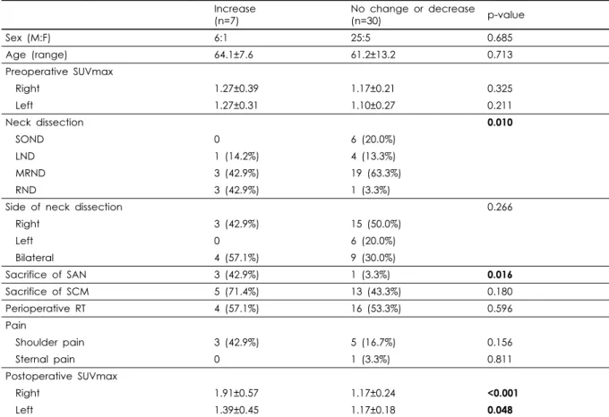
Table 1. Patients’ characteristics according to visual analysis of FDG uptake change of the SCJ
Age Sex Neck dissection Side SAN SCM RT or RAI Side of uptake
Male 72 RND Right Sacrificed Resected Postop. RT Right
Female 69 LND Right Preserved Preserved Postop. RAI Right
Male 69 RND Bilateral Sacrificed(right) Preserved Postop. RAI- Bilateral
Male 58 RND Right Sacrificed Resected Postop. RAI Right
Male 53 MRND Bilateral Preserved Resected(right) Postop. RT Right
Male 55 MRND Right Preserved Resected Postop. RT Left
Male 66 MRND Right Preserved Resected Postop. RT Right
SAN; spinal accessory nerve, SCM; sternocleidomastoid muscle, RT; radiation therapy, RAI; radioactive iodine therapy, PTC;
papillary thyroid cancer, LND; lateral neck dissection, MRND; modified radical neck dissection, RND; radical neck dissection Table 2. Visual analysis by PET/CT in 7 patients presenting with FDG uptake change of the SCJ
changes in shoulder instability, but not an indicator for clin- ical manifestation of local pain (Fig. 1).
However, quantitative assessment failed to show any cor- relation between perioperative parameters and increased up- take around SCJ, which suggests that minimal quantitative changes may occur without clinical significance in patients after ND. The quantitative analysis of FDG PET/CT re- vealed that changes in SCJ occurred in 54.1% (n=20) of patients who underwent ND. This exceeded our expectation, as only nine patients complained of symptoms associated
with SCJ. The incidence of increased uptake (n=7), accord- ing to qualitative assessment, was roughly similar to the number of patients presenting clinical symptoms. Three among seven patients with increased uptake had shoulder or sternal pain. Hence, quantitative assessment may not be necessary if post-ND patients with minimal changes around SCJ do not show any significant changes by qualitative analysis of pre- and postoperative PET/CT imaging.
Because SCJ-related problems after ND, such as sterno- costoclavicular hyperostosis, osteitis condensans, Friedrich’s
Increase (n=20)
No change or decrease
(n=17) p-value
Sex (M:F) 17:3 14:3 0.587
Age (range) 60.6±14.7 62.7±9.1 0.598
Preoperative SUVmax
Right 1.17±0.29 1.20±0.20 0.755
Left 1.11±0.35 1.16±0.18 0.554
Neck dissection 0.299
SOND 2 (10.0%) 4 (23.5%)
LND 4 (20.0%) 1 (5.9%)
MRND 10 (50.0%) 12 (70.6%)
RND 4 (20.0%) 0
Side of neck dissection 0.972
Right 10 (50.0%) 8 (47.1%)
Left 3 (15.0%) 3 (17.6%)
Bilateral 7 (35.0%) 6 (35.3%)
Sacrifice of SAN 4 (20.0%) 0 0.073
Sacrifice of SCM 10 (50.0%) 8 (47.1%) 0.560
Perioperative RT 10 (50.0%) 10 (58.8%) 0.419
Pain
Shoulder pain 6 (30.0%) 2 (11.8%) 0.174
Sternal pain 0 1 (5.9%) 0.459
Postoperative SUVmax
Right 1.48±0.51 1.12±0.21 0.010
Left 1.31±0.29 1.10±0.16 0.009
SOND; supraomohyoid neck dissection, LND; lateral neck dissection, MRND; modified radical neck dissection, RND; radical neck dissection, SAN; spinal accessory nerve, SCM; sternocleidomastoid muscle, RT; radiation therapy.
Table 3. Comparison between uptake + and uptake – group in quantitative measurement
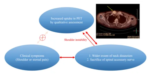
Fig. 1. Increased uptake of SCJ in postoperative PET by qualitative assessment and its association with operative findings.
Increased uptake in PET around SCJ may result from shoulder instability after neck dissection, especially those with wider extent and sacrifice of spinal accessory nerve. There was no significant correlation between clinical symptom and increased uptake, which indicated that uptake in SCJ of postoperative PET is an indicator for subclinical changes in shoulder instability, but not an indicator for clinical manifestation of local pain. PET image showed FDG uptake was diffusely appeared along the articular surface (arrow) (M/55, Tongue cancer, MRND type II (SCM sacrifice)).
disease, and Tietze’s syndrome10,11), are often benign and self-limiting, it is highly probable for physicians to overlook SCJ-related problems. In addition, a shoulder disability
caused by ND affects not only SCJ but acromioclavicular joint. Nevertheless, increased uptake around SCJ is more frequently observed than other joints associated with shoulder
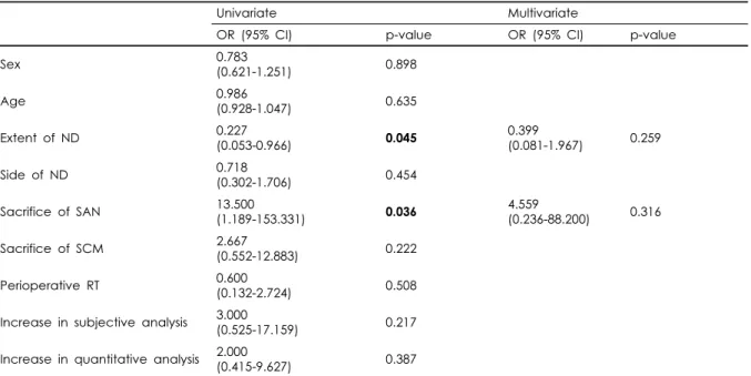
Univariate Multivariate
OR (95% CI) p-value OR (95% CI) p-value
Sex 0.783
(0.621-1.251) 0.898
Age 0.986
(0.928-1.047) 0.635
Extent of ND 0.227
(0.053-0.966) 0.045 0.399
(0.081-1.967) 0.259
Side of ND 0.718
(0.302-1.706) 0.454
Sacrifice of SAN 13.500
(1.189-153.331) 0.036 4.559
(0.236-88.200) 0.316
Sacrifice of SCM 2.667
(0.552-12.883) 0.222
Perioperative RT 0.600
(0.132-2.724) 0.508
Increase in subjective analysis 3.000
(0.525-17.159) 0.217
Increase in quantitative analysis 2.000
(0.415-9.627) 0.387
ND; neck dissection, SAN; spinal accessory nerve, SCM; sternocleidomastoid muscle, RT; radiation therapy.
Table 4. Uni- and multi-variate analysis for the correlation between postoperative shoulder or sternal pain and perioperative parameters
problem. In this regard, physicians might underestimate the clinical significance and importance of SCJ-related prob- lems post-ND. Hence, if patients show increased uptake by qualitative analysis of pre- and postoperative PET/CT imag- ing, a careful follow-up is highly recommended to detect any SCJ-related problems, since these patients may have subclinical changes around SCJ.
In this study, SAN sacrifice was shown to be one of the factors that were significantly related to the changes in SCJ.
This finding is in line with previous reports. It has been reported that postoperative stability of SCJ is mainly de- pendent on the integrity of SAN12), and denervation change of the trapezius muscle innervated by the SAN plays a key role in SCJ-related problems.13) Cho et al. suggested that the volume ratio of the trapezius muscles measured on a CT scan may predict the development of shoulder dysfunc- tion after ND.14) However, a relatively large intra- and/or inter-observer variability is unavoidable for this method, as they adopted a freehanded drawing technique to measure the trapezius muscle volumes on CT scans.
On the other hand, it has been reported that SCM is re- sponsible for SCJ-related problems post-ND. Cantlon and Gluckman suggested that sternocleidomastoid section ren- dered the clavicle relatively more prominently and that shoulder drooping widened and deepened the supra- clavicular fossa, further increasing the joint prominence af-
ter radical ND.15) It seems that the SCM section can also contribute to SCJ-related problems by decreasing the blood supply to SCJ. However, in this current study, no statisti- cally significant correlation was found between the SCM section and the changes in SCJ on FDG PET/CT.
Our study showed no significant correlation between peri- operative radiation therapies and FDG uptake changes in SCJ. According to a review by Ward and Langdon, radiation therapy might be partly responsible for SCJ-related prob- lems after ND, causing avascular necrosis, resulting in stress fracture in the clavicular head.16) Radiation-induced osteo- necrosis is related to the absorbed radiation dose, and the probability of radiation-induced bony changes depends on many factors, including total dose and dose per fraction.
Tolerance doses causing a maximum severe complication rate of 50% within the first five years after irradiation range from 20Gy to 30Gy and 65Gy to 70Gy for single and frac- tionated doses, respectively. Generally, adjuvant radiation to manage head and neck cancer exposes minimal amounts of radiation to SCJ, or SCJ is excluded altogether from the radiation planning. Therefore, the probability of radia- tion-induced changes around SCJ seems to be low in pa- tients after ND.
Lastly, osseous metastases should be considered for those with increased FDG uptake around SCJ, although the in- cidence of this is highly rare. Osseous metastases are often
encountered in patients who have been followed-up using FDG PET/CT. Metastatic bony lesion is usually represented by a discrete focal uptake with a high SUV value. Contrary to the features of osseous metastases, diffused FDG uptake was observed along the articular surface, and SUV value was low with an approximate mean SUVmax of 1.57 in quan- titative analysis.6) Accordingly, the possibility of osseous metastasis or active joint inflammation could be excluded from the differential diagnosis.
This study has some limitations to consider. The number of included patients was relatively small. In addition, the retrospective nature of this study may have resulted in po- tential bias. However, to the best of our knowledge, this is the first study to evaluate postoperative changes around SCJ using FDG PET/CT imaging. Although rare, the clin- ical significance of SCJ-related problems after DN is not negligible. Subjective analysis by an experienced nuclear physician may be enough to detect possible subclinical shoulder instability and ongoing changes in SCJ. Although the clinical symptom is not correlated with the changes on PET/CT scan, we believe that patients with a wide extent of ND and sacrifice of SAN may benefit more from should- er rehabilitation if there is an increase in FDG uptake, de- termined qualitatively, around SCJ on PET/CT scan.
References
1) Cantlon GE, Gluckman JL. Sternoclavicular joint hypertrophy following radical neck dissection. Head Neck Surg. 1983;5:
218-221.
2) Halfpenny W, Goodger N. Early fracture of clavicle following neck dissection. J Laryngol Otol. 2000; 114:714-715.
3) Lee KH, Cho A, Choi YJ, Lee SW, Ha YJ, Jung SJ, et al. The role of (18) F-fluorodeoxyglucose-positron emission tomography in the assessment of disease activity in patients with takayasu arteritis. Arthritis Rheum. 2012; 64:866-755.
4) Soussan M, Augier A, Brillet PY, Weinmann P, Valeyre D.
Functional imaging in extrapulmonary sarcoidosis: FDG-PET/CT
and MR features. Clin Nucl Med. 2014; 39:146-159.
5) Cohen C, Mekinian A, Uzunhan Y, Fauchais AL, Dhote R, Pop G, et al. 18F-fluorodeoxyglucose positron emission tomog- raphy/computer tomography as an objective tool for assessing disease activity in Sjögren's syndrome. Autoimmun Rev. 2013;
12:1109-1114.
6) Hong YH, Kong EJ. (18F)Fluoro-deoxy-D-glucose uptake of knee joints in the aspect of age-related osteoarthritis: a case-con- trol study. BMC Musculoskeletal Disord. 2013; 22:141-149.
7) Cappiello J, Piazza C, Giudice M, De Maria G, Nicolai P.
Shoulder disabil-ity after different selective neck dissections (levels II–IV versus levels II–V): a comparative study.
Laryngoscope. 2005;115:259–263.
8) Carr SD, Bowyer D, Cox G. Upper limb dysfunction following selectiveneck dissection: a retrospective questionnaire study.
Head Neck. 2009;31:789–792.
9) Cheng PT, Hao SP, Lin YH, Yeh AR. Objective comparison of shoulderdysfunction after three neck dissection techniques. Ann Otol Rhinol Laryngol. 2000;109(8 Pt 1):761–766.
10) Berthelot JM, Mabille A, Nomballais MF, Maugars Y, Robert R, Prost A. Osteitis condensans of the clavicle: does fibrocartilage play a role? A report of two cases with spontaneous clinical and roentgenographic resolution. Rev Rhum Engl Ed. 1995;62:
501-506.
11) Kurguzov OP, Solomka Ia A, Kuznetsov NA. Tietze's syndrome.
Khirurgiia. 1991; 9:161-167.
12) McArdle PJ, Kalbassi R, Ilankovan V. Stability of the sternocla- vicular joint. A retrospective study. Br J Oral Maxillofac Surg.
2003; 41:12-15.
13) Ewing MR, Martin H. Disability following radical neck dis- section; an assessment based on the postoperative evaluation of 100 patients. Cancer. 1952; 5:873-883.
14) Cho JG, Lee N, Park MW, Baek SK, Kwon SY, Jung KY, et al.
Measurement of the trapezius muscle volume: A new assessment strategy of shoulder dysfunction after neck dissection for the treatment of head and neck cancers. Head Neck. 2015;5:
619-623.
15) Cantlon GE, Gluckman JL. Sternoclavicular joint hypertrophy following radical neck dissection. Head Neck Surg. 1983;5:
218-221.
16) Ord RA, Langdon JD. Stress fracture of the clavicle. A rare late complication of radical neck dissection. J Maxillofac Surg. 1986;
14:281-284.