This is an Open Access article distributed under the terms of the Creative Commons Attribution Non-Commercial License (http: //creativecommons.org/licenses/by- nc/4.0/) which permits unrestricted non-commercial use, distribution, and reproduction in any medium, provided the original work is properly cited.
© 2019 THE KOREAN SOCIETY OF MYCOLOGY.
Accepted: November 14, 2019 Revised: November 11, 2019 Received: June 07, 2019 https://doi.org/10.4489/KJM.20190044 Kor. J. Mycol. 2019 September, 47(4): 393-406
OPEN ACCESS pISSN : 0253-651X eISSN : 2383-5249
RESEARCH ARTICLE
무 탄저병을 일으키는 Colletotrichum속의 종 동정 및 병원성
최효원1,*, 김점순2 , 홍성기1 , 이영기1
1국립농업과학원 작물보호과, 2유해생물팀
Identification and Pathogenicity of
Colletotrichum Species Causing Anthracnose of Korean Radish ( Rhaphanus sativus) in
Korea
Hyo-Won Choi1,*, Jeomsoon Kim2, Sung Kee Hong1, Young Kee Lee1
1Crop Protection Division
2Microbial Safety Team, National Institute of Agricultural Sciences, Wanju 55365, Korea
*Corresponding author: [email protected]
ABSTRACT
In August 2017, anthracnose symptoms were observed on the petioles and leaf veins of Korean radish (Rhaphanus sativus) in Hongcheon, Jeongseon, and Pyeongchang of the Gangwon province, Korea. Many grayish to dark-brown spots of 1-2 mm in diameter, appeared on the lower surface and leaf veins of the radish leaves. The spots gradually enlarged and coalesced to form dark-brown irregular lesions. Ten Colletotrichum isolates were obtained from the affected tissues of the Korean radish. Out of them, eight isolates were identified as C. higginsianum and two isolates were identified as C. truncatum based on morphological characteristics and multi-locus molecular phylogenetic analysis using the internal transcribed spacers and intervening 5.8S rDNA (ITS), partial beta-tubulin gene (TUB2), partial actin gene (ACT), and partial chitin synthase-1 gene (CHS1). The pathogenicity test was carried out on wounded and unwounded Korean radish (cv. Siraegimu and Osarimu), and Chinese cabbage (cv. Chuno and Smart) by inoculating with a spore suspension. All isolates except one C. truncatum isolate developed typical symptoms on both wounded and unwounded Korean radish. In Chinese cabbage, only the plants inoculated with C.
higginsianum isolates developed symptoms regardless of the wound. This is the first report of anthracnose caused by C. truncatum on Korean radish in Korea.
Keywords: Anthracnose, Colletotrichum higginsianum, Colletotrichum truncatum, Korean radish
서론
우리나라의 4대 채소 중 하나인 무(Raphanus sativus L.)는 배추과에 속하는 뿌리채소 작물로 주로 김치, 단무지 등의 요리 재료로 사용되며, 외국에서는 꼬투리 부분을 식용으로 섭취하기도 하고 일부 품종은 사료 또는 유지 작물로 재배되고 있다[1].
국내에서 무에 발생하는 병은 무름병, 시들음병 등 22종이 보고되어 있으며, 이 중 균류에 의 한 병은 14종이 기록되어 있다[2]. 무 탄저병은 잎과 잎줄기에 약간 움푹 들어간 병반을 형성 하며 갈색 내지 흑색의 테두리를 가진 부정형의 회색 반점으로 나타나는 것이 특징이며, 심 하게 발생할 경우 잎이 황화되고 결국 떨어지는 등의 피해를 준다. 국내의 경우, 무 탄저병균 으로 Colletotrichum higginsianum만이 알려져 있으며, 같은 배추과 작물에 속하는 배추에는 C.
higginsianum과 C. dematium이 관여하는 것으로 보고된 바 있다[3]. 반면, 일본에서는 무 탄저병에 C. higginsianum외에 C. dematium이 관여하며[4], 중국에서는 Chinese flowering cabbage 탄저병에 C.
higginsianum과 C. truncatum이 관여한다고 보고하였다[5]. 전 세계적으로 무 탄저병 발생에 의한 수량감소 등의 심각한 피해는 알려진 바 없으나, 병 발생시 적절한 방제를 실시하지 않거나 탄저 병 발생에 유리한 다습한 조건이 지속되면 그 피해가 예상된다.
본 연구에서는 탄저병 증상이 발생한 무 잎으로부터 Colletotrichum균을 분리하였고, 균학적 특 성과 multilocus sequence typing 분석으로 2종의 균을 동정하였으며, 무와 배추 각 2개 품종을 대상 으로 병원성 검정을 수행한 결과를 보고하고자 한다.
재료 및 방법
병원균 분리 및 보존
2017년 8월, 강원도 고랭지의 무 재배 지역인 홍천군, 정선군, 평창군의 재배포장에서 병든 시료 를 채집하였다. 병원균 분리를 위하여 채집한 잎과 줄기에 형성된 병징의 경계부위를 3×3 mm 크 기로 잘라 그 조각을 1% 차아염소산나트륨 (NaOCl) 용액으로 1분간 표면살균하고, 멸균수로 3회 세척한 후 물기를 제거하여 물한천 배지(water agar, WA)에 치상하였다. 3-5일 후, 시료 절편에서 자라난 균총으로부터 Colletotrichum균을 단포자 분리하였다. 순수 분리된 균주는 감자한천배지 (potato dextrose agar, PDA) 사면배지에 이식하여 배양하고, 10°C에 보존하면서 실험에 사용하였다.
형태적 특성 조사
수집 균주의 균학적 특성을 조사하기 위하여 이전에 보고된 Colletotrichum균의 형태적 특성 조 사 방법에 근거하여 synthetic nutrient poor agar(SNA)배지, PDA 배지, oatmeal agar(OA)배지에서의 형태적, 배양적 특성을 조사하였다[6]. 각 균주를 PDA 배지에 접종하고 25˚C에서 5일간 배양한 후, 직경 5 mm의 cork borer로 균총을 잘라 새로운 SNA, PDA, OA 배지에 접종하였다. 이후 SNA plate는 22˚C, 12h/12h near ultraviolet(NUV)/암조건인 배양기에서 14일간 배양한 후, 분생포자과 (conidiomata), 분생포자 및 부착기의 특징을 조사하였다. 또한 PDA와 OA plate는 25˚C, 암조건에 서 7일간 배양하였고, 이후 균총 생장속도와 모양 등을 조사하여 기술된 종과 비교하였다[6, 7].
분리균주의 DNA 추출 및 염기서열 분석
단포자 분리한 균주의 genomic DNA를 추출하기 위하여 10개 균주를 각각 potato dextrose broth (PDB) 배지에 접종하고, 25°C에서 7일간 정치배양하였다. 균사체를 miracloth로 회수하고 동결 건조시킨 후, 곱게 마쇄하여 Choi 등[8]의 방법으로 genomic DNA를 추출하고, -20°C에 보관하면 서 실험에 사용하였다. DNA 염기서열 분석은 Damm 등[6]의 방법을 참고하여 internal transcribed spacers and intervening 5.8S rDNA (ITS), partial beta-tubulin gene (TUB2), partial actin gene (ACT), partial chitin synthase-1 gene (CHS-1) 부위를 대상으로 실시하였다. 각 부위의 PCR 증폭은 각각 ITS 1F/ITS 4[9], T1/BT4R[10], ACT-512F/ACT-783R[11], CHS-354R/CHS-79F[11] 프라이머쌍을 사용하였다. PCR 반응액은 100 ng/μL의 template DNA, 10×Taq buffer, 2 mM dNTPs, 10 pmole/
μL의 각 프라이머쌍, 0.5 unit의 Taq DNA polymerase를 총량 50 μL로 만들었다. PCR 증폭된 밴 드는 1% agarose gel에 전기영동한 후 염색하여 관찰하였고, 밴드가 확인된 PCR 산물은 Wizard SV Gel & PCR Clean-up System kit (Promega, San Luis Obispo, CA, USA)를 사용하여 정제한 후, direct sequencing을 통해 염기서열을 분석하였다. 분석된 염기서열은 Clustal W 소프트웨어를 이 용하여 정렬하였고, nucleotide의 유사도를 계산하고 MEGA 6.0 프로그램을 이용하여 neighbor-
joining 분석법으로 계통수를 작성하였다. Colletotrichum higginsianum의 경우, 계통수 작성을 위한 reference 균주로 C. destructivum 종 복합체에 속하는 C. fuscum, C. bryoniicola, C. tabaci, C. ocimi, C.
destructivum 균주와 outgroup으로 Colletotrichum sp. CBS 107.40을 사용하였다. C. truncatum은 굽은 형태의 포자를 가진 Colletotrichum종인 C. lineola, C. dematium, C. circinans, C. phaseolorum 등을 비 교하였고, outgroup으로 C. lindemuthianum CBS 151.28을 사용하였다.
병원성 검정
수집 균주의 병원성을 확인하기 위하여 10개 분리 균주를 무와 배추 잎에 접종하여 병원성 검정 을 수행하였다. 국내 시판 품종 중 무는 시래기무와 오사리무, 배추는 추노배추와 스마트배추를 대상으로 하였다. 종자를 파종하고 온실에서 두 달간 건전하게 생육시킨 무와 배추의 잎을 사용 하였다. 생육상태가 비슷한 무와 배추 잎을 따내어 70% 에탄올을 분무하여 잎 표면을 소독하고 멸균수로 3회 세척한 뒤 무균대에서 건조시켜 준비하였다. 접종원은 무에서 분리한 10개 균주를 모두 사용하였는데, 각 균주를 PDA 배지에 10일간 배양한 후, 멸균수를 붓고 포자를 긁어 2겹의 miracloth로 걸러내었고, 포자현탁액 농도를 1×10⁶ spores/mL로 조정하였다. 상처 접종은 멸균한 바늘을 이용하여 상처를 낸 후 포자현탁액 10 μL를 접종하였고, 무상처 접종은 상처없이 포자현 탁액을 접종하였으며, 대조구는 상처와 무상처 부위에 10 μL의 멸균수를 떨어뜨렸다. 이후 멸균 수에 적신 키친타올을 깐 플라스틱 박스에 접종한 무와 배추 잎을 넣고 실온에 두었다. 접종 7일 후, 무와 배추 잎에 형성된 병반의 길이를 측정하여 병 발생 정도를 조사하였으며, 병든 부위에서 병원균의 재분리 여부를 확인하였다.
결과
병징
주로 잎에 발생하는데 초기에는 회색 내지 짙은 갈색의 작은 점무늬 증상이 잎자루와 주맥에 형 성된다(Fig. 1). 병이 계속해서 진전되면 점무늬가 서로 합쳐지면서 짙은 갈색 내지 검정색의 불규 칙한 병반이 형성되고 약간 움푹 들어가며, 심해지면 결국 잎이 말라 죽게 된다. 습한 상태가 지속 되면 자연 병반에서도 분생포자반(acervuli)이 관찰되기도 한다.
형태적 특성
고랭지 무 재배포장에서 수집한 병든 잎에서 총 10개의 균주를 순수 분리하였으며, 분리지역 등 의 정보는 Table 1에 기술하였다. 10개 균주 중 8개 균주는 분생포자의 형태가 원통형(cylindrical) 이었고, 다른 2개 균주는 낫 모양(falcate)의 굽은 형태를 나타내었다(Fig. 2A, 3A). 원통형의 포자를
가진 첫번째 그룹 8개 균주는 모두 동일한 형태의 배양적 특성을 가졌으며, 7일간 배양한 균총의 직경은 PDA 배지에서 약 60.7 mm, OA 배지에서는 약 42.3 mm로 PDA 배지에서 생장속도가 더 빨
랐다(Fig. 2D, 2E, Table 2). 반면 낫 모양의 포자를 가진 2개 균주는 PDA 배지에서 진한 올리브색을 띠었고, 7일간 배양한 결과 PDA 배지에서는 36.3 mm, OA 배지에서는 48.3 mm의 크기를 나타내 어 OA 배지에서의 생장속도가 더 빠른 것으로 조사되었다(Fig. 3D, 3E, Table 3). 원통형 포자를 형 성하는 균주는 SNA 배지에서 옅은 오렌지색의 분생포자반(acerveuli)을 형성하였고(Fig. 2C), 분생 포자는 격벽이 없고 약간 휘어지거나 직선형의 원통모양을 나타냈으며, 크기는 11.1-18.1 × 4.2- 6.2 μm이었다(Fig. 2A). 또한 이들 균주의 부착기(appressoira)는 갈색을 띠며, 전체적인 테두리 부 분이 타원형이고, 간혹 불규칙적인 모양을 보였다(Fig. 2B). 낫 모양의 포자를 가진 균주의 분생 포자반은 크림색을 나타냈고(Fig. 3C), 분생포자는 낫 모양이며 휘어졌고, 격벽이 없으며, 크기 는 20.4-28.0 × 3.3-4.4 μm이었다(Fig. 3A). 부착기는 옅은 갈색이며, 테두리 부분은 전체적으로 둥 글고 타원형이었으며, 간혹 불규칙적인 모양을 나타냈다(Fig. 3B). 이와 같은 형태적 특성에 의해 C06210 균주는 C. destructivum species complex에 속하는 종으로, C06209 균주는 C. truncatum으로 동정하였다[6,7].
Fig. 1. Dark brown to black irregular spots on petioles and leaflets of Korean radish anthracnose caused by Colletotrichum species. A, symptoms on petioles and leaflets in Jeongseon; B, weak symptoms on petioles in Pyeongchang.
Table 1. List of Colletotrichum spp. isolates collected from diseased radish leaves and GenBank accession numbers in this study
Species Isolates Locations GenBank Accession Number
ITS TUB2 ACT CHS-1
Colletotrichum higginsianum C06201 Hongcheon
(37.72N 128.39E) MH820152 MK048416 MK118042 MK118052 C06202 Hongcheon
(37.72N 128.39E) MH820153 MK048417 MK118043 MK118053 C06203
(KACC48667) Pyeongchang
(37.63N 128.54E) MH820154 MK048418 MK118044 MK118054 C06204 Pyeongchang
(37.63N 128.54E) MH820155 MK048419 MK118045 MK118055 C06205 Pyeongchang
(37.60N 128.50E) MH820156 MK048420 MK118046 MK118056 C06207 Pyeongchang
(37.60N 128.53E) MH820158 MK048422 MK118048 MK118058 C06208 Jeongseon
(37.43N 128.85E) MH820159 MK048423 MK118049 MK118059 C06210
(KACC48666) Jeongseon
(37.49N 128.85E) MH820161 MK048425 MK118051 MK118061
C. truncatum C06206 Pyeongchang
(37.60N 128.53E) MH820157 MK048421 MK118047 MK118057 C06209
(KACC48665) Jeongseon
(37.49N 128.87E) MH820160 MK048424 MK118050 MK118060 ITS, internal transcribed spacers and intervening 5.8S nrDNA; TUB2, partial beta-tubulin gene; ACT, partial actin gene;
CHS-1, partial chitin synthase-1 gene.
Fig. 2. Morphological characteristics of Colletotrichum higginsianum isolated from infected radish leaves. A, conidia; B, appressoria; C, acervuli; D, colony on PDA; E, colony on OA.
(Scale bars: A, B =10μm, C = 200μm).
Fig. 3. Morphological characteristics of Colletotrichum truncatum isolated from infected radish leaves. A, conidia; B, appressoria; C, acervuli; D, colony on PDA; E, colony on OA.
(Scale bars: A, B =10μm, C = 200μm).
염기서열 분석
형태적 특성에 의해 각각 C. destructivum species complex와 C. truncatum으로 확인된 무 분리 균의 정확한 동정을 위하여 ITS, TUB2, ACT, CHS-1 유전자의 염기서열을 분석하였다. 그 결 과, C. destructivum species complex에 속하는 균주는 C. higginsianum으로 C. truncatum으로 동정 된 균주는 형태적 특성에 의한 결과와 일치하는 것으로 나타났다(Fig. 4, 5). 국내 무에서 분리한 C. higginsianum은 reference인 C. higginsianum Abr 3-1, Abo 1-1, Abr 2-2, MAFF 305635균주와 ITS
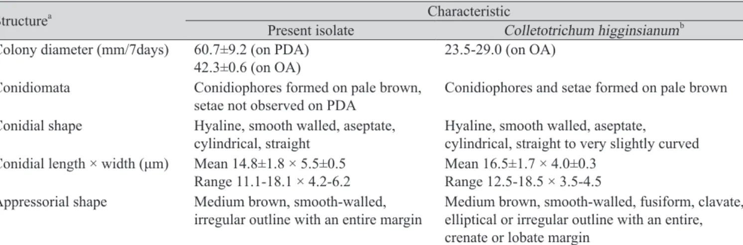
Table 2. Comparison of morphological characteristics between the present isolate (C06210) obtained from infected radish leaves and Colletotrichum higginsianum described previously
Structurea Characteristic
Present isolate Colletotrichum higginsianumb Colony diameter (mm/7days) 60.7±9.2 (on PDA)
42.3±0.6 (on OA) 23.5-29.0 (on OA) Conidiomata Conidiophores formed on pale brown,
setae not observed on PDA Conidiophores and setae formed on pale brown Conidial shape Hyaline, smooth walled, aseptate,
cylindrical, straight Hyaline, smooth walled, aseptate,
cylindrical, straight to very slightly curved Conidial length × width (μm) Mean 14.8±1.8 × 5.5±0.5
Range 11.1-18.1 × 4.2-6.2 Mean 16.5±1.7 × 4.0±0.3 Range 12.5-18.5 × 3.5-4.5 Appressorial shape Medium brown, smooth-walled,
irregular outline with an entire margin Medium brown, smooth-walled, fusiform, clavate, elliptical or irregular outline with an entire, crenate or lobate margin
aColony characters and growth rates were investigated at 7 days after incubation on PDA and OA. The characteristics of conidia and appressoria were investigated after growth on PDA at 10 days
bC. higginsianum described by Damm et al. (2014).
Table 3. Comparison of morphological characteristics between the present isolate (C06209) obtained from infected radish leaves and Colletotrichum truncatum described previously
Structurea Characteristic
Present isolate Colletotrichum truncatum b Colony diameter (mm/7days) 36.3±1.2 (on PDA)
48.3±0.6 (on OA) 19.0 (on OA)
Conidiomata Acervular,conidiophores and setae formed
directly on hyphae Acervular, conidiophores and setae formed directly on hyphae
Conidial shape Hyaline, smooth-walled to verruculose, aseptate, usually slightly curved, ending at the round and truncate base
Hyaline, smooth-walled to verruculose, aseptate, usually slightly curved, ending at the round and truncate base Conidial length × width (μm) Mean 23.1±1.4 × 4.0±0.3
Range 20.4-28.0 ×3.3-4.4 Mean 21.8±1.9 × 3.8±0.3 Range 16.5-26.0 × 3-4.5 Appressorial shape Light to medium brown,
entire edge to lobed,
outline roundish to ellipsoidal or clavate
Light to medium brown, entire edge to lobed,
outline roundish to ellipsoidal or clavate
aColony characters and growth rates were investigated at 7 days after incubation on PDA and OA. The characteristics of conidia and appressoria were investigated after growth on PDA at 10 days
bC. truncatum described by Damm et al. (2009).
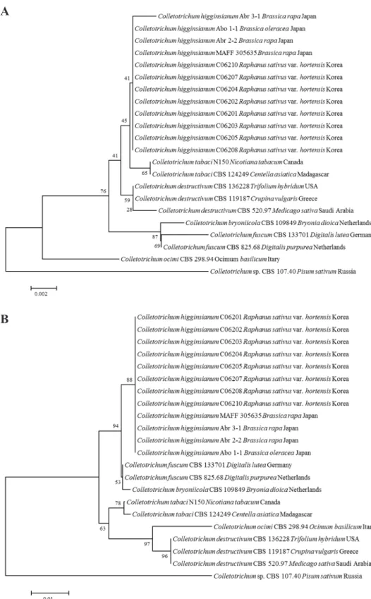
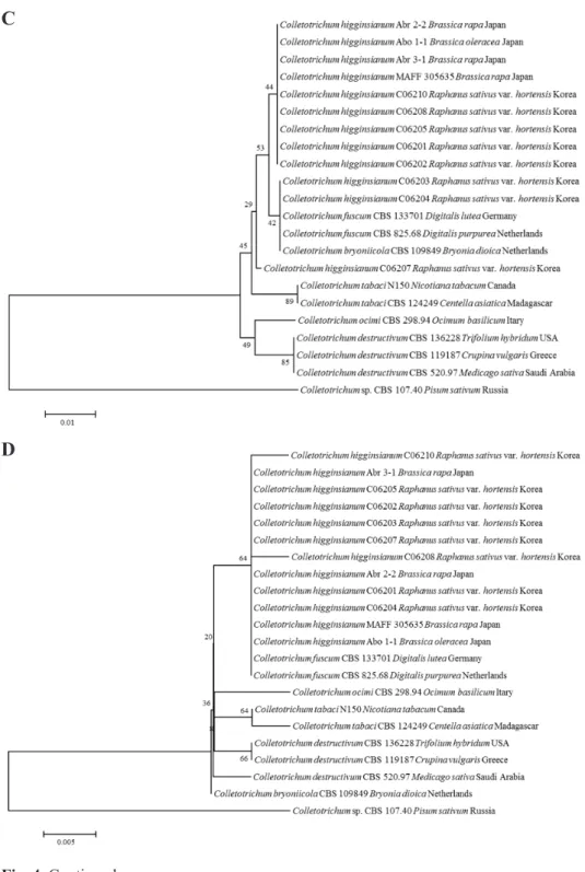
Fig. 4. Phylogenetic tree based on the internal transcribed spacers and intervening 5.8S nrDNA; A, partial beta-tubulin; B, partial actin; C, partial chitin synthase-1; D, genes sequence data for Colletotrichum higginsianum isolated from infected radish leaves and reference species(including C. destructivum species complex). Sequence data were obtained from the NCBI GenBank database. The tree was generated using neighbor-joining analysis
A
B
는 99.8-100%, TUB2는 100%, ACT은 99.6-100%, CHS-1은 99.6-100%의 상동성이 있었다. 그러 나 이 중 ACT, CHS-1 유전자의 경우, C. fuscum reference인 CBS 133701, CBS 825.68 균주와의 상 동성도 99.6-100%로 나타나 C. higginsianum의 종 구분을 위한 마커로는 효율적이지 않은 것으 로 판단된다(Fig. 4C, 4D). 한편, C. truncatum 역시 4개의 유전자 염기서열 분석에서 reference 균
Fig. 4. Continued.
D C
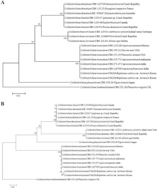
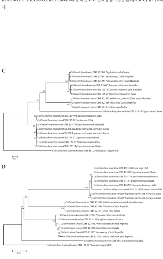
주인 CBS 371.67 등 6균주와 동일한 그룹을 형성하였고, 각각의 상동성은 ITS 부위는 99.8-100%, TUB2는 99.4%, ACT은 98.8-100%, CHS-1은 98.4-99.2%를 나타냈다. 사용된 4개 유전자 부위 는 C. truncatum의 종 구분에는 비교적 효율적이었으나, ITS와 TUB2의 경우는 C. dematium과 C.
lineola를 구분하지 못하는 것으로 나타났다(Fig. 5C, 5D). 본 연구에서 분석된 각 균주의 염기서
Fig. 5. Phylogenetic tree based on the internal transcribed spacers and intervening 5.8S nrDNA; A, partial beta-tubulin; B, partial actin; C, partial chitin synthase-1; D genes sequence data for Colletotrichum truncatum isolated from infected radish leaves and reference species(including curved Colletotrichum species). Sequence data were obtained from the NCBI GenBank database. The tree was generated using neighbor-joining analysis and Kimura 2-parameter model. Bar represents the number of nucleotide substitutions per site.
A
B
열은 NCBI GenBank에 등록하였고, accession number는 Table 1에 기술하였다. 또한, 형태적 특성 과 염기서열분석에 의해 최종적으로 동정된 무 분리균 중 C06203, C06210, C06209 균주는 각각 KACC48667, KACC48666, KACC48665의 등록번호로 한국농업미생물센터(KACC)에 기탁되었 다.
Fig. 5. Continued.
C
D
병원성 검정
분리 균주의 병원성을 확인하기 위하여 건전하게 키운 무와 배추 잎을 대상으로 상처 접종과 무 상처 접종을 구분하고, 포자현탁액을 떨어뜨려 접종하였다. 접종 7일 후, 멸균수를 접종한 대조구 에서는 병징이 관찰되지 않았으나, C. higginsianum을 접종한 무와 배추 잎에서는 추노배추 무상 처구의 1개 균주(C06201)를 제외하면, 상처 여부와 상관없이 모든 접종구에서 병징이 관찰되었 다(Table 4). 그러나 C. truncatum 접종구에서는 1개 균주(C06209)가 무에만 병을 일으켰으며, 2개 균주 모두 배추에는 상처와 무상처구에서 모두 병징이 관찰되지 않았다(Table 4). 접종구에서 나 타난 병징에서 모두 C. higginsianum과 C. truncatum이 재분리되는 것으로 확인되었다.
고찰
본 연구에서는 국내 고랭지 무 재배지에서 발생한 탄저병 증상으로부터 2종의 Colletotrichum 을 분리하였고, 이 2종은 형태적 특성과 4개의 DNA 염기서열분석에 의해 C. higginsianum과 C.
truncatum으로 동정되었고, 이 중 C. higginsianum은 무와 배추에 모두 병원성을 나타냈으나 C.
truncatum은 무에만 병을 일으키는 것으로 확인되었다. 지금까지 국내에는 무탄저병균으로 C.
higginsianum만 보고되어 있었으며, 배추에서는 2001년 C. higginsianum 외에 C. dematium이 모 잘록병과 탄저병을 일으킨다고 보고되었다[3]. 일본에서는 무 탄저병에 C. higginsianum과 C.
dematium이 관여하는 것으로 보고된 바 있으며[4], 중국에서는 배추과에 속하는 Chinese flowering cabbage (Brassica parachinensis)의 탄저병균으로 C. higginsianum 외에 C. truncatum이 새롭게 보고 된 바 있다[5]. 일본에서 C. higginsianum과 C. dematium에 의한 무 탄저병의 포장에서의 병징은 매 우 비슷하여 구분이 쉽지 않다고 하였으며, 이는 국내 무 탄저병에서도 유사한 경향이었다. 그러 Table 4. Pathogenicity of Colletotrichum higginsianum and C. truncatum isolates to the leaves of Korean radish and Chinese cabbage by artificial inoculation
Species Isolate Pathogenicitya to Korean radish Pathogenicity to Chinese cabbage cv. Siraegimu cv. Osarimu cv. Chuno cv. Smart Unwounded Wounded Unwounded Wounded Unwounded Wounded Unwounded Wounded
C. higginsianum C06201 + + + + - ++ ++ ++
C06202 ++ ++ ntb ++ ++ +++ +++ ++
C06203 ntb ++ ntb + ++ +++ ++ ++
C06204 ++ ++ + ++ ++ ++ ++ ++
C06205 ++ ++ + ++ ++ ++ ++ ++
C06207 + ++ ++ ++ ++ ++ + +
C06208 ++ + + +++ ++ ++ ++ +
C06210 +++ ++ + +++ ++ + + ++
C. truncatum C06206 - - - - - - - -
C06209 ++ ++ ++ ++ - - - -
Control - - - - - - - -
aPathogenicity was rated based on the lesion formation seven days after inoculation. +, 1-5 mm of lesion length; ++, 5.1-10.0 mm of lesion length; +++, 10.1mm < of lesion length; -, no symptom.
bnt : not tested
나 일본의 무 탄저병의 경우, C. higginsianum에 비해 C. dematium이 더 심하게 병을 일으킨다고 보 고하였고, 중국의 Chinese flowering cabbage 탄저병의 경우에는 병징에 차이는 없지만, 병이 발생 하는 시기에 차이가 있다고 보고하였다[5,6]. 따라서 국내에서도 무 탄저병균 2종의 발생시기 차 이 혹은 지역적 차이 등의 조사를 위한 자세한 병 발생 연구가 필요할 것으로 생각된다.
Colletotirhcum의 종 동정은 전통적으로 포자의 형태와 크기, 강모(setae)의 유무, 균사 생장속도 및 부착기의 형태 등 형태적 특성에 의해 이루어졌다[12]. 그러나 이와 같은 형태적 동정은 기준 이 되는 특성이 종 간에 서로 겹치거나, 환경 조건에 의해 형태나 크기의 변화가 있기 때문에 정 확한 동정이 어려운 것으로 알려져 있다[12]. 따라서 최근에는 형태적 특성과 분자생물학적 특성 을 함께 이용한 Colletotrichum species complex 연구가 활발히 수행되었다[13,14]. 본 연구에서도 2 종의 Colletotricum균의 형태적 특성에 의한 동정은 species complex 수준으로만 가능했으며, 정확 한 동정은 유전자 염기서열 분석에 의해 이루어졌다. 최근에는 종 동정을 위해 염기서열분석을 할 때, ITS region 이나 translation elongation factor 1 alpha 등 특정한 1개 부위가 아니라 2~3개 이상 여러 유전자 부위를 대상으로 하고 있는데, 이는 1개 부위에 의한 유전정보로 종을 구분하기에 충 분하지 않기 때문이다[13]. 본 연구에서 사용한 ACT, CHS-1 유전자의 경우도 C. higginsianum과 C.
fuscum reference 균주 간 상동성이 매우 높아 종간 구분이 어려웠고, C. dematium과 C. lineola 역시 ITS나 TUB2 부위에 의해 구분되지 않는 것으로 나타나 Colletotrichum의 종 구분을 위해서는 다양 한 마커를 사용해야 한다는 것을 뒷받침한다.
한편, C. truncatum은 기주범위가 매우 넓은 종으로 알려져 있으며, 주로 콩과 또는 가지과 작물 에 탄저병을 일으킨다[15]. Damm 등[7]에 의하면 C. truncatum은 네덜란드에서 Brassica sp. 작물에 탄저병을 일으킨다고 하였으며, 중국의 Chinese flowering cabbage에도 탄저병을 일으킨다고 하 였으나[5] 무에서 탄저병을 일으킨다고 보고된 바는 없다. 즉, 본 연구에서 C. truncatum에 의한 무 탄저병은 국내뿐 아니라 세계적으로도 처음으로 확인된 것이다. 특히 우리나라 무 탄저병 시료 에서 분리된 C. truncatum은 CHS-1 유전자 분석에 의해 미국의 리마콩 분리균인 C. truncatum CBS 151.35 균주와 98.4%의 비교적 낮은 상동성을 나타냈기 때문에 종 내에서도 변이가 있을 것으로 추정되며, 기주별 수집 균주를 대상으로 한 유전적 다양성 연구가 필요할 것으로 판단된다. 또한 본 연구에서 무에서만 병원성이 확인되었기 때문에, 그 외의 배추과 작물 및 넓은 기주범위를 감 안한 다양한 작물을 대상으로 병원학적 특성 검정이 수행되어야 할 것으로 생각된다.
지금까지 국내에서 무 탄저병에 의한 심각한 피해는 보고된 바 없으나, 최근 이상기상 및 재 배환경을 고려할 때, 병 발생에 유리한 조건이 되면 그 피해가 예상되는 병으로 판단된다. 따라 서 정확한 병원균 동정을 통해 다양한 원인균을 파악하고, 병원균의 특성을 고려한 저항성 품종 의 육종이나 효과적인 약제의 선발 등 효율적인 관리방안을 마련하는 준비가 필요할 것으로 생 각된다. 이상과 같이 우리나라 무 탄저병에는 기존에 알려진 C. higginsianum외에 새로운 종인 C.
truncatum이 관여하며, 이 종을 국내에서 처음으로 무탄저병균으로 보고하고자 한다.
적요
2017년 8월경, 강원도 홍천군, 정선군, 평창군 소재 무 재배 포장에서 잎자루와 잎의 주맥에 형성 된 탄저병 병징이 관찰되었다. 회색 내지 짙은 갈색을 띤 1-2 mm 크기의 점무늬가 아래 잎과 잎
의 주맥에 형성되었다. 이러한 점무늬는 점차 확대되고 합쳐지면서 갈색을 띤 불규칙한 병반으 로 나타났다. 병든 무 잎에서 10개의 Colletotrichum균을 분리하였다. 분리 균주의 형태적 특성 과 internal transcribed spacers and intervening 5.8S rDNA (ITS), partial beta-tubulin gene (TUB2), partial actin gene (ACT) 및 partial chitin synthase-1 gene (CHS-1) 부위를 사용한 다자위 분자 계통 분석 (multilocus molecular phylogenetic analysis)에 의해 8개 균주는 Colletotrichum higginsianum, 2개 균주 는 C. truncatum으로 동정되었다. 무(시래기무, 오사리무)와 배추(품종: 추노, 스마트)를 대상으로 상처와 무상처로 구분하여 포자현탁액을 떨어뜨려서 접종하는 방법을 사용하여 병원성 검정을 수행하였다. 무에서는 C. truncatum 1개 균주를 제외하고 상처와 무상처 접종에서 모두 병징을 나 타냈고, 배추의 경우는 C. higginsianum 접종구에서만 상처의 유무에 관계없이 병이 발생하였다.
국내에는 기존에 C. higginsianum은 무와 배추의 탄저병균으로 보고되었으며, C. truncatum에 의한 무 탄저병의 발생은 본 연구에서 국내 최초로 보고한다.
ACKNOWLEDGEMENTS
This study was supported by a grant (Project No. PJ012640) from Rural Development Administration, Republic of Korea.
REFERENCES
1. Seo MS, Chung JH, Park BS, Kim JS. Analysis of surgars content by genotypes in 82 radish(Raphanus sativus L.). Kor J Plant Res 2018;31:453-65.
2. The Korean Society of Plant Pathology. List of plant diseases in Korea. 5th ed. Seoul: Korean Society of Plant Pathology; 2009.
3. Park K, Kim CH. Occurrence of anthracnose on cabbage caused by Colletotrichum dematium.
Mycobiology 2001;29:61-2.
4. Sato T, Muta T, Imamura Y, Nojima H, Moriwaki J, Yaguchi Y. Anthracnose of Japanese radish caused by Colletotrichum dematium. J Gen Plant Pathol 2005;71:380-3.
5. He Y, Chen Q, Shu C, Yang M, Zhou E. Colletotrichum truncatum, a new cause of anthracnose on Chinese flowering cabbage (Brassica parachinensis) in China. Trop Plant Pathol 2016;41:183-92.
6. Damm U, O'Connell RJ, Groenewald JZ, Crous PW. The Colletotrichum destructivum species complex – hemibiotrophic pathogens of forage and field crops. Stud Mycol 2014;79:49-84.
7. Damm U, Woudenberg JHC, Cannon PF, Crous PW. Colletotrichum species with curved conidia from herbaceous hosts. Fungal Divers 2009;39:45-87.
8. Choi HW, Kim JM, Hong SK, Kim WG, Chun SC, Yu SH. Mating types and optimum culture conditions for sexual state formation of Fusarium fujikuroi isolates. Mycobiology 2009;37:247-50.
9. White TJ, Bruns T, Lee S, Taylor J. Amplification and direct sequencing of fungal ribosomal RNA genes for phylogenetics. In: PCR Protocols: A guide to methods and application. San Diego:Academic Press; 1990. p. 315-22.
10. Woudenberg JHC, Aveskamp MM, Gruyter J, Spiers AG, Crous PW. Multiple Didymella teleomorphs are linked to the Phoma clematidina morphotype. Persoonia 2009;22:56-62.
11. Carbone I, Kohn LM. A method for designing primer sets for speciation studies in filamentous ascomycetes. Mycologia 1999;91:553-6.
12. Sutton BC. The genus Glomerella and anamorph Colletotrichum. In: Bailey JA, Jear MJ, editors. Colletotrichum: biology, pathology and control. Wallingford :CAB International; 1992.
p. 1-26.
13. Hyde KD, Cai L, Cannon PF, Crouch JA, Crous PW, Damm U, Goodwin PH, Chen H, Johnston PR, Jones EBG, et al. Colletotrichum-names in current use. Fungal Divers 2009;39:147-82.
14. Cannon PF, Damm U, Johnston PR, Weir BS. Colletotrichum-current status and future directions. Stud Mycol 2012;73:181-213.
15. Ford R, Banniza S, Photita W, Taylor PWJ. Morphological and molecular discrimination of Colletotrichum truncatum causing anthracnose on lentil in Canada. Australa Plant Path 2004;33:559-69.