0022-538X/10/$12.00 doi:10.1128/JVI.02484-09
Copyright © 2010, American Society for Microbiology. All Rights Reserved.
Suppressor of Cytokine Signaling 3 Suppresses Hepatitis C Virus Replication in an mTOR-Dependent Manner 䌤
Run-Xuan Shao, Leiliang Zhang, Lee F. Peng, Eileen Sun, Woo Jin Chung, Jae Yong Jang, Wei-Lun Tsai, Guibenson Hyppolite, and Raymond T. Chung*
Gastrointestinal Unit, Department of Medicine, Massachusetts General Hospital, Harvard Medical School, Boston, Massachusetts
Received 25 November 2009/Accepted 27 March 2010
We and others have observed that hepatic levels of suppressor of cytokine signaling 3 (SOCS3) are significantly higher in persons with chronic hepatitis C, particularly those who are nonresponders to interferon (IFN) treatment, than in healthy individuals. However, the relationship between SOCS3 and hepatitis C virus (HCV) replication remains unclear. Given its putative role, we hypothesized that SOCS3 is permissive for viral replication. We therefore used the OR6 cell line, which harbors a genotype 1b full-length HCV replicon, and the genotype 2a full-length HCV strain JFH1 infection system to analyze the effects of SOCS3 overexpression and short hairpin RNA (shRNA)-mediated knockdown on HCV replica- tion. We further analyzed the role of mTOR in the effects of SOCS3 by treating selected cells with rapamycin. OR6 cells and JFH1-infected Huh7.5.1 cells expressed significantly less SOCS3 than control cells. Furthermore, inhibition of HCV replication with the HCV protease inhibitor BILN 2061 restored SOCS3 protein levels. SOCS3 overexpression in OR6 cells and JFH1-infected Huh7.5.1 cells resulted in significantly lower HCV replication than that in the control cells, despite SOCS3-related inhibition of STAT1 phosphorylation and type I IFN signaling. In contrast, JFH1-infected cells with stable SOCS3 knockdown expressed higher levels of HCV proteins and RNA than did control cells. SOCS3-targeting shRNA also knocked down mTOR and phospho-mTOR. The mTOR inhibitor rapamycin reversed the inhibitory effects of SOCS3. In independent investigations, SOCS3 unexpectedly suppressed HCV repli- cation in an mTOR-dependent manner. These findings suggest that increased SOCS3 levels consistently observed in chronic IFN nonresponders may reflect a compensatory host antiviral response to persistent infection and that manipulation of SOCS3/mTOR may offer benefit against HCV infection.
Hepatitis C virus (HCV) is a small, enveloped plus-strand RNA virus in the genus Hepacivirus and the family Flaviviridae.
HCV has a 9.6-kb genome that encodes structural (core, E1, and E2) and nonstructural (NS2 to NS5B) proteins (1). The HCV life cycle and host-virus interactions that determine the outcome of infection have been difficult to study because small- animal models of HCV infection are not available and cell culture models were not developed until recently. The devel- opment of subgenomic and genomic replicons provided a ma- jor breakthrough for the understanding of HCV replication and virus-cell interactions. Recently, several groups have cre- ated a robust genotype 2a full-length HCV replication system (23, 27). Huh7.5.1 cells transfected with in vitro-transcribed genomic RNA from HCV strain JFH1 will successfully secrete virions. Therefore, transfection of Huh7.5.1-derived cells with JFH1 RNA allows for the recovery of a viable JFH1 virus that can then be serially passaged and used for infection-based experimentation.
The family of suppressors of cytokine signaling (SOCS; also known as JAB and SSI) consists of eight members: SOCS1 through SOCS7 and the cytokine-inducible Src homology 2 domain-containing protein (CIS) (14, 25). SOCS1, SOCS2,
SOCS3, and CIS mRNAs are induced by a variety of cytokines and growth factors such as gamma interferon (IFN-␥), inter- leukin-2 (IL-2), IL-3, IL-6, erythropoietin, and prolactin, but the correlation varies among tissue types (14, 22). SOCS family members modulate signaling by several mechanisms, which include inactivation of the Janus kinases (JAKs) by blocking access to both signal transducer and activator of transcription (STAT)- and nuclear factorB (NF-B)-mediated pathways.
SOCS3 is one of the negative regulators of cytokine signal- ing that function via the JAK-STAT pathway (5, 17, 20).
SOCS1 induces STAT3 phosphorylation, while SOCS3 strongly interacts with activated cytokine receptors, such as gp130, to negatively regulate STAT3 phosphorylation (19).
SOCS3 uses its Src homology 2 domain to bind the cytokine receptor with high affinity to attenuate the activities of JAKs via the kinase-inhibitory region in order to abolish STAT3 phosphorylation (4, 22).
Bode et al. have reported that SOCS3 can be induced by HCV core protein and suppress JAK-STAT signaling to block the IFN-induced formation of ISGF3 in cell cultures (2). We and others have observed that levels of hepatic SOCS3 are significantly higher in persons with chronic hepatitis C (CHC), particularly those who are nonresponders to IFN treatment (7, 13, 24), than in healthy individuals. Patients with high tumor necrosis factor alpha (TNF-␣) levels have a poor response to IFN-␣ therapy; it has been proposed previously that this out- come may occur via induction of SOCS3 proteins that interfere
* Corresponding author. Mailing address: GI Unit, Warren 1007, Massachusetts General Hospital, 55 Fruit Street, Boston, MA 02114. Phone: (617) 724-7562. Fax: (617) 643-0446. E-mail: rtchung
@partners.org.
䌤Published ahead of print on 7 April 2010.
6060
on January 26, 2016 by Keimyung University Medical Library http://jvi.asm.org/ Downloaded from
with the interaction between the IFN-␣ receptor and its sig- naling proteins (24).
Researchers in a previous study have found that HCV core protein can induce SOCS3 expression and inhibit phos- pho-STAT1 expression (2). The authors hypothesized that elevation of SOCS3 levels and inhibition of phospho-STAT1 impair the antiviral activity of IFN. Thus, high levels of SOCS3 in the CHC patients may be a reason for a decrease or lack of responsiveness to IFN therapy (7, 24). However, these hypotheses have not been confirmed experimentally.
MATERIALS AND METHODS
Cell culture and virus.Huh7.5.1 cells (27) were grown in Dulbecco’s modified Eagle’s medium (DMEM) supplemented with 10% fetal bovine serum (FBS).
Infectious JFH1 and JFH1-GND mutant constructs were obtained from Takaji Wakita and used for infection as described previously (23). A multiplicity of infection (MOI) of 0.01 focus-forming units (FFU)/cell was used for infection in this study. OR6 cells (stably harboring a replicon comprising full-length genotype 1b HCV RNA and coexpressing Renilla luciferase) (8) were grown in 10% FBS supplemented with 500g/ml of Geneticin G418 (Promega, Madison, WI). To prepare cured cells, OR6 cells were treated with 50 ng/ml pegylated IFN (peg- IFN) for 2 weeks. After this treatment period, the cured OR6 cells were grown in DMEM with 10% FBS for another 2 weeks. A Renilla luciferase assay and Western blotting for HCV core protein confirmed the absence of HCV replica- tion in cured OR6 cells.
Plasmids and transfection.pCDNA3-Myc and pCDNA3-Myc-hSOCS3 were generous gifts from the laboratory of Jie Chen (12), and the full-length HCV core construct pCAG-C191 was a kind gift from Tetsuro Suzuki (National In- stitute of Infectious Diseases, Japan) (21). Huh7.5.1, OR6, or cured OR6 cells grown on 12-well plates at 60 to 70% confluence were transfected with 2g of pCDNA3-Myc or pCDNA3-Myc-hSOCS3 by using FuGene HD according to the manufacturer’s protocol. For OR6 or cured OR6 cells, Western blotting or reverse transcription-quantitative PCR (RT-qPCR) was performed 72 h after transfection. For Huh7.5.1 cells infected with a JFH1 inoculum, Western blotting or RT-PCR was performed 48 h after transfection. Huh7.5.1 cells were trans- fected with 2g of the empty vector pCAG or pCMV or a vector expressing HCV core protein (pCAG-C191), structural proteins (core protein, E1, and E2), NS3 and NS4A, NS4B, NS5A, or NS5B by the same methods. Western blotting was performed 72 h posttransfection.
Western blotting.Cells were lysed using a radioimmunoprecipitation assay (RIPA) buffer containing 0.1% sodium dodecyl sulfate (SDS), 0.5% NP-40, 10 mM Tris-HCl (pH 7.4), 1 mM EDTA, and 150 mM NaCl. Whole-cell lysates were sonicated, boiled at 95°C for 5 min, and chilled on ice for 10 min. Proteins were separated by SDS-PAGE with NuPAGE Novex precast 4 to 12% bis-Tris gradient gels (Invitrogen, Carlsbad, CA) and transferred onto polyvinylidene difluoride (PVDF) membranes. The primary antibodies included mouse anti- STAT1, rabbit antibody to STAT1 phosphorylated at Tyr701, rabbit anti-mTOR, and rabbit antibody to mTOR phosphorylated at Ser2448 (Cell Signaling Tech- nology, Inc., Beverly, MA); mouse anti-HCV core protein and NS5B (Affinity BioReagents Inc., Golden, CO); mouse anti-HCV NS3, NS5A, NS4A, and NS4B (Virogen, Inc., Watertown, MA); mouse anti-HCV E1 and E2 (Austral Biologi- cals, San Ramon, CA); mouse anti-SOCS3 (for endogenous SOCS3; Abcam, Inc., Cambridge, MA); rabbit anti-SOCS3 (for exogenous SOCS3; AnaSpec, Inc., San Jose, CA); and mouse anti-beta-actin (Sigma, Inc., St. Louis, MO). The secondary antibodies were horseradish peroxidase (HRP)-conjugated enhanced chemiluminescence (ECL) donkey anti-rabbit IgG or HRP-conjugated ECL sheep anti-mouse IgG (Amersham Biosciences, Piscataway, NJ). The ECL West- ern blotting detection kit (Amersham Biosciences, Piscataway, NJ) was used to detect chemiluminescent signals.
JFH1 and JFH1-GND mutant RNA synthesis and transfection.In vitro syn- thesis of JFH1 and JFH1-GND mutant HCV RNAs and transfection of cells with RNAs were performed as described previously (23, 27).
Immunoprecipitation assay.Huh7.5.1 cells were grown in 10-cm plates. JFH1- and mock-infected cells were lysed at 5 days postinfection by using a RIPA buffer containing 0.1% SDS, 0.5% NP-40, 10 mM Tris-HCl (pH 7.4), 1 mM EDTA, and 150 mM NaCl and supplemented with a protease inhibitor cocktail (10l/ml lysis buffer; Sigma, St. Louis, MO). Protein A/G beads were incubated with mouse anti-SOCS3 (Abcam, Inc., Cambridge, MA) at 4°C with rolling for 2 h. After incubation, the beads were washed three times with phosphate-buffered saline (PBS). Cell lysates were incubated with anti-SOCS3 banding beads at 4°C with
rolling for 2 h. After incubation, the beads were washed three times with PBS.
The precipitates were then boiled for 5 min in Laemmli sample buffer and run on an SDS–4 to 12% PAGE gel. Western blotting was performed as described above. Mouse anti-SOCS3 (Abcam, Inc., Cambridge, MA) and mouse antiubiq- uitin (Cell Signaling Technology, Inc., Beverly, MA) were used as primary anti- bodies.
Renilla luciferase assay and luciferase reporter gene assay.OR6 cells were seeded at 5,000 cells/well into 96-well plates. After 24 h, cells were transfected with pCDNA3-Myc or pCDNA3-Myc-hSOCS3 by using FuGene HD according to the manufacturer’s protocol. HCV replication in OR6 cells was evaluated by monitoring Renilla luciferase activity with an assay system from Promega (Mad- ison, WI) (8). Gene expression was monitored by using a dual-luciferase reporter assay system (Promega, Madison, WI). To monitor IFN signaling directed by the IFN-stimulated response element (ISRE), cells were cotransfected with the plasmids pISRE-luc (500 ng/well), expressing firefly luciferase, and pRL-TK (50 ng/well), expressing Renilla luciferase, along with the appropriate plasmid (pCDNA3-Myc or pCDNA3-Myc-hSOCS3; 2g/well) and relative luciferase activity was assessed. Relative luciferase activity was calculated by dividing the firefly luciferase value by the Renilla luciferase value.
Cell viability assay.Huh7.5.1 cells or OR6 cells were seeded into 96-well plates. Cells were treated according to different experimental designs. Cell via- bility was monitored using the CellTiter-Glo luminescent cell viability assay kit according to the protocol of the manufacturer (Promega, Madison, WI).
Real-time PCR.HCV RNA and SOCS3 and beta-actin mRNAs were mea- sured according to the method of Castet et al. (3). Briefly, total cellular and viral RNAs were isolated postinfection using RNeasy minicolumns (Qiagen) with on-column DNase digestion, reverse transcribed by random priming with the high-capacity cDNA reverse transcription kit (Applied Biosystems, Foster City, CA), and then quantitated by real-time PCR using the DyNAmo HS SYBR green quantitative PCR (qPCR) kit (Finnzymes, Espoo, Finland).
Establishment of a Huh7.5.1 cell line with stable knockdown of SOCS3 ex- pression.Sequences targeting the SOCS3 gene were selected to generate a short hairpin RNA (shRNA) and the following sense oligonucleotide: 5⬘-TCGGGAG TTCCTGGACCAGTA-3⬘ (28). The short interfering RNA (siRNA) expression vector for SOCS3, pcPUR⫹U6-SOCS3i, was obtained from Takara Holdings Inc. (Japan) and designed according to the instructions of the manufacturer (iGENE Therapeutics, Tsukuba, Japan) using the pcPUR⫹U6i cassette vector (iGENE Therapeutics), which was described previously (10, 16). The pcPUR⫹U6-GFPi cassette vector served as the control. These vectors were introduced into Huh7.5.1 cells to establish stable knockdown of SOCS3. Briefly, Huh7.5.1 cells were transfected with the targeting or the control vector by using FuGene HD according the manufacturer’s protocol, and clones or cell pools resistant to puromycin (Sigma, Steinheim, Germany) were selected as stable transfectants. Knockdown of SOCS3 was confirmed by Western blotting.
Statistics.Data analysis was carried out using Student’s t test with pooled variance. Data were expressed as an average of at least quadruplicate values, unless stated otherwise. The significance of differences was calculated by two-way analysis of variance (ANOVA). In all analyses, values of P of ⬍0.05 were considered statistically significant.
RESULTS
HCV replication decreases SOCS3 protein expression.Our previous studies have found that among patients with chronic HCV infection, nonresponders to peg-IFN and riba- virin exhibit higher pretreatment hepatic SOCS3 expression levels than responders (13). In view of these findings, we hypothesized that SOCS3 has permissive effects on HCV replication. To establish the relationship between HCV rep- lication and SOCS3, we used cells harboring an HCV geno- type 1b full-length replicon (OR6 cells) and JFH1-infected cells to analyze SOCS3 levels. As Fig. 1A shows, Huh7 cells and cured OR6 cells exhibited high-level expression of SOCS3, while the HCV replicon-expressing OR6 cells had nearly undetectable levels of SOCS3. Elevation of the low SOCS3 levels occurred after treatment of OR6 cells with the HCV protease inhibitor BILN 2061 (Fig. 1B). Furthermore, a time course of JFH1 infection of Huh7.5.1 cells showed an increase in HCV core protein expression with a correspond-
on January 26, 2016 by Keimyung University Medical Library http://jvi.asm.org/ Downloaded from
ing decrease in SOCS3 levels (Fig. 1C). Treatment of the JFH1-infected Huh7.5.1 cells with BILN 2061 restored SOCS3 protein levels and decreased expression of HCV core protein (Fig. 1D).
SOCS3 is degraded in a ubiquitination- and proteasome- dependent manner.It has been reported previously that HCV core protein increases SOCS3 mRNA levels (2) and that
SOCS3 mRNA levels are increased in chimpanzees infected with HCV for 3 days (7). In contrast, in our experiments, we observed decreased levels of SOCS3 protein in HCV replicon- expressing cells whereas, in JFH1-infected cells, SOCS3 levels did not decline in the first 3 days despite high levels of HCV RNA and core protein expression. To explore these differ- ences, we also examined the kinetics of SOCS3 mRNA expres- FIG. 1. HCV replication decreases SOCS3 protein expression. Huh7 cells, OR6 cells cured of the HCV replicon, OR6 cells, Huh7.5.1 cells, and JFH1-infected Huh7.5.1 cells (analyzed at 6 and 12 h and 1, 2, 3, 5, and 7 days postinfection) were cultured and lysed for Western blotting analyses as described in Materials and Methods. We treated selected cells with peg-IFN at 100 U/ml for 10 min (see panels B and D) before lysis. To inhibit HCV replication, we treated cells with BILN 2061 at a 1.6M final concentration for 5 days and then harvested the cells (see panels B and D). Huh7.5.1 cells were infected with JFH1 for 3 days before treatment with BILN 2061 (see panel D). (A) HCV replication decreases SOCS3 levels in OR6 cells carrying an HCV replicon. (B) Inhibition of HCV replication restores SOCS3 levels in replicon-carrying OR6 cells. P-STAT1, phospho-STAT1. (C) HCV infection decreases SOCS3 levels. Three independent experiments were performed, and the results were analyzed. (Top) Representative Western blot. (Middle and bottom) SOCS3 (middle) or core protein (bottom) expression levels in the Western blot were quantified by densitometry analysis, and the ratio to beta-actin was calculated by setting the value for mock infection (for SOCS3 [middle]) or 24 h postinfection (for core protein [bottom]) at 1. Results are expressed as means⫾ standard deviations (SD) (n⫽ 3). SOCS3 expression levels at 5 and 7 days (d) postinfection were compared to those in mock-infected cells (*, P⬍ 0.001). Core expression levels at 2, 3, 5, and 7 days postinfection did not change significantly. (D) Inhibition of HCV replication restores SOCS3 levels in JFH1-infected cells.
on January 26, 2016 by Keimyung University Medical Library http://jvi.asm.org/ Downloaded from
sion from 1 to 28 days following infection with HCV JFH1.
Interestingly, we found that SOCS3 mRNA levels were signif- icantly higher in JFH1-infected cells than in mock-infected cells (Fig. 2A) and that SOCS3 mRNA kinetics mirrored HCV JFH1 infection kinetics (Fig. 2B). We also compared SOCS3 mRNA levels in OR6 cells and cured OR6 cells lacking the HCV replicon and found no significant difference between the two populations (Fig. 2C). These data suggest discordance between SOCS3 mRNA and protein levels. To elucidate the basis for this observed discordance, we examined SOCS3 levels in both OR6 cells and JFH1-infected cells after treatment with the proteasome inhibitor MG132. Treatment with MG132 blocked the inhibitory effect of HCV on SOCS3 protein levels in both replicon-harboring OR6 cells (Fig. 2D) and JFH1-infected cells (Fig. 2E). These data suggest that the decreases in SOCS3 protein levels in JFH1-infected cells and OR6 cells are attributable to proteasome-dependent degradation of SOCS3 in the presence of HCV. To test the hypothesis that the degradation of SOCS3 is dependent on ubiquitination, we immunoprecipitated SOCS3 and used
Western blotting to detect ubiquitinated SOCS3. Figure 2F shows that JFH1 infection significantly increased the ubiquitin- ation of SOCS3 compared to mock infection. These results suggest that JFH1 degrades SOCS3 through a ubiquitination- and proteasome-dependent pathway.
HCV individual protein constructs and transfection with a replication-defective viral RNA do not alter SOCS3 expres- sion.To determine how HCV influences SOCS3 protein ex- pression, we overexpressed HCV core, E1, E2, NS3, NS4A, NS4B, NS5A, and NS5B proteins individually in Huh7.5.1 cells by using expression constructs. Figure 3A shows that SOCS3 levels were not altered by HCV core, E1, E2, NS3, NS4A, NS4B, NS5A, and NS5B protein overexpression compared to SOCS3 levels in cells transfected with empty vectors (pCAG and pCMV). We also transfected cells with JFH1-GND mu- tant RNA and JFH1 RNA. As Fig. 3B shows, replication- defective JFH1-GND mutant RNA did not alter SOCS3 levels.
These findings indicate that individually expressed HCV core, E1, E2, NS3, NS4A, NS4B, NS5A, and NS5B proteins do not FIG. 2. SOCS3 is degraded in a ubiquitination- and proteasome-dependent manner. Huh7.5.1 cells were infected with JFH1 virus or no-JFH1 virus mock infection medium. At days 1, 2, 3, 5, 7, 14, 21, and 28 postinfection, cells were collected, total RNA was isolated, and HCV RNA and SOCS3 mRNA were quantified using RT-qPCR as described in Materials and Methods. Cured OR6 cells and replicon- carrying OR6 cells were also treated using the same method. Beta-actin mRNA quantification was used for normalization. RNA levels are expressed as ratios of HCV RNA to beta-actin mRNA, and data are presented as means⫾ SD. Each experiment was performed in triplicate.
To determine whether SOCS3 proteins were degraded during HCV replication, the proteasome inhibitor MG132 (20M) was added for 24 h to cured OR6 cells, replicon-carrying OR6 cells, and JFH1- and mock-infected cells (at 5 days postinfection). To determine whether SOCS3 protein is degraded through ubiquitination-mediated disposal by the proteasome, cells at 5 days post-JFH1 and mock infection were subjected to immunoprecipitation (IP) using mouse anti-SOCS3 and proteins were detected by Western blotting (WB) using SOCS3 and ubiquitin antibodies. (A) Time course of SOCS3 mRNA levels in JFH1-infected and mock-infected Huh7.5.1 cells.ⴱ, P ⬍ 0.003; ⴱⴱ, P ⬍ 0.05.
(B) Time course of intracellular HCV RNA levels in JFH1-infected or mock-infected Huh7.5.1 cells. Results were normalized with respect to beta-actin RNA. (C) SOCS3 mRNA levels in OR6 cells and OR6 cells cured of the HCV replicon. (D) Addition of the proteasome inhibitor MG132 blocks the degradation of SOCS3 in OR6 cells. (E) Addition of the proteasome inhibitor MG132 blocks the degradation of SOCS3 in JFH1-infected cells. (F) JFH1 infection increased ubiquitinated SOCS3.
on January 26, 2016 by Keimyung University Medical Library http://jvi.asm.org/ Downloaded from
induce SOCS3 and, further, that HCV replication appears to be required to induce SOCS3.
SOCS3 overexpression downregulates HCV replication.To determine whether SOCS3 influences HCV replication, we overexpressed SOCS3 in replicon-harboring OR6 cells and JFH1-infected Huh7.5.1 cells. Using Western blotting, a Re- nilla luciferase assay, and RT-qPCR to assess HCV replication in the replicon-harboring OR6 cells, we found that SOCS3 overexpression downregulated core protein levels (Fig. 4A) and decreased HCV replication as measured by the Renilla luciferase assay (Renilla luciferase activity ratio with respect to cell viability for SOCS3-overexpressing cells, 1.6, and for con- trol cells, 3.3; P⫽ 0.0012) (Fig. 4B) and HCV RNA as mea- sured by RT-qPCR (HCV/actin RNA ratio in SOCS3-overex- pressing cells, 2.1, and in control cells, 5.1; P⫽ 0.028) (Fig.
4C). In addition, JFH1-infected Huh7.5.1 cells overexpressing SOCS3 exhibited decreased HCV core protein levels (Fig. 5A) and HCV RNA levels (HCV/actin RNA ratio in SOCS3-over- expressing cells, 1.0, and in control cells, 2.4; P⫽ 0.043) (Fig.
5B). Interestingly, SOCS3 overexpression also decreased phos- pho-STAT1 expression in both OR6 cells (Fig. 4A) and JFH1-
infected cells (Fig. 5A) and also decreased ISRE activity (Fig.
4D and 5C). These results suggest that SOCS3-mediated downregulation of HCV replication occurs despite apparent inhibition of conventional type I IFN signaling. They collec- tively implicate an alternative antiviral pathway induced by SOCS3.
Knockdown of SOCS3 increases HCV replication.Our data strongly support the unexpected finding that overexpression of SOCS3 results in a corresponding decrease in HCV rep- lication. However, to further confirm our findings, we used shRNA targeting SOCS3 to knock down protein expression in Huh7.5.1 cells. Based on our findings, we anticipated that knockdown of SOCS3 would enhance HCV replication. In- deed, we observed significantly higher levels of HCV core and NS3 proteins in the knockdown cells than in the control cells (Fig. 6A). Similarly, over a 5-day time course, we ob- served significantly higher HCV RNA levels in three cell lines with stable shRNA knockdown of SOCS3 than in con- trol cells (Fig. 6B).
Previous studies have investigated the relationship be- tween HCV RNA replication and phosphatidylinositol 3-ki- nase (PI3K) and mTOR signaling. PI3K has been shown to be activated by a direct interaction between NS5A and the p85 regulatory subunit of PI3K, as well as by the enhanced expression of N-Ras in HCV replicon-harboring cells. N- Ras-mediated PI3K activation results in mTOR activation, and when mTOR is knocked down, HCV abundance is en- hanced (15). Ishida et al. also found HCV RNA replication to be suppressed by the mTOR substrate P70 S6K (9). Thus, the evidence supports the possibility that activation of the mTOR signaling pathway suppresses HCV replication. To determine whether SOCS3 levels correlate with mTOR lev- els in the context of HCV infection, we assayed mTOR and phospho-mTOR levels in JFH1-infected Huh7.5.1 cells treated with shRNA targeting SOCS3. Indeed, we found that knockdown of SOCS3 also knocked down both mTOR and phospho-mTOR levels (Fig. 6A).
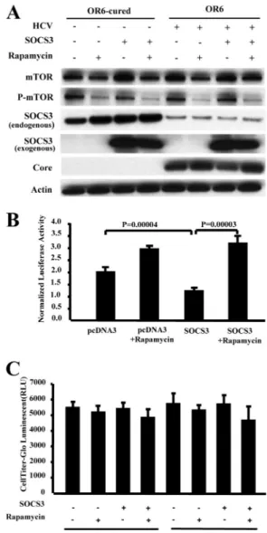
SOCS3 downregulates HCV replication through the mTOR pathway.Given our finding that knockdown of SOCS3 also decreased mTOR expression, we suspected that SOCS3 may downregulate HCV replication through the mTOR pathway.
To test this hypothesis, we overexpressed SOCS3 in the presence of the mTOR inhibitor rapamycin. We found that inhibition of mTOR could reverse the inhibitory effects of SOCS3 on HCV replication in the replicon-carrying OR6 cells and JFH1-infected Huh7.5.1 cells (Fig. 7A and B and 8A and B).
DISCUSSION
We and others have found SOCS3 to be expressed at high levels in patients nonresponsive to IFN treatment (7, 13, 18, 24). Persico et al. also found that HCV genotype 1b-infected patients exhibit higher-level expression of SOCS3 than ge- notype 2a-infected patients (18). They hypothesized that SOCS3 overexpression may explain why HCV genotype 1b- infected patients do not respond to IFN and suggested that reducing SOCS3 gene expression may be an approach to treating HCV infection (18). However, the hypothesis was based on clinical observation and lacked substantiation by FIG. 3. HCV individual protein constructs and transfection with a
replication-defective viral RNA do not alter SOCS3 expression.
(A) Transfection of Huh7.5.1 cells with the empty vectors pCAG and pCMV or pCMV-driven vectors expressing full-length HCV core pro- tein (pCAG-C191), structural proteins (core, E1, and E2 proteins), NS3 and NS4A, NS4B, NS5A, and NS5B was performed for 72 h before cells were lysed for Western blotting. Lanes: 1, sample from cells expressing pCAG empty vector; 2, sample from cells expressing pCMV empty vector; 3, sample from cells expressing full-length HCV core construct pCAG-C191; 4, sample from cells expressing pCMV- HCVst (encoding core, E1, and E2 proteins); 5, sample from cells expressing pCMV NS3-4A; 6, sample from cells expressing pCMV NS4B; 7, sample from cells expressing pCMV NS5A; and 8, sample from cells expressing pCMV NS5B. (B) At the different indicated times posttransfection with JFH1 and mutant JFH1-GND RNAs, Huh7.5.1 cells were lysed using RIPA buffer and Western blotting was performed as described in Materials and Methods.
on January 26, 2016 by Keimyung University Medical Library http://jvi.asm.org/ Downloaded from
biological experimentation. In this study, we used OR6 cells harboring an HCV genotype 1b replicon (8) and the HCV genotype 2a strain JFH1 infection model (23) to investigate the relationship between SOCS3 and HCV replication. We found that HCV replication does not decrease SOCS3 mRNA levels but rather decreases SOCS3 protein levels in a proteasome-dependent manner. Interestingly, we found that SOCS3 overexpression actually suppresses HCV repli- cation and does so despite SOCS3’s inhibition of classical type I IFN signaling. We confirmed the reciprocal relation- ship between SOCS3 and HCV replication by knocking down SOCS3, which leads to a significant increase in HCV replication as assayed through Western blotting and real- time PCR. Furthermore, SOCS3 levels were restored using the HCV protease inhibitor BILN 2061.
In addition, we found that knockdown of SOCS3 also down- regulated mTOR and phospho-mTOR protein levels and that the mTOR inhibitor rapamycin reversed the antiviral effects of SOCS3. Taken together, these data indicate that SOCS3 down- regulates HCV replication in an mTOR-dependent manner.
HCV is remarkably successful in its ability to establish
persistent infection that, unless interrupted by IFN-based therapy, will continue for the lifetime of the individual and present opportunities for further transmission within the human population. This success is linked to the abilities of HCV to evade and antagonize the immune response of the host and to resist the antiviral actions of IFN therapy. HCV evades the host response through a complex combination of processes that include signaling interference and continual viral genetic variation. These evasion strategies support per- sistent infection and the spread of HCV (6). It is the hepatic host response that imposes initial immune defenses against HCV infection. The host response is triggered when a pathogen-associated molecular pattern (PAMP) presented by the infecting virus is recognized and engaged by specific PAMP receptor factors expressed in the host cell, initiating signals that ultimately induce the expression of antiviral effector genes.
Several groups have speculated that elevated SOCS3 ex- pression inhibits phospho-STAT1 expression, which impairs the IFN defense pathway (7, 13, 18, 24). In addition, it has been reported previously that HCV core protein induces FIG. 4. SOCS3 downregulates HCV replication in replicon-carrying OR6 cells. OR6 cells were transfected with the pCDNA3-Myc or pCDNA3-Myc-hSOCS3 plasmid, and 72 h later the cells were lysed for Western blotting, Renilla luciferase assay, and RT-qPCR analyses as described in Materials and Methods. To determine whether SOCS3 influences the IFN signaling pathway, cells were cotransfected with the ISRE-luciferase reporter plasmid and the pCDNA3-Myc or pCDNA3-Myc-hSOCS3 plasmid. Luciferase assays were performed as described in Materials and Methods. Peg-IFN at a dose of 100 U/ml was added 24 h before cells were harvested (see panels A, C, D, and E). The cells were seeded into 96-well plates, and cell viability assays were performed as described in Materials and Methods. (A) Western blotting revealed that overexpression of SOCS3 decreased HCV core protein levels. To distinguish between endogenous and exogenous SOCS3, we used mouse anti-SOCS3 for endogenous SOCS3 and rabbit anti-SOCS3 for exogenous SOCS3. (B) Renilla luciferase assay results show that overexpression of SOCS3 decreases HCV RNA replication. OR6 cells (5,000 cells/well) were plated into a 96-well plate for 1 day prior to being transfected with pCDNA3-Myc or pCDNA3-Myc-hSOCS3. Results were normalized with respect to cell viability. Data are the means⫾ SD of results from three independent experiments. (C) HCV RNA levels as measured by RT-qPCR. Results were normalized with respect to beta-actin RNA. Data are the means⫾ SD of results from three independent experiments. (D) Luciferase assays results show that overexpression of SOCS3 decreases ISRE-luciferase activity. Results were normalized with respect to cell viability. Data are the means⫾ SD of results from three independent experiments. (E) Results of cell viability assays. RLU, relative light units.
on January 26, 2016 by Keimyung University Medical Library http://jvi.asm.org/ Downloaded from
SOCS3 expression in cell lines, resulting in impaired IFN and specifically STAT1 signaling (2, 11, 26). However, we found that Huh7.5.1 cells expressing HCV core protein did not have altered SOCS3 levels. Interestingly, SOCS3 over- expression in cells harboring an HCV genotype 1 replicon and in JFH1-infected cells did result in inhibition of IFN- induced STAT1 phosphorylation and also blocked ISRE activity, as shown by ISRE reporter assays (Fig. 4D and 5C).
Despite this block of IFN-induced phospho-STAT1 and ISRE activities, overexpression of SOCS3 still clearly pro- duced an inhibitory effect on HCV replication, suggesting that the antiviral actions of SOCS3 are mediated by an independent pathway. We found that SOCS3 stimulation of mTOR, which is independent of IFN signaling (9), may overcome any classical IFN blockade and produce a net antiviral effect against HCV.
While it is formally possible that higher levels of SOCS3 are required for more robust inhibition of the JAK-STAT path-
way, we still observed an antiviral effect at these levels of SOCS3 expression. Our data thus point to an unexpected an- tiviral action of SOCS3 in HCV infection, one mediated by the mTOR pathway. Furthermore, in a bona fide HCV infection model, there does not appear to be impairment of the antiviral actions of IFN by SOCS3.
What about the reciprocal effect of HCV on SOCS3 lev- els? We consistently observed a decrease of SOCS3 protein levels with prolonged HCV infection in multiple model sys- tems. The apparent dissociation of protein levels from in- creased mRNA levels suggests an alteration in the posttran- scriptional stability of SOCS3 mRNA or possibly increased degradation of SOCS3 protein. The finding that the protea- some inhibitor MG132 restored SOCS3 protein levels in the presence of HCV supports the explanation that HCV infec- tion promotes degradation of SOCS3 protein through a ubiquitination-dependent, proteasomally mediated path- way. That this is an HCV-dependent phenomenon is rein- FIG. 5. SOCS3 downregulates HCV replication in JFH1-infected cells. Twenty-four hours after transfection with the pCDNA3-Myc or pCDNA3-Myc-hSOCS3 plasmid, Huh7.5.1 cells were infected with JFH1. At 72 h postinfection, Western blotting and qPCR were performed as described in Materials and Methods. To determine whether SOCS3 influences the IFN signaling pathway, cells were cotransfected with the ISRE-luciferase reporter plasmid and the pCDNA3-Myc or pCDNA3-Myc-hSOCS3 plasmid. Luciferase assays were performed as described in Materials and Methods. Peg-IFN at a dose of 100 U/ml was added to the cells for 24 h before cells were collected. The cells were seeded into 96-well plates, and cell viability assays were performed as described in Materials and Methods. (A) Overexpression of SOCS3 decreases HCV core proteins levels, as assayed by Western blotting. To distinguish between endogenous and exogenous SOCS3, we used mouse anti-SOCS3 for endogenous SOCS3 and rabbit anti-SOCS3 for exogenous SOCS3. (B) Overexpression of SOCS3 decreases HCV RNA replication, as exhibited by HCV RNA levels measured by RT-qPCR. Results were normalized with respect to beta-actin RNA (data are the means⫾ SD of results from three independent experiments). (C) Overexpression of SOCS3 decreases ISRE-luciferase activity.
Results were normalized with respect to pRL-TK Renilla luciferase. Data are the means ⫾ SD of results from three independent experiments. (D) Results of cell viability assays.
on January 26, 2016 by Keimyung University Medical Library http://jvi.asm.org/ Downloaded from
forced by our finding that administration of the direct anti- viral agent BILN 2061 restored levels of SOCS3. This decrease in SOCS3 also appears to depend on HCV repli- cation, since expression of individual HCV core, E1, E2, NS3, NS4A, NS4B, NS5A, and NS5B proteins and the rep- lication-defective JFH1-GND RNA did not reproduce these effects. Thus, HCV replication appears to be associated with decreased SOCS3 levels, which given the net antiviral effect of SOCS3, could support the persistent infection state.
How can these experimental findings be reconciled with the consistent observation of higher SOCS3 levels in pa- tients who fail to respond to IFN (7, 12, 17, 23)? These apparently paradoxical findings suggest that, like IFN-stim- ulated genes, many of which express proteins at increased levels in nonresponder patients, some antiviral genes may be upregulated in a compensatory manner in response to im- paired antiviral defenses elsewhere in the pathway, with a net block of antiviral effector genes. Alternatively, other yet to be elucidated antagonistic IFN-stimulated genes may al-
ter the net antiviral state. Exploration of these hypotheses awaits identification of the key IFN-stimulated genes re- sponsible for clearance of HCV.
In conclusion, we have demonstrated that SOCS3 sup- presses HCV replication in an mTOR-dependent manner and that this IFN-independent mechanism represents the dominant pathway for SOCS3 downregulation of HCV rep- FIG. 6. SOCS3 knockdown increases HCV replication in JFH1-
infected cells. Stable knockdown by shRNA targeting SOCS3 (shSOCS3) in Huh7.5.1 cells was established as described in Materials and Methods. We infected cells with inoculum from JFH1-infected Huh7.5.1 cells and transfected cells with the pU6shGFP control vector (carrying shRNA targeting green fluorescent protein [GFP]), the pU6 empty vector, and pU6shSOCS3. (A) Seventy-two hours postinfection, cells were collected for Western blotting. Lanes: 1 and 5, samples from cells expressing pU6shGFP; 6, samples from cells carrying the pU6 empty vector; 2 and 7, samples from stable pU6shSOCS3-expressing cell line 1; 3 and 8, samples from stable pU6shSOCS3-expressing cell line 2; and 4 and 9, samples from stable pU6shSOCS3-expressing cell line 3. The results show that knockdown of SOCS3 increases HCV core and NS3 protein levels. Knockdown of SOCS3 expression also down- regulates mTOR protein expression. P-mTOR, phospho-mTOR.
(B) After infection for 1, 2, 3, and 5 days, cells were collected, RNA was purified, and HCV RNA was measured by qPCR. Results were normalized with respect to beta-actin RNA. Data are the means⫾ SD of results from three independent experiments.*, P⬍ 0.004;**, P⬍ 0.002. HCV RNA replication was significantly increased in SOCS3 knockdown cells compared to control GFP knockdown cells.
FIG. 7. mTOR inhibition reverses the inhibitory effects of SOCS3 on HCV replication in replicon-harboring OR6 cells. The replicon-carrying OR6 cells and cured OR6 cells were transfected with the pCDNA3-Myc or pCDNA3-Myc-hSOCS3 plasmid for 24 h and then treated with rapa- mycin at a 100 nM final concentration. Seventy-two hours after the addi- tion of the pCDNA3-Myc or pCDNA3-Myc-hSOCS3 construct, the cells were collected and lysed for Western blotting and Renilla luciferase assay analyses. The cells were seeded into 96-well plates, and cell viability assays were performed as described in Materials and Methods. (A) The mTOR inhibitor rapamycin reverses SOCS3’s inhibition of HCV core protein levels, as assayed by Western blotting. To distinguish between endogenous and exogenous SOCS3, we used mouse anti-SOCS3 for endogenous SOCS3 and rabbit anti-SOCS3 for exogenous SOCS3. (B) Rapamycin reverses SOCS3’s inhibition of HCV RNA replication. The Renilla lucif- erase assay results were normalized with respect to cell viability. Data are the means⫾ SD of results from three independent experiments. (C) Re- sults of cell viability assays.
on January 26, 2016 by Keimyung University Medical Library http://jvi.asm.org/ Downloaded from
lication. Efforts to enhance SOCS3 and mTOR function may be a useful adjunctive strategy to control HCV infection.
ACKNOWLEDGMENTS
We thank the following investigators and institutes for supplying the reagents listed here: Nobuyuki Kato and Masanori Ikeda, Okayama
University Graduate School of Medicine, Okayama, Japan (OR6 cell line); Francis Chisari, Scripps Institute, La Jolla, CA (Huh7.5.1 cell line); Takaji Wakita, Tokyo Metropolitan Institute for Neuroscience, Tokyo, Japan (infectious HCV JFH1 DNA constructs); and Jie Chen, University of Illinois, Urbana, IL (pCDNA3-Myc and pCDNA3-Myc- hSOCS3 plasmids). We thank Wenyu Lin and Andrew W. Tai for scientific discussions related to this article and Jorge Mendez-Navarro and Nikolaus Jilg for their helpful editing of the manuscript.
This work was supported by National Institutes of Health grants AI069939-01 (to R.T.C.) and DK078772 (to R.T.C.).
REFERENCES
1. Bartenschlager, R., L. Ahlborn-Laake, J. Mous, and H. Jacobsen. 1994.
Kinetic and structural analyses of hepatitis C virus polyprotein processing.
J. Virol. 68:5045–5055.
2. Bode, J. G., S. Ludwig, C. Ehrhardt, U. Albrecht, A. Erhardt, F. Schaper, P. C. Heinrich, and D. Haussinger.2003. IFN-alpha antagonistic activity of HCV core protein involves induction of suppressor of cytokine signaling-3.
FASEB J. 17:488–490.
3. Castet, V., C. Fournier, A. Soulier, R. Brillet, J. Coste, D. Larrey, D.
Dhumeaux, P. Maurel, and J. M. Pawlotsky.2002. Alpha interferon inhibits hepatitis C virus replication in primary human hepatocytes infected in vitro.
J. Virol. 76:8189–8199.
4. Cui, Q., W. Jiang, Y. X. Wang, C. Lv, J. J. Luo, W. Zhang, F. Lin, Y. X. Yin, R. Cai, P. Wei, and C. Qian.2008. Transfer of suppressor of cytokine signaling 3 by an oncolytic adenovirus induces potential antitumor activities in hepatocellular carcinoma. Hepatology 47:105–112.
5. Endo, T. A., M. Masuhara, M. Yokouchi, R. Suzuki, H. Sakamoto, K. Mitsui, A. Matsumoto, S. Tanimura, M. Ohtsubo, H. Misawa, T. Miyazaki, N.
Leonor, T. Taniguchi, T. Fujita, Y. Kanakura, S. Komiya, and A. Yoshimura.
1997. A new protein containing an SH2 domain that inhibits JAK kinases.
Nature 387:921–924.
6. Gale, M., Jr., and E. M. Foy. 2005. Evasion of intracellular host defence by hepatitis C virus. Nature 436:939–945.
7. Huang, Y., J. J. Feld, R. K. Sapp, S. Nanda, J. H. Lin, L. M. Blatt, M. W.
Fried, K. Murthy, and T. J. Liang.2007. Defective hepatic response to interferon and activation of suppressor of cytokine signaling 3 in chronic hepatitis C. Gastroenterology 132:733–744.
8. Ikeda, M., K. Abe, H. Dansako, T. Nakamura, K. Naka, and N. Kato. 2005.
Efficient replication of a full-length hepatitis C virus genome, strain O, in cell culture, and development of a luciferase reporter system. Biochem. Biophys.
Res. Commun. 329:1350–1359.
9. Ishida, H., K. Li, M. Yi, and S. M. Lemon. 2007. p21-activated kinase 1 is activated through the mammalian target of rapamycin/p70 S6 kinase pathway and regulates the replication of hepatitis C virus in human hepatoma cells.
J. Biol. Chem. 282:11836–11848.
10. Jazag, A., H. Ijichi, F. Kanai, T. Imamura, B. Guleng, M. Ohta, J. Imamura, Y. Tanaka, K. Tateishi, T. Ikenoue, T. Kawakami, Y. Arakawa, M. Miyagishi, K. Taira, T. Kawabe, and M. Omata.2005. Smad4 silencing in pancreatic cancer cell lines using stable RNA interference and gene expression profiles induced by transforming growth factor-beta. Oncogene 24:662–671.
11. Kawaguchi, T., T. Yoshida, M. Harada, T. Hisamoto, Y. Nagao, T. Ide, E.
Taniguchi, H. Kumemura, S. Hanada, M. Maeyama, S. Baba, H. Koga, R.
Kumashiro, T. Ueno, H. Ogata, A. Yoshimura, and M. Sata.2004. Hepatitis C virus down-regulates insulin receptor substrates 1 and 2 through up- regulation of suppressor of cytokine signaling 3. Am. J. Pathol. 165:1499–
1508.
12. Kim, J. H., J. E. Kim, H. Y. Liu, W. H. Cao, and J. Chen. 2008. Regulation of interleukin-6-induced hepatic insulin resistance by mammalian target of rapamycin through the STAT3-SOCS3 pathway. J. Biol. Chem. 283:708–715.
13. Kim, K. A., W. Lin, A. W. Tai, R. X. Shao, E. Weinberg, C. B. Borges, A. K.
Bhan, H. Zheng, Y. Kamegaya, and R. T. Chung.2009. Hepatic SOCS3 expression is strongly associated with non-response to therapy and race in HCV and HCV/HIV infection. J. Hepatol. 50:705–711.
14. Luedde, T., T. Wuestefeld, and C. Trautwein. 2001. A new player in the team:
SOCS-3 socks it to cytokine signaling in the regenerating liver. Hepatology 34:1254–1256.
15. Mannova, P., and L. Beretta. 2005. Activation of the N-Ras–PI3K–Akt–
mTOR pathway by hepatitis C virus: control of cell survival and viral repli- cation. J. Virol. 79:8742–8749.
16. Miyagishi, M., and K. Taira. 2002. U6 promoter-driven siRNAs with four uridine 3⬘ overhangs efficiently suppress targeted gene expression in mam- malian cells. Nat. Biotechnol. 20:497–500.
17. Naka, T., M. Narazaki, M. Hirata, T. Matsumoto, S. Minamoto, A. Aono, N.
Nishimoto, T. Kajita, T. Taga, K. Yoshizaki, S. Akira, and T. Kishimoto.
1997. Structure and function of a new STAT-induced STAT inhibitor. Na- ture 387:924–929.
18. Persico, M., M. Capasso, E. Persico, M. Svelto, R. Russo, D. Spano, L.
Croce, V. La Mura, F. Moschella, F. Masutti, R. Torella, C. Tiribelli, and A.
Iolascon.2007. Suppressor of cytokine signaling 3 (SOCS3) expression and FIG. 8. mTOR inhibition reverses the inhibitory effects of SOCS3 on
HCV replication in JFH1-infected cells. Huh7.5.1 cells were transfected with the pCDNA3-Myc or pCDNA3-Myc-hSOCS3 plasmid. Twenty-four hours after transfection, the cells were mock infected or infected with JFH1 inoculum and treated with 100 nM rapamycin. Seventy-two hours after the addition of the pCDNA constructs, the cells were lysed for Western blotting and RT-qPCR. The cells were seeded into 96-well plates, and cell viability assays were performed as described in Materials and Methods. (A) Western blotting results show that mTOR inhibitor rapamycin reverses SOCS3’s inhibition of HCV core protein levels. To distinguish between endogenous and exogenous SOCS3, we used mouse anti-SOCS3 for endogenous SOCS3 and rabbit anti-SOCS3 for exoge- nous SOCS3. (B) The mTOR inhibitor rapamycin reverses SOCS3’s in- hibition of HCV RNA replication. HCV RNA was measured by qPCR, and results were normalized with respect to beta-actin. Data are the means⫾ SD of results from three independent experiments. ⴱ, P ⫽ 0.04;
ⴱⴱ, P ⫽ 0.004. (C) Results of cell viability assays.
on January 26, 2016 by Keimyung University Medical Library http://jvi.asm.org/ Downloaded from
hepatitis C virus-related chronic hepatitis: insulin resistance and response to antiviral therapy. Hepatology 46:1009–1015.
19. Seki, E., Y. Kondo, Y. Iimuro, T. Naka, G. Son, T. Kishimoto, J. Fujimoto, H. Tsutsui, and K. Nakanishi.2008. Demonstration of cooperative contri- bution of MET- and EGFR-mediated STAT3 phosphorylation to liver re- generation by exogenous suppressor of cytokine signalings. J. Hepatol. 48:
237–245.
20. Starr, R., T. A. Willson, E. M. Viney, L. J. Murray, J. R. Rayner, B. J.
Jenkins, T. J. Gonda, W. S. Alexander, D. Metcalf, N. A. Nicola, and D. J.
Hilton.1997. A family of cytokine-inducible inhibitors of signalling. Nature 387:917–921.
21. Suzuki, R., K. Tamura, J. Li, K. Ishii, Y. Matsuura, T. Miyamura, and T.
Suzuki.2001. Ubiquitin-mediated degradation of hepatitis C virus core pro- tein is regulated by processing at its carboxyl terminus. Virology 280:301–
309.
22. Torisu, T., N. Sato, D. Yoshiga, T. Kobayashi, T. Yoshioka, H. Mori, M. Iida, and A. Yoshimura.2007. The dual function of hepatic SOCS3 in insulin resistance in vivo. Genes Cells 12:143–154.
23. Wakita, T., T. Pietschmann, T. Kato, T. Date, M. Miyamoto, Z. Zhao, K.
Murthy, A. Habermann, H. G. Krausslich, M. Mizokami, R. Bartenschlager,
and T. J. Liang.2005. Production of infectious hepatitis C virus in tissue culture from a cloned viral genome. Nat. Med. 11:791–796.
24. Walsh, M. J., J. R. Jonsson, M. M. Richardson, G. M. Lipka, D. M. Purdie, D. Clouston, and E. E. Powell.2006. Non-response to antiviral therapy is associated with obesity and increased hepatic expression of suppressor of cytokine signalling 3 (SOCS-3) in patients with chronic hepatitis C, viral genotype 1. Gut 55:529–535.
25. Yasukawa, H., A. Sasaki, and A. Yoshimura. 2000. Negative regulation of cytokine signaling pathways. Annu. Rev. Immunol. 18:143–164.
26. Yoshida, T., T. Hanada, T. Tokuhisa, K. Kosai, M. Sata, M. Kohara, and A.
Yoshimura.2002. Activation of STAT3 by the hepatitis C virus core protein leads to cellular transformation. J. Exp. Med. 196:641–653.
27. Zhong, J., P. Gastaminza, G. Cheng, S. Kapadia, T. Kato, D. R. Burton, S. F.
Wieland, S. L. Uprichard, T. Wakita, and F. V. Chisari. 2005. Robust hepatitis C virus infection in vitro. Proc. Natl. Acad. Sci. U. S. A. 102:9294–
9299.
28. Zimmerer, J. M., G. B. Lesinski, S. V. Kondadasula, V. I. Karpa, A. Lehman, A. Raychaudhury, B. Becknell, and W. E. Carson III. 2007. IFN-alpha- induced signal transduction, gene expression, and antitumor activity of im- mune effector cells are negatively regulated by suppressor of cytokine sig- naling proteins. J. Immunol. 178:4832–4845.