• 검색 결과가 없습니다.

Enzymatic Conjugation of RGD Peptides on the Surface of Fibroin Microspheres

N/A
N/A
Protected

Academic year: 2021

Share "Enzymatic Conjugation of RGD Peptides on the Surface of Fibroin Microspheres"

Copied!
6
0
0

로드 중.... (전체 텍스트 보기)

전체 글

(1)

Article https://doi.org/10.14478/ace.2019.1110

1. Introduction 1)

Biomedical polymers were frequently functionalized with Arg-Gly-Asp (RGD) peptides to provide cell adhesion sites since the RGD motif is found in many extracellular matrix proteins and extensively studied[1].

RGD modified polymers promoted adhesion, proliferation, and collagen secretion of human fibroblasts[2], and even provide an enhanced anti- tumor activity[3]. RGD peptides were covalently attached to polymers via functional groups like hydroxyl-, amino-, or carboxyl groups[4].

Because of the presence of reactive functional groups, protected RGD peptides were used to modify the polymers activated by peptide cou- pling reagents[5]. Unprotected aqueous RGD peptides can be directly mixed to activated polymers with a less hydrolyzable coupling agent, such as N-hydroxysuccinimide esters[6]. Selected pairs of functional groups were developed to form stable covalent bonds without the need for an activating agent such as thiol-containing RGD peptides with maleimide functionalized surfaces[7].

In this study, RGD peptides were enzymatically coupled on the sur- face of fibroin microspheres. Fibroin, an insoluble protein polymer of silk fiber, is available in quantity and proven to be biocompatible since being used as a suture for decades[8]. Thus, fibroin has been used to fabricate biocompatible materials such as film, hydrogel, coating mate- rials, and including fibroin microspheres in a previous report[9]. Since the fibroin microspheres were made by condensation of insoluble pro-

† Corresponding Author: Kangwon National University,

Department of Biotechnology and Bioengineering, Chuncheon 24341, Korea Tel: +82-33-250-6276 e-mail: [email protected]

pISSN: 1225-0112eISSN: 2288-4505 @ 2020 The Korean Society of Industrial and Engineering Chemistry. All rights reserved.

tein polymer chains without any cross-linking or surface modification, numerous reactive N-terminal amine and C-terminal carboxyl groups could be exposed on the surface of fibroin microspheres. The presence of terminal functional groups prompted us to investigate the enzymatic coupling of RGD peptides on the surface of fibroin microspheres.

Protease catalyzed solid-phase peptide synthesis was first demon- strated using thermolysin that directly synthesized several dipeptides from soluble N-protected amino acids onto solid support[10]. A smaller protease, papain in molecular weight, seems a better candidate since it is more accessible to the functional groups on the solid support. Papain has been widely used for enzymatic coupling of peptides[11]. Therefore, papain was used to catalyze peptide bond formation on to the surface of fibroin microspheres.

In this study, RGD peptides were designed to carry cysteine residues at both sides of the sequence, glycine as a spacer, and two phenyl- alanine residues at the C-terminal. The RGD peptides were enzymati- cally attached to the surface of the fibroin microspheres using papain.

Anchorage-dependent BALB/c 3T3 cells were used to evaluate the ef- fect of RGD functionalized fibroin microspheres on cell attachment and proliferation under non-adherent culture conditions.

2. Materials and Methods

2.1. Materials

Freeze-dried fibroin was provided by FineCo., Ltd. (Chuncheon, Korea). Papain (P3375), decane, propidium iodide (PI), Span 80, Tween 80, and other reagents were purchased from Sigma-Aldrich (St. Louis, MO, USA). The cell line BALB/c 3T3 used was purchased from ATCC (American Type Culture Collection CCL163; Rockville, MD, USA),

Enzymatic Conjugation of RGD Peptides on the Surface of Fibroin Microspheres

Hyun Sang Jeon, Jin Sil Lee, and Won Hur

Department of Biotech & Bioengineering, Kangwon National University, Chuncheon 24341, Korea (Received December 30, 2019; Revised January 7, 2020; Accepted January 8, 2020)

Abstract

Biomaterials are frequently functionalized with Arg-Gly-Asp (RGD) peptides to provide cell adhesion sites. In this study, RGD peptides were enzymatically coupled on to the surface of fibroin microspheres. Papain exhibited a strong preference for dansyl phenylalanine for the peptide formation with fibroin microspheres. Thus, RGD1 peptide was designed to carry cys- teine to both sides of the sequence, glycine as a spacer and two residues of phenylalanine at the C-terminal (CRGDCGFF).

The enzymatic modification facilitated by an increasing amount of substrate and by the presence of organic solvent, dime- thylsulfoxide at 25% (v/v). Microspheres coupled with RGD1, showed a significantly different precipitation property and an increased apparent volume, possibly due to the steric hindrance of RGD peptides on the surface. Transmission electron micro- scopy also confirmed the presence of cysteine residues in RGD1 coupled on the surface of microspheres stained with gold nanoparticles. RGD1-microspheres significantly facilitated the growth of murine fibroblast 3T3 cells even under non-adhesion culture conditions.

Keywords: Microspheres, RGD, Surface modification, Papain, RGD-microspheres

(2)

and ultra-low attachment plates from Corning (3471; Corning, NY, USA) were used for cell culture. Peptides were synthesized by Anygen (Daejon. Korea).

2.2. Preparation and modification of fibroin microspheres Aqueous fibroin mixture (8 mL) was added to an organic phase composed of Span 80 (2.2 mL), Tween 80 (1.8 mL), and decane (16 mL), and then homogenized (Polytron PT2100; Kinematica, Luceme, Switzerland) at 30,000 rpm for 60 s. The resulting translucent emulsion was dried in a rotary vacuum evaporator (JP/N 1000S-W, Eyela, Tokyo, Japan) at 40 ℃ for 30 min. The microspheres that formed in the emul- sion were harvested by centrifugation at 4,000 rpm for 5 min, washed with ethanol several times, and dried at room temperature.

For the surface modification of microspheres, dansyl amino acids or RGD peptide 6 mg, microspheres 10 mg, and papain 5 mg were added to 1 mL of a phosphate buffer solution with 25% (v/v) DMSO in brown microtubes and incubated at room temperature for 2 h. Micro- spheres were washed with distilled water three times, resuspended in 1 mL of anhydrous ethanol, and dried.

Dansylated microspheres were washed, resuspended in phosphate buffer (PBS, pH 7.4), transferred to a multiwall plate and analyzed by measuring fluorescence at 355 nm / 460 nm using a fluorescence mi- croplate reader (Fluoroskan-FL Ascent, Thermo Electron Corp, USA).

Fluorescence intensity was expressed as an arbitrary unit (AU) and converted into moles of the primary amine group based on the AU of dansyl glycine fluorescence intensity.

2.3. Electron microscopy and analysis

The dried microspheres were observed by field-emission scanning electron microscopy (FESEM; Hitachi S-4800; Hitachi, Japan) at an accelerating voltage of 5.0 kV, and also by transmission electron mi- croscopy (TEM; JEM-2010; JEOL, Japan). Microspheres were added to 1 mL of gold nanoparticle solution and boiled for 5 min, followed by washing with distilled water three times. The specimen stained with gold nanoparticles placed on a grid and observed under TEM.

Ellman’s assay was also performed to determine the cysteine content of RGD microspheres[12]. Ellman's reagent solution 50 µL was added to 250 µL of the reaction buffer containing 1 mg of microspheres and reacted for 15 min. Samples were centrifuged at 5,000 rpm for 5 min, and 1 mL of the supernatant was taken and measured for absorbance at 412 nm with a UV spectrophotometer (Thermo Spectronic 10S, Rochester, USA).

2.4. Cell culture and co-incubation with RGD microspheres BALB/c 3T3 cells were cultured at 37 ℃ in a humidified atmos- phere maintained at 5% CO

2

using Dulbecco’s modified Eagle’s me- dium (DMEM; Lonza, Walkersville, MD, USA) with 4.5 g/L glucose and l-glutamine supplemented with 10% bovine calf serum (BCS; Lonza, USA). Fibroin microsphere suspension (1 mg/mL) was sterilized by autoclaving. Cells were dissociated using trypsin/EDTA (trypsin 0.25%, EDTA 1 mM in PBS; Lonza) and counted.

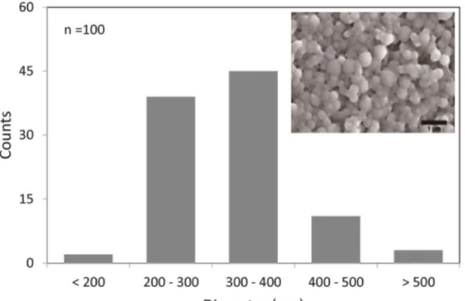
Figure 1. The size distribution of fibroin microspheres (inset: an SEM micrograph).

3. Results and Discussion

3.1. Estimation of primary amines on fibroin microspheres Insoluble microspheres were prepared by condensing fibroin peptides in W/O emulsion under vacuum dehydration conditions, as reported in the previous study[9]. SEM micrograph showed that the microspheres were roughly spherical and mostly between 200~500 nm in diameter (Figure 1). The microspheres might carry many functional groups such as primary amines and lysine residues because those were made of par- tially fragmented fibroin molecules. It prompted us to measure the pos- sible number of functional groups on fibroin microspheres using dansyl chloride, a reactive fluorescent molecule. Dansylation of fibroin micro- spheres resulted in a shift of fluorescence intensity by 46.0 AU/mg.

Based on the fluorescence intensity of dansyl glycine (64.26 AU/µmol) measured under identical conditions, the number of dansylated func- tional groups was estimated to be 4.3 × 10

17

per mg of microspheres (Table 1). Providing the specific gravity of fibroin microspheres is 1.2 g/cm

3

, the corresponding density of dansylation is 5.2 × 10

8

/µm

3

, and the average space distance between adjacent dansylated functional groups is 1.25 nm.

The result suggests that the dansyl-reactive functional groups were present densely on the surface of fibroin microspheres. It is because that dansyl chloride readily reacts with primary amines at N-terminal and lysine residue, secondary amine in proline, and also with tyrosine [13]. The dense distribution of dansylated functional groups can be at- tributed to an exceptionally high content of tyrosine in fibroin, which reached about 10%[14]. The density of surface functional groups is an essential characteristic of micro or nanoparticles that affect the proper- ties and applications[4]. Accordingly, fibroin microsphere can be an excellent platform biomaterial for surface modification because of suf- ficient functional groups.

3.2. Enzymatic peptide formation on the surface of fibroin micro- spheres

A protease-mediated peptide coupling reaction was performed to modify the functional groups on the surface of fibroin microspheres.

Papain was used to couple different dansyl amino acids on to fibroin

(3)

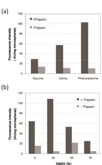
Figure 2. Fluorescence intensities of microspheres modified with di- fferent dansyl amino acids using papain (a). Fluorescence intensities of microspheres conjugated with dansyl phenylalanine using papain in varying DMSO content (b).

microspheres using papain (Figure 2). Microspheres reacted with dan- syl phenylalanine (3 mg/mL) showed the highest increase in the fluo- rescence intensity, followed by dansyl valine and dansyl glycine. No significant increase in fluorescence intensity was observed in those with other dansyl amino acids. It suggests that dansyl phenylalanine can be the most suitable substrate for papain in reverse proteolysis.

Papain-mediated peptide coupling was also performed with varying concentrations of dansyl phenylalanine under the same conditions. The fluorescence intensities of modified microspheres with dansyl phenyl- alanine at the concentration of 3, 6, 9 mg/mL increased by 9.8, 18.4,

Figure 3. Confocal microscopic images of microspheres reacted with dansyl phenylalanine with (a) and without (b) papain. Densitometric profile of fluorescence intensity of papain catalyzed microspheres with dansyl phenylalanine (c). A schematic diagram of papain-catalyzed coupling of a dansyl amino acid with microspheres (d).

20.1 folds. Since the reaction efficiency decreased significantly at 9 mg/mL, dansyl phenylalanine concentration was fixed to 6 mg/mL in further reactions. It corresponds to 0.007 mmol/L and two folds of the primary amines on the microspheres in the reaction mixtures. To facili- tate the reverse proteolysis reaction, DMSO was added to the reaction mixture at 0, 25, 50, 75% (v/v) and reacted under identical conditions.

The resulting fluorescence intensities were 64.4, 108.6, 53.3, and 25.2 AU/mg [Figure 2(b)]. DMSO 25% showed the highest fluorescence. In the above experiments, phenylalanine showed the highest substrate spe- cificity of the reverse reaction of papain.

Enzymatic coupling of dansyl phenylalanine was confirmed using CLSM at an excitation wavelength of 334 nm with a confocal scanning microscope. Green fluorescence was observed in the microspheres ob- tained after the reaction with dansyl phenylalanine using papain, in comparison to those without papain [Figure 3(a), (b)]. The fluorescence intensity of a stacked CLSM micrograph was measured using Image J [Figure 3(c)]. The fluorescence intensity profile has a broad peak with a plateau on the top. These results verified papain-catalyzed cou- pling of dansyl phenylalanine fibroin microspheres, as shown schemati-

Item Values Units

Fluorescence intensity of dansyl glycine 64.26 AU/µmol

Fluorescence intensity of dansyl microspheres 46.0 AU/mg

Mol of dansylated functional groups per microspheres 0.716 µmol/mg

Number of dansylated functional groups per microspheres 4.31 × 10

17

/mg

Density of dansylated functional groups 5.2 × 10

8

/µm

3

Average distance between adjainst functional groups 1.25 nm

Table 1. Estimation of the Number of N-terminal Amines on Fibroin Microspheres

(4)

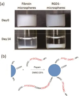
Figure 4. Precipitation profile of RGD1-modified microsphere suspen- sions (1 mg/mL) in glass vials for 14 days (a) and a schematic model of papain catalyzed coupling of RGD1 peptide on to fibroin micro- spheres (b).

cally in Figure 3(d).

The results agree with the finding that phenylalanine has been used as a preferred acyl-donor substrate than other amino acids in pa- pain-catalyzed coupling to leucine amide[15] and synthesizing ho- mo-oligomers[16]. Although papain cleaves peptide bonds of basic amino acids, leucine, or glycine at the P1 positions, it also has a strong preference for phenylalanine at the P2 position for hydrolysis reaction [17]. Diazomethyl ketones of z-Phe and z-Phe-Phe are known as active site-directed inhibitors of papain[18], suggesting that phenylalanine and its dimer could bind to the active site of papain. Thus, peptides end with phenylalanine or its dimer seem to be a likely candidate as a sub- strate for papain catalyzed peptide coupling reaction.

3.3. Enzymatic modification of fibroin microspheres with RGD peptides

RGD peptides were designed to carry cysteine residues at both sides of the sequence, glycine residues as a spacer, and two phenylalanine residues at the C-terminal, of which amino acid sequences are CRGD- CGFF (RGD1). Papain-catalyzed coupling reactions of the RGD1 pep- tide on to fibroin microspheres were performed. The resulting yield of the RGD1-microspheres was 57%. A milky suspension of RGD1-micro- spheres was prepared at a concentration of 1 mg/mL in distilled water, which showed a significantly different precipitation profile [Figure 4(a)]. The RGD1-microspheres precipitated and formed a thick layer of sediments. They exhibited an increased apparent volume than unmodi-

Figure 5. Scanning electron microscope images (top), transmission electron microscope (TEM) images (middle) of fibroin microspheres, and RGD1-microspheres. TEM images of fibroin microspheres and RGD1-microspheres stained with gold nanoparticles (bottom).

fied microspheres, possibly due to the steric hindrance of the RGD1 peptides on the surface. The sediments were easily resuspended by gentle shaking of the vials. It is also possible that tandemly repeated RGD1 peptides were tethered on the surface of fibroin microspheres, as shown schematically in Figure 4(b).

The RGD1-microspheres were dried, sputter-coated with gold, and observed with SEM (Figure 5). The micrograph of RGD1-microspheres appears blurred compared to spherical fibroin microspheres. TEM mi- crographs present similarly sized microspheres between 300~400 nm in diameter both in RGD1-microspheres and fibroin microspheres.

RGD1-microspheres were stained using gold nanoparticles that could react with thiol groups in cysteine residues of RGD peptides to form S-Au bonds. In the TEM micrograph of RGD1-microspheres, black spots of similar size to gold nanoparticles were observed in the sur- rounding dark areas. Ellman’s assay exhibited that the sulfur content of RGD1-microspheres was 1.3% that was significantly higher than that of microspheres (0.08%). These results indicate that RGD1-peptides were coupled to the functional groups on microspheres.

Although RGD1 peptides were covalently coupled on to the surface

of microspheres, this study was not able to identify which function

groups were involved with RGD1 peptide bond formation. RGD1 pep-

tides probably coupled to the free N-terminals of fibroin peptide chains

and the primary amine of lysine residue due to phenylalanine specific

peptide formation by papain. However, the possibility cannot be ex-

cluded that the carboxyl group of RGD1-peptides could form an ester

with tyrosine because papain also carries esterase activity[19].

(5)

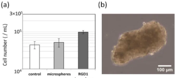
Figure 6. Counts of 3T3 cell aggregates cultured in the presence of fibroin microspheres or RGD1-microspheres under non-adhesion con- ditions (a) and a microscope image of 3T3 cell aggregate from culture medium containing RGD1-microspheres (b).

3.4. Cell culture enhancement

RGD1-functionalized microspheres were used to culture fibroblast 3T3 cells under non-adhesion conditions. Suspension of multicellular aggregates was obtained by repeated pipetting of culture seeded with 3T3 cells with and without microspheres for 72 h. The 3T3 cells ag- gregates were largely ovoid or spheroidal in shape with a rough outer surface. Larger and darker aggregates were obtained in the presence of RGD1-microspheres, of which the number of dissociated cells was 1.0

× 10

5

cells/mL. The cell counts were significantly higher than those with and without fibroin microspheres [Figure 6(a)]. The longitudinal length of 3T3 cell aggregates with RGD1-microspheres was mostly in the range between 200~400 µm [Figure 6(b)].

A solid surface or specified extracellular matrices are essential for the attachment and survival of anchorage-dependent 3T3 cells[20]. The effectiveness of RGD in promoting cell attachment to a wide variety of biomaterials was confirmed in many publications, both in vitro and in vivo[21]. This study also demonstrated that RGD functionalized mi- crospheres enhanced fibroblast culture even under non-adhesion conditions. However, the detailed effects of RGD1-microspheres on proliferation and other cellular responses remain mostly unexplored.

Although RGD1 peptide contains two cysteine residues, the effect of RGD cyclization also remains uninvestigated. Recently, high density, clustering, and/or cyclization of RGD peptides elicited beneficial cell responses in vivo, such as increased affinity for the platelets and more substantial bone regeneration[21]. Fibroin microspheres can be suitable base material for the functionalization with various complex RGD structures, due to abundant surface functional groups and enzymatic peptide coupling method of this study.

4. Conclusions

Papain catalyzed peptide formation was facilitated by dansyl phenyl- alanine with fibroin microspheres and by the presence of organic sol- vent DMSO at 25% (v/v). Thus, peptide RGD1 was designed to carry cysteine to both sides of the sequence, glycine as a spacer and phenyl- alanine at the C-terminal (CRGDCGFF). Microspheres, coupled with RGD1, showed a significantly different precipitation property and an increased apparent volume, possibly due to the steric hindrance of the RGD peptides on the surface. Transmission electron microscopy also

confirmed the presence of cysteine residues in RGD peptides coupled on the surface of microspheres stained with gold nanoparticles. Accor- dingly, peptide RGD1 was enzymatically coupled to on the surface of fibroin microspheres. The RGD1-microspheres significantly facilitated the growth of murine fibroblast 3T3 cells even under non-adhesion culture conditions. Here, we demonstrated the utility of enzymatic functionalization of a RGD peptide on to the surface of fibroin micro- spheres.

Acknowledgement

This research was supported by the National Research Foundation of Korea (NRF-2016R1D1A1B01011660).

References

1. S. Bellis, Advantages of RGD peptides for directing cell associa- tion with biomaterials, Biomaterials, 32, 4205-4210 (2011).

2. E. Ruoslahti and M. D. Pierschbacher, New perspectives in cell adhesion: RGD and integrins, Science, 238, 491-497 (1987).

3. F. Wang, Y. Li, Y. Shen, A. Wang, S. Wang, and T. Xie, The functions and applications of RGD in tumor therapy and tissue en- gineering, Int. J. Mol. Sci., 14, 13447-13462 (2013).

4. U. Hersel, C. Dahmen, and H. Kessler, RGD modified polymers:

Biomaterials for stimulated cell adhesion and beyond, Biomate- rials, 24(24), 4385-4415 (2003).

5. L. A. Carpino, H. Shroff, S. A. Triolo, E. M. E. Mansour, H.

Wenschuh, and F. Albericio, The 2,2,4,6,7-pentamethyldihydroben- zofuran-5-sulfonyl group (Pbf) as arginine side-chain protectant, Tetrahedron Lett., 34, 7829-7832 (1993).

6. D. L. Hern and J. A. Hubbell, Incorporation of adhesion peptides into nonadhesive hydrogels useful for tissue resurfacing, J. Biomed.

Mater. Res., 39, 266-276 (1998).

7. S. S. Lateef, S. Boateng, T. J. Hartman, C. A. Crot, B. Russell, and L. Hanley, GRGDSP peptide-bound silicone membranes with- stand mechanical flexing in vitro and display enhanced fibroblast adhesion, Biomaterials, 23, 3159-3168 (2002).

8. C. Vepari and D. J. Kaplan, Silk as a biomaterial, Prog. Polym.

Sci., 32(8-9), 991-1007 (2007).

9. E. J. Kim, S. Y. Lee, and W. Hur, Preparation of core-shell micro- capsules using nanodispersed fibroin, J. Appl. Polym. Sci., 121(6), 3460-3465 (2011).

10. R. V. Ulijn, B. Baragaña, P. J. Halling, and S. L. Flitsch, Pro- tease-catalyzed peptide synthesis on solid support, J. Am. Chem.

Soc., 124(37), 10988-10989 (2002).

11. M. Schuster, B. Munoz, W. Yuan, and C. H. Wong, Papain-cata- lyzed synthesis of peptide isosteres, Tetrahedron Lett., 34(8), 1247-1250 (1993).

12. C. K. Riener, G. Kada, and H. J. Gruber, Quick measurement of protein sulfhydryls with Ellman’s reagent and with 4, 4´-dithiodi- pyridine, Anal. Bioanal. Chem., 373(4-5), 266-276 (2002).

13. J. M. Walker, The dansyl method for identifying N-terminal amino acids. In: J. M. Walker (ed.), Basic Protein and Peptide Protocols, 321-328, Humana Press (1994).

14. C. Z. Zhou, F. Confalonieri, N. Medina, Y. Zivanovic, C. Esnault,

T. Yang, J. Janin, M. Duguet, R. Perasso, and Z. G. Li, Fine orga-

(6)

nization of Bombyx mori fibroin heavy chain gene. Nucleic Acids Res., 28(12), 2413-2419 (2000).

15. K. Morihara and T. Oka, Peptide bond synthesis catalyzed by sub- tilisin, papain, and pepsin, J. Biochem., 89(2), 85-395 (1981).

16. G. Anderson and P. L. Luisi, Papain-induced oligomerization of α- amino acid esters, Helv. Chim. Acta, 62, 488-494 (1979).

17. F. Lecaille, C. Serveau, F. Gauthier, and G. Lalmanach, Revisiting the S2 specificity of papain by structural analogs of Phe, FEBS Lett., 445(2-3), 311-314 (1999).

18. R. Leary, D. Larsen, H. Watanabe, and E. Shaw, Diazomethyl ke- tone substrate derivatives as active-site-directed inhibitors of thiol

proteases Papain, Biochemistry, 16(26), 5857-5861 (1977).

19. Y. V. Mitin, K. Braun, and P. Kuhl, Papain catalyzed synthesis of glyceryl esters of N-protected amino acids and peptides for the use in trypsin catalyzed peptide synthesis, Biotechnol. Bioeng., 54(3), 287-290 (1997).

20. C. H. O’Neill, P. N. Riddle, and P. W. Jordan, The relation be- tween surface area and anchorage dependence of growth in ham- ster and mouse fibroblasts. Cell, 16(4), 909-918 (1979).

21. S. L. Bellis, Advantages of RGD peptides for directing cell associ-

ation with biomaterials, Biomaterials, 32(18), 4205-4210 (2011).

수치

Figure 1. The size distribution of fibroin microspheres (inset: an SEM  micrograph).
Table 1. Estimation of the Number of N-terminal Amines on Fibroin Microspheres
Figure 5. Scanning electron microscope images (top), transmission  electron microscope (TEM) images (middle) of fibroin microspheres,  and RGD1-microspheres
Figure 6. Counts of 3T3 cell aggregates cultured in the presence of  fibroin microspheres or RGD1-microspheres under non-adhesion con-  ditions (a) and a microscope image of 3T3 cell aggregate from culture  medium containing RGD1-microspheres (b).

참조

관련 문서

MP Yousef Al-Fadalah, Supervisor of Parliament Business Environment Committee declared that the Public Institution for Housing Care intends to dedicate nearly 150

The main objective of the Bi Regional Center for SMES Development is oriented towards generating the conditions of cooperation among the countries which

q March 28, 1906: County Magistrate Shim Heung-taek of Uldo-gun[Uldo County] heard this news from the Japanese survey team, he reported to the Korean central government

In a statement to Kuwait News Agency (KUNA) on the sidelines of a meeting of the Arab Parliament's Foreign Affairs Political and National Security

The meeting was attended by Assistant Foreign Minister for GCC Affairs, Ambassador, Nasser Al-Muzayyen, and Deputy Assistant Foreign Minister for the Office of the

“ Sheikh Nasser has a written message from HH the Amir, Sheikh Sabah Al-Ahmad Al-Jaber Al-Sabah to the Chinese President, Chi Gen Beng related to enhancing mutual

On his part, CEO of Express Roads Authority, Saud Al-Naqqi said that the heavy rains of the previous day led to clogging parts of the express

Kuwait will celebrate on Sunday the fourth anniversary of the UN honoring and proclamation of His Highness the Amir, Sheikh Sabah Al-Ahmad Al-Jaber Al-Sabah as