• 검색 결과가 없습니다.

Fractional Flow Reserve Versus Angiography in Left Circumflex Ostial Intervention After Left Main Crossover Stenting

N/A
N/A
Protected

Academic year: 2021

Share "Fractional Flow Reserve Versus Angiography in Left Circumflex Ostial Intervention After Left Main Crossover Stenting"

Copied!
4
0
0

로드 중.... (전체 텍스트 보기)

전체 글

(1)

304

Print ISSN 1738-5520 / On-line ISSN 1738-5555 Copyright © 2011 The Korean Society of Cardiology ORIGINAL ARTICLE

DOI 10.4070/kcj.2011.41.6.304

Open Access

Fractional Flow Reserve Versus Angiography in Left Circumflex Ostial Intervention After Left Main Crossover Stenting

Chang-Wook Nam, MD

1

, Seung-Ho Hur, MD

1

, Bon-Kwon Koo, MD

2

, Joon Hyung Doh, MD

3

, Yun-Kyeong Cho, MD

1

, Hyoung-Seob Park, MD

1

, Hyuck-Jun Yoon, MD

1

, Hyungseop Kim, MD

1

, In-Sung Chung, MD

1

,

Yoon-Nyun Kim, MD

1

, William F. Fearon, MD

4

, Seung-Jae Tahk, MD

5

, and Kwon-Bae Kim, MD

1

1

Department of Internal Medicine, Keimyung University College of Medicine, Dongsan Medical Center, Daegu,

2

Department of Internal Medicine, Seoul National University College of Medicine, Seoul,

3

Department of Internal Medicine, Inje University College of Medicine, Ilsan Paik Hospital, Goyang, Korea

4

Stanford University Medical Center, Cardiovascular Medicine, California, USA

5

Department of Internal Medicine, Ajou University School of Medicine, Suwon, Korea

ABSTRACT

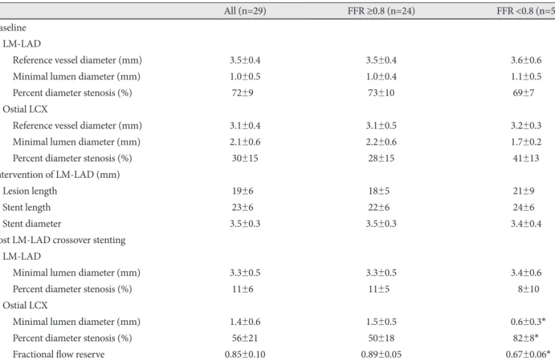
Background and Objectives: Discrepancy between angiographic percent (%) diameter stenosis and fractional flow reserve (FFR) exists in non-left main bifurcation lesions. The aim of this study was to compare angiographic stenosis severity and FFR in jailed ostial left circumflex artery (LCX) lesions after left main (LM)-to-left anterior descending artery (LAD) cross- over stenting. Subjects and Methods: Twenty-nine (n=29) patients with distal LM or ostial LAD lesions treated by LM-to- LAD crossover stenting were consecutively enrolled. After successful stenting, FFR was measured at the jailed LCX. Addi- tional intervention was performed in lesions with FFR <0.8. Results: The mean reference diameter of LCX was 3.1±0.4 mm, and percent diameter stenosis after crossover stenting was 56±21%. Angiographically significant stenosis (>50%) at the osti- al LCX occurred in 59% (17/29) of cases. Among them, only five (29%) lesions had functional significance, and underwent additional procedure. During follow-up, three patients in the deferral group and two patients in the additional intervention group had target lesion revascularization. Conclusion: There was a discrepancy between angiographic percent diameter stenosis and FFR in jailed LCX lesions after LM crossover stenting. (Korean Circ J 2011;41:304-307)

KEY WORDS: Coronary disease; Physiology; Stents; Angiography.

Received: June 19, 2010

Revision Received: September 3, 2010 Accepted: October 14, 2010

Correspondence: Seung-Ho Hur, MD, Division of Cardiology, Depart- ment of Internal Medicine, Keimyung University College of Medicine, Dongsan Medical Center, 216 Dalseong-ro, Jung-gu, Daegu 700-712, Korea

Tel: 82-53-250-7949, Fax: 82-53-250-7034 E-mail: shur@dsmc.or.kr

• The authors have no financial conflicts of interest.

cc

This is an Open Access article distributed under the terms of the Cre- ative Commons Attribution Non-Commercial License (http://creativecom- mons.org/licenses/by-nc/3.0) which permits unrestricted non-commer- cial use, distribution, and reproduction in any medium, provided the origi- nal work is properly cited.

Introduction

Recent studies evaluating percutaneous coronary interven- tion (PCI) with drug-eluting stent (DES) for left main (LM) disease have shown promising results.

1)2)

However, the decis-

ion to intervene the jailed left circumflex coronary artery (LCX) after an LM stent extending into the left anterior descending coronary artery (LAD) is difficult in some cases if based on angiogram alone. Previous studies suggested that measure- ment of fractional flow reserve (FFR) across jailed side branch lesions prevents unnecessary complex interventions in bi- furcation lesions.

3-5)

However, the safety and efficacy of the same strategy for jailed LCX after LM-to-LAD crossover stent- ing when a larger amount of myocardium may be in jeopardy remains unvalidated. The aim of this study was to compare an- giographic stenosis severity and FFR in jailed ostial LCX le- sions after LM-to-LAD crossover stenting.

Subjects and Methods

Patient population

Patients who underwent LM-to-LAD crossover stenting

(2)

Chang-Wook Nam, et al. 305

and met the following criteria were prospectively enrolled from 3 centers in the Republic of Korea: 1) significant de novo coronary artery disease in distal LM or ostial LAD and, 2) reference vessel diameter of LCX over 2.5 mm by visual estimation. Patients were excluded if any one of the follow- ing conditions was present: predilation of LCx before main branch stent implantation, significant non-ostial LCx lesion, infarct-related artery or angiographically visible thrombus, primary myocardial disease, left ventricular hypertrophy, con- traindications to adenosine, or intolerance to aspirin or clo- pidogrel. The study protocol was approved by the Instituti- onal Review Board and all patients gave informed consent prior to study enrollment.

Study procedure

PCI was performed according to standard interventional te- chniques. Before the index procedure, all patients received oral aspirin (a loading dose of 200 mg) and clopidogrel (a load- ing dose of 300-600 mg before the procedure). Oral antipl- atelet therapy during the study period followed guidelines recommending a combination of aspirin (100 mg daily; inde- finitely) and clopidogrel (75 mg daily; more than 12 months).

Intravenous boluses of heparin (100 U/kg) were administer- ed before intervention and the dosage was adjusted to main- tain an activated clotting time exceeding 250 seconds during the procedure.

6)

All coronary angiograms were analyzed us- ing standard definitions and measurements by Quantitative Coronary Angiography (QCA; Quantcor QCA, version 4.0, Pie Medical Imaging, Maastricht, the Netherlands) by an ex- perienced physician blinded to the FFR values.

Fractional flow reserve measurements

FFR is defined as the ratio between distal coronary pres- sure and aortic pressure, both measured simultaneously at ma- ximal hyperemia. After successful insertion of LM-to-LAD crossover stenting, FFR was measured down the jailed LCX using a 0.014-inch pressure guide wire (PressureWire, Radi Medical Systems, Uppsala, Sweden). The pressure wire was passed through the struts of the stent and positioned more than 10 mm distal to the LCX ostium to assess ostial-narrow- ing severity. Hyperemia was induced with intracoronary bo- lus administration (80 µg) or intravenous continuous infus- ion (140 µg/kg/min) of adenosine.

7)

Lesions with an FFR less than 0.80 were considered to be functionally significant ste- nosis. In such cases, additional procedures, such as kissing balloon or provisional stenting, were performed at the phy- sician’s discretion.

Clinical events

Major adverse cardiac events were defined as a composite of death, myocardial infarction, and any repeat revasculariz- ation. Death was defined as all-cause mortality. Myocardial in-

farction was defined as greater than or equal to 3-fold eleva- tion of creatine kinase-MB level, or new Q-waves in greater than or equal to 2 contiguous electrocardiogram leads. Tar- get vessel revascularization (TVR) included target lesion re- vascularization and bypass surgery of the pertinent lesion.

TVR was only based on the presence of symptoms and/or signs of ischemia. Stent thrombosis was defined according to the Academic Research Consortium guidelines.

Statistical analysis

Categorical variables are expressed as proportions. Continu- ous variables are expressed as means and standard deviations.

Correlations between the FFR and percent diameter stenosis were evaluated using the Pearson correlation analysis with the Statistical Package for the Social Sciences (SPSS) statistical so- ftware, version 15.0 (SPSS, Inc., Chicago, IL, USA).

Results

Twenty-nine (n=29) patients were enrolled in this study.

The baseline characteristics and procedural results are sum- marized in Table 1 and 2. The mean age of the patients was 64±9 years and 69% were male. On the baseline coronary angiogram, 6 patients had distal LM disease and 23 patients had ostial LAD disease without significant distal LM disease.

Intravenous adenosine was used in 7 cases (24.1%). The mean reference vessel diameter of the ostial LCX was 3.1±0.4 mm.

The baseline mean percent diameter stenosis (%DS) of ostial LCX was 30±15%. After LM-to-LAD crossover stenting, mean %DS of ostial LCX was 56±21%. FFR of ostial LCX was greater than or equal to 0.80 in 24 patients (0.89±0.05), less than 0.80 in 5 patients (0.67±0.06). Among 5 patients, 3 achieved adequate results (FFR >0.90) after kissing balloon inflation, and the others by provisional T-stenting. When Table 1. Patient baseline characteristics

Characteristic n=29

Age (years) 64±9

Male gender, n (%) 20 (69)

History, n (%)

Diabetes 05 (17)

Hypertension 13 (45)

Hypercholesterolemia 10 (35)

Current smoking 07 (24)

Previous myocardial infarction 01 (3.4)

Previous PCI 02 (7)

Unstable angina, n (%) 10 (35)

Left ventricular ejection fraction (%) 61±8

Multi vessel disease, n (%) 23 (79)

Plus-minus values are mean±SD. PCI: percutaneous coronary in-

tervention

(3)

306 FFR vs. Angiography in Jailed LCX

QCA was reanalyzed in 17 lesions with DS greater than in ostial LCX, the results were similar to that presented in Table 2. The angle between LAD and LCX was 124 degree in the FFR ≥0.8 group, and 114 degree in the FFR <0.8 group (p=

0.35).

A plot of FFR as a function of %DS of the ostial LCX after stent deployment is shown in Fig. 1. Although there was a ne- gative correlation between %DS and FFR (r=-0.469, p=0.01), no lesion with ≤50 %DS had FFR <0.80. Among 17 lesions

with >50 %DS in ostial LCX, only 5 lesions (29%) were func- tionally significant. Even in 8 lesions with >70 %DS, 3 lesions remained functionally non-significant. The best cut-off value of %DS to predict functional significance (i.e., FFR <0.80) was 82% (sensitivity 60%, specificity 58%; area under the curve 0.958, 95% confidence interval 0.889-1.028).

Clinical follow-up was performed in all patients. Mean dur- ation was 20±9 months (minimum 4, maximum 30). During the follow-up period, 3 patients in the deferral group under- went TLR. Two cases were related to LM-to-LAD stent rest- enosis, and the other was due to the progression of ostial LCX lesion. Two patients in the additional procedure group expe- rienced adverse events (1 non-cardiac death, 1 TLR for LM- to-LAD stent restonsis). No stent thrombosis was observed in these patients.

Discussion

The major findings of the current study is that there is a dis- crepancy between angiographic percent diameter stenosis and FFR in jailed LCX lesions, and the FFR-guided jailed LCX PCI strategy could reduce the need for additional PCI.

Despite the emergence of DES, coronary bifurcation les- ions still have a higher restenosis rate, particularly at the os- tium of the side branch in comparison with simple lesions.

8)

Table 2. Procedural results

All (n=29) FFR ≥0.8 (n=24) FFR <0.8 (n=5)

Baseline LM-LAD

Reference vessel diameter (mm) 3.5±0.4 3.5±0.4 03.6±0.6

Minimal lumen diameter (mm) 1.0±0.5 1.0±0.4 01.1±0.5

Percent diameter stenosis (%) 72±90 73±10 0.69±7

Ostial LCX

Reference vessel diameter (mm) 3.1±0.4 3.1±0.5 03.2±0.3

Minimal lumen diameter (mm) 2.1±0.6 2.2±0.6 01.7±0.2

Percent diameter stenosis (%) 30±15 28±15 0.41±13

Intervention of LM-LAD (mm)

Lesion length 19±60 18±50 0.21±9

Stent length 23±60 22±60 0.24±6

Stent diameter 3.5±0.3 3.5±0.3 03.4±0.4

Post LM-LAD crossover stenting LM-LAD

Minimal lumen diameter (mm) 3.3±0.5 3.3±0.5 03.4±0.6

Percent diameter stenosis (%) 11±60 11±50 0.08±10

Ostial LCX

Minimal lumen diameter (mm) 1.4±0.6 1.5±0.5 00.6±0.3*

Percent diameter stenosis (%) 56±21 50±18 .082±8*

Fractional flow reserve 0.85±0.10 0.89±0.05 0.67±0.06*

Plus-minus values are means±SD. *p<0.05. FFR: fractional flow reserve, LM: left main, LAD: left anterior descending coronary artery, LCX:

left circumflex coronary artery

Fig. 1. Correlation between fractional flow reserve and percent diameter stenosis of ostial circumflex coronary artery after left main to left anterior descending coronary artery stenting.

1.0 0.9 0.8 0.7 0.6

r=-0.469 p=0.01

Percent diamater stenosis

Fr ac tio na l fl ow re se rv e

25 50 75 100

(4)

Chang-Wook Nam, et al. 307

It is important to assess the functional significance of jailed LCX lesions as 2 stent strategies, because LM diseases have been associated with worse outcomes than simple crossover stenting.

9)

Although one stent technique is preferable in this lesion type, the frequency of ostial LCX intervention after LM-to-LAD crossover stenting in this lesion type was >40%

in a previous study,

1)

which might have been related to larger plaques and carina shifting-related wide angle in LM bifur- cation lesions.

Assessment of the severity of bifurcation lesions and the performance of PCI in bifurcation remain challenging. An- giographic assessment of the severity of bifurcation lesions is hampered by the inherent limitations of angiography, such as overlap of adjacent vessels, angulation, and foreshortening of the origin of side branches.

10-12)

Intravascular ultrasound can provide delicate information about the anatomical chan- ge of bifurcation lesion. However, it may yield inaccurate in- formation in this situation because of veiled ostium by stent strut. Previous studies by Koo et al.

13)

have demonstrated the feasibility and safety of physiological assessment with FFR of jailed side branches, and found a discrepancy between angi- ographic and functional evaluation of these lesions.

5)

We per- formed this study to apply the same strategy to jailed LCX lesions.

In the current study, angiographically significant stenosis (>50%) at the ostial LCX after LM-to-LAD crossover stenting occurred in 59% (17/29) of cases. Among them, only 5 (29%) patients had functionally significant obstruction of LCX by pressure wire criteria. Adequate coronary flow was preserv- ed in 71% (12/17) patients with significant stenosis of ostial LCX after LM-to-LAD crossover stenting. Thus, the real func- tional state of ostial LCX can be widely different from that appears on angiography. In the clinical context, there were 3 cases of TLR in deferred lesions. Two cases were related to LM-to-LAD stent restenosis. Only 1 TLR case (4%) was deve- loped in 24 deferred LCX lesions. There was no cardiac death, stent thrombosis or MI related to lesions in which PCI was de- ferred according to FFR. Unfortunately, this prospective pilot study was not powered to detect a difference in clinical out- come. With larger numbers one can anticipate that the need for revascularization of the ostial LCX after LM-to-LAD crossover stenting may be reduced, if the additional proce- dure is guided by FFR. However, further studies are needed to evaluate the efficacy of this strategy.

Limitations

There are several limitations to this study. First, this study in- cluded small number of patients. Second, a selection bias may have been introduced, because not all patients with distal LM or ostial LAD disease at three centers were evaluated for this study. Third, it was a non-randomized study. Because we did not compare with routine performance of additional proce-

dures, it is difficult to confirm the benefits of our procedure.

Finally, as we included only mild to moderate LCX ostial le- sion with short lesion lengths, our results cannot be directly ap- plied to more severe or diffuse lesions.

Conclusions

There was a discrepancy between angiographic percent dia- meter stenosis and FFR in jailed LCx lesions after LM-to- LAD stenting.

Acknowledgments

The authors thank Dr. Roberto Patarca for his editorial input.

This study was supported by a grant from the Korean Society of Circula- tion (2007), Republic of Korea. Dr. Yoon-Nyun Kim declares receipt of research grant No. RTI04-01-01 from the Regional Technology Innovation Program of the Ministry of Knowledge Economy (MKE), the Republic of Korea.

REFERENCES

1) Kim YH, Park SW, Hong MK, et al. Comparison of simple and com- plex stenting techniques in the treatment of unprotected left main co- ronary artery bifurcation stenosis. Am J Cardiol 2006;97:1597-601.

2) Kim W, Kim YJ, Lee WJ, et al. Lesion location: its impacts on the pro- cedural and postprocedural outcomes of unprotected left main coro- nary stenting. Korean Circ J 2007;37:419-24.

3) Lim MJ, Kern MJ. Utility of coronary physiologic hemodynamics for bifurcation, aorto-ostial, and ostial branch stenoses to guide treatment decisions. Catheter Cardiovasc Interv 2005;65:461-8.

4) Koo BK, Kang HJ, Youn TJ, et al. Physiologic assessment of jailed side branch lesions using fractional flow reserve. J Am Coll Cardiol 2005;46:633-7.

5) Bellenger NG, Swallow R, Wald DS, et al. Haemodynamic signific- ance of ostial side branch nipping following percutaneous interven- tion at bifurcations: a pressure wire pilot study. Heart 2007;93:249-50.

6) King SB 3rd, Smith SC Jr, Hirshfeld JW Jr, et al. 2007 Focused Up- date of the ACC/AHA/SCAI 2005 Guideline Update for Percutaneous Coronary Intervention: a report of the American College of Cardiolo- gy/American Heart Association Task Force on Practice Guidelines:

2007 Writing Group to Review New Evidence and Update the ACC/

AHA/SCAI 2005 Guideline Update for Percutaneous Coronary In- tervention, Writing on Behalf of the 2005 Writing Committee. Circu- lation 2008;117:261-95.

7) Suh JW, Koo BK, Jo SH, et al. Optimal dosage and method of adminis- tration of adenosine for measuring the coronary flow reserve and the fractional flow reserve in Koreans. Korean Circ J 2006;36:300-7.

8) Colombo A, Moses JW, Morice MC, et al. Randomized study to ev- aluate sirolimus-eluting stents implanted at coronary bifurcation le- sions. Circulation 2004;109:1244-9.

9) Toyofuku M, Kimura T, Morimoto T, et al. Three-year outcomes af- ter sirolimus-eluting stent implantation for unprotected left main cor- onary artery disease: insights from the j-Cypher registry. Circulation 2009;120:1866-74.

10) White CW, Wright CB, Doty DB, et al. Does visual interpretation of the coronary angiogram predict the physiologic importance of a coro- nary stenosis. N Engl J Med 1984;310:819-24.

11) Marcus ML, Skorton DJ, Johnson MR, Collins SM, Harrison DG, Kerter RE. Visual estimate of percent diameter coronary stenosis: a battered gold standard. J Am Coll Cardiol 1988;11:882-5.

12) Ziaee A, Parham WA, Herrmann SC, Stewart RE, Lim MJ, Kern MJ.

Lack of relation between imaging and physiology in ostial coronary artery narrowings. Am J Cardiol 2004;93:1404-7.

13) Koo BK, Park KW, Kang HJ, et al. Physiological evaluation of the

provisional side-branch intervention strategy for bifurcation lesions

using fractional flow reserve. Eur Heart J 2008;29:726-32.

수치

Fig. 1. Correlation between fractional flow reserve and percent  diameter stenosis of ostial circumflex coronary artery after left  main to left anterior descending coronary artery stenting.

참조

관련 문서

systemic circulation, in the right ventricle and oxygenated blood from the lungs, or pulmonary circulation, in the left ventricle, as in birds and mammals.. Two vessels,

The index is calculated with the latest 5-year auction data of 400 selected Classic, Modern, and Contemporary Chinese painting artists from major auction houses..

Average of the indexed values of the following data: (1) Total value of the indexed score for disability-adjusted life years (the number of years lost due to illness,

In summary, in order to detect malicious flows in a target network traffic, the flow correlation index between the target network traffic and the seed flow is calculated,

region above the central cavity in KSNP steam generator, and the main 

- Example) Suppose that a new product or facility is proposed and that the criterion for success is a 10 percent rate of return on the investment for a 5-year life of the

Because the free surface and interface are out of phase, between them a quasi- bottom exists where there is no vertical flow.. The LHS and RHS are imaginary

The main body of the study examines the linguistic origin and meaning of prāṇa as it is very important in yoga training and the meaning of prāṇāyāma in