Serotype Distribution and Virulence Profile of Salmonella enterica Serovars Isolated from Food Animals and Humans in Lagos Nigeria
Ajayi Abraham1*, Smith Stella2, Bode-Sojobi Ibidunni3, Kalpy Julien Coulibaly4, Jolaiya Tolulope Funbi1, and Adeleye Adeyemi Isaac1
1Department of Microbiology, University of Lagos Akoka, Nigeria
2Molecular Biology and Biotechnology Department, Nigerian Institute of Medical Research Lagos, Nigeria
3Department of Medical Microbiology, Lagos University Teaching Hospital, Idi-Araba, Lagos, Nigeria
4Centre Nationale de Référence de Salmonella, Laboratoire de Bactériologie et Virologie Institut Pasteur de Côte d'Ivoire, France
Received: August 16, 2018 / Revised: October 11, 2018 / Accepted: October 26, 2018
Introduction
Salmonella remains a pathogen of concern in public health as it is a leading cause of intestinal illness around the world. Salmonella causes febrile illness and acute gastroenteritis in individuals of all ages depending on the
serotype responsible for the infection [1, 2]. Non-typhoidal Salmonella (NTS) serovars are the major cause of Salmo- nella gastroenteritis which is usually self-limiting, how- ever this could result in invasive systemic infection in infants, the elderly and immunocompromised people most especially in sub-Saharan Africa [3, 4]. Though Salmonella serovars are genetically related, they display wide variation in host specificity, disease manifestation and virulence. Salmonella comprise of two major species, Salmonella enterica and Salmonella bongori with the Distribution of Salmonella enterica serovars and their associated virulence determinants is wide-spread among food animals, which are continuously implicated in periodic salmonellosis outbreaks globally. The aim of this study was to determine and evaluate the diversity of five Salmonella serovar virulence genes (invA, pefA, cdtB, spvC and iroN) isolated from food animals and humans. Using standard microbiological techniques, Salmonella spp. were isolated from the feces of humans and three major food animals. Viru- lence determinants of the isolates were assayed using PCR. Clonal relatedness of the dominant serovar was determined via pulsed-field gel electrophoresis (PFGE) using the restriction enzyme, Xbal. Seventy one Salmonella spp. were isolated and serotyped into 44 serovars. Non-typhoidal Salmonella (NTS; 68) accounted for majority (95.8%) of the Salmonella serovars. Isolates from chicken (34) accounted for 47.9% of all isolates, out of which S. Budapest (14) was predominant (34.8%). However, the dominant S. Budapest serovars showed no genetic relatedness. The invA gene located on SPI-1 was detected in all isolates. Fur- thermore, 94% of the isolates from sheep harbored the spvC genes. The iroN gene was present in 50%, 100%, 88%, and 91% of isolates from human, chicken, sheep, and cattle, respectively. The pefA gene was detected in 18 isolates from chicken and a single isolate from sheep. Notably, having diverse Salmonella serovars containing plasmid encoded virulence genes circulating the food chain is of public health significance;
hence, surveillance is required.
Keywords: Pathogenicity Islands, PFGE, Salmonella, serotype, virulence factors
*Corresponding author Tel: +234-80-5417-5996
E-mail: [email protected]
© 2019, The Korean Society for Microbiology and Biotechnology
former divided into six subspecies comprising S. enterica subsp. enterica (I), S. enterica subsp. salamae (II), S.
enterica subsp. arizonae (IIIa), S. enterica subsp. dia- rizonae (IIIb), S. enterica subsp. houtenae (IV) and S.
enterica subsp. indica (VI). Subspecies I consist of over 1,367 serovars, majorly infecting warm blooded animals including humans [5, 6]. Virulence in Salmonella is associated with a combination of plasmid and chromo- somal factors. The invA gene which is said to facilitate attachment and invasion of M-cells is chromosomally located. Hence it is presumed to be present in most, if not all Salmonella enterica serovars. The invA gene belong to Salmonella pathogenicity island one (SPI-1) which is associated with the type three secretion system (TIIIS) [7, 8]. Certain Salmonella serovars in subspecies I carry a vast low-copy number plasmids that contain virulence genes. spv (RABCD) essential for bacterial multiplication in the reticuloendothelial system is located on a 7.8 kb region of Salmonella virulence plasmid. Also plasmid borne are the fimbriae (pef) genes that mediate Salmo- nella intestinal adhesion. The pef locus accommodates four genes pef (BACDI) [9−11]. The food chain plays a sig- nificant role in Salmonella infections thus several foods could serve as a vehicle of transmission [12]. Major out- breaks of Salmonellosis have been linked to the con- sumption of contaminated foods mainly foods of animal origin such as beef, pork, poultry and milk [13−15]. Sero- typing and virulotyping are useful tools in Salmonella epidemiology [16], hence this study explores the serovar distribution and virulence gene relationship of Salmo- nella enterica serovars isolated from three major food animals and humans in Lagos.
Materials and Methods
Study design and sample collection
This study comprised of two groups: (1) apparently healthy food handlers and (2) three major food animals (Chicken, sheep and cattle). Three hundred and fifty eight five stool samples were collected from group one, while three hundred and six stool samples were collected from the viscera (intestine) of the food animals that make up group two at the point of slaughter. Samples were collected in sterile universal bottles and transported in a thermo box at 4℃ to the laboratory for immediate processing.
Isolation and identification of isolates
Five grams of each faecal sample were enriched in 25 ml of selenite F broth (Oxoid, UK) and incubated at 37℃ for 18−24 h. This was followed by plating unto Sal- monella-Shigella (SS) agar (Oxoid) and incubated at 37℃ for 24 h. Presumptive colonies were further purified by subculturing on nutrient agar (UK). Pure colonies were identified using biochemical tests [17].
Serotyping
Serotyping was done by slide agglutination test to characterize O and H antigens according to White Kauffmann-Le Minor scheme [18] using commercially available antisera (Bio-Rad F-92430 Marnes La Coquette, France).
Pulse field gel electrophoresis (PFGE)
Pulse field gel electrophoresis was carried out in line with PulseNet standardized laboratory protocol with slight modification. Isolates of Salmonella Budapest with the control strain Braenderup [H9812] were inoculated into Brain Heart Infusion (BHI) broth and incubated at 37℃ for 6 h and then cultured unto Tryptic Soy Agar (Oxoid) with 5% defibrinated sheep blood and incubated overnight at 37℃. Cell suspension was made by transfer- ring colonies of isolates with a sterile cotton swab into cell suspension buffer (100 mM Tris: 100 mM EDTA, pH 8.0). Cell suspensions were adjusted to 6.8 and 7.0 using a DENSIMAT (Biomerieux SA, France) and kept in ice. Agarose plugs were prepared by adding 20 µl of 20 mg/ml proteinase K (Promaga Madison, USA) to 200 µl of cell suspension and 280 µl of a 1% agarose LFTM (Amresco, USA)/1% Sodium Dodecyl Sulphate (SDS) ultrapure grade (Amresco) mixture held at 55℃ was added and mixed gently then applied carefully to plug molds. Lysing was accomplished by treating agarose plugs in cell lysis buffer (50 mM Tris: 50 mM EDTA, pH 8.0 + 1% sarcosyl) with 30 µl proteinase K (20 mg/ml) for 2 h at 55℃ and then temperature was reduced to 37℃
overnight. Plugs were washed twice with pre-heated ster- ile deionized water and six times with pre-heated TE buf- fer. All washing was done for 15 min at 55℃ in a shaker.
Restriction digest was done by 25 µl of Xbal 10 U/µl (Thermo Fisher Scientific Inc., USA) at 37℃ overnight.
One percent agarose (LFTM Amresco) in 0.5 X Tris-Borate
EDTA buffer was used in casting agarose gel and PFGE was performed in a CHEF DR III system (Bio-Rad, USA) with running parameter set as initial switch time 2.2 s, final switch time 63.8 s, Voltage, 6 V, included angle 120° and running time 19 h. After electrophoresis gel was stained with ethidium bromide for 30 min and de-stained for 25 min then viewed using Gel DocTM EZ imager (Bio- Rad) and analyzed with GelJ .v.2.0 [19] using the Dice coefficient and unweighted pair group method (UPGMA) a dendrogram was generated.
DNA extraction
Phenol-chloroform-isoamyl alcohol method of DNA extraction according to Adi et al. [20] was adapted with slight modifications.
Detection of virulence genes by polymerase chain reac- tion (PCR)
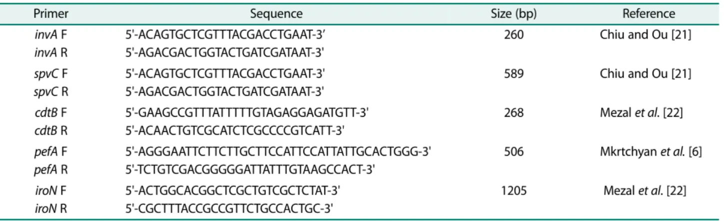
Five virulence genes were assayed for in all the 71 Salmonella isolates using primers (Table 1) targeting invA, spvC, iroN, pefA and cdtB. A 50 µl PCR reaction was used which contained 28.8 µl of nuclease free water, 3.5 µl MgCl2 (25 mM), 10 µl 5x PCR buffer, 1 µl dNTPs (10 mM), 0.75 µl of each forward and reverse primers (10 µM), 0.2 µl One Taq DNA polymerase (5,000 U/ml) (New England Biolabs) and 5 µl DNA template. Multi- plex PCR was used for invA/spvC and pefA/iroN while simplex PCR was used for cdtB. PCR programming con- ditions included:
invA/spvC
Thirty five cycles of Initial denaturation 94℃ for 2 min, denaturation 94℃ for 30 s, annealing at 56℃ for 30 s,
elongation 72℃ for 2 min and final elongation as 72℃ for 10 min.
pefA/iroN
Thirty five cycles of Initial denaturation 94℃ for 2 min, denaturation 94℃ for 30 s, annealing at 65℃ for 30 s, elongation 72℃ for 1 min and final elongation as 72℃ for 10 min.
cdtB
Thirty five cycles of Initial denaturation 94℃ for 2 min, denaturation 94℃ for 30 s, annealing at 60℃ for 30 s, elongation 72℃ for 1 min and final elongation as 72℃ for 10 min.
PCR products were separated on a 1.5% agarose gel at 120 V and a 100 bp DNA ladder (New England Biolabs) was used as molecular weight maker.
Ethical approval
Ethical approval for this study was obtained from the Human Research and Ethical Committee (HREC) of the Lagos University Teaching Hospital with code number ADM/DCST/HREC/APP/1118 and Nigerian Institute of Medical Research- Institutional Review Board, with proj- ect number IRB/12/180.
Results
Isolation and identification
A total of 71 Salmonella isolates were recovered from human, cattle, sheep and chicken faeces which was sero- typed into 44 serovars. Salmonella Budapest had the highest level of occurrence (31.8%) followed by Salmonella
Table 1. Primers used in the detection of virulence genes.
Primer Sequence Size (bp) Reference
invA F invA R
5'-ACAGTGCTCGTTTACGACCTGAAT-3’
5'-AGACGACTGGTACTGATCGATAAT-3'
260 Chiu and Ou [21]
spvC F spvC R
5'-ACAGTGCTCGTTTACGACCTGAAT-3' 5'-AGACGACTGGTACTGATCGATAAT-3'
589 Chiu and Ou [21]
cdtB F cdtB R
5'-GAAGCCGTTTATTTTTGTAGAGGAGATGTT-3' 5'-ACAACTGTCGCATCTCGCCCCGTCATT-3'
268 Mezal et al. [22]
pefA F pefA R
5'-AGGGAATTCTTCTTGCTTCCATTCCATTATTGCACTGGG-3' 5'-TCTGTCGACGGGGGATTATTTGTAAGCCACT-3'
506 Mkrtchyan et al. [6]
iroN F iroN R
5'-ACTGGCACGGCTCGCTGTCGCTCTAT-3' 5'-CGCTTTACCGCCGTTCTGCCACTGC-3'
1205 Mezal et al. [22]
Essen (15.9%). Thirteen Salmonella Budapest were iso- lated from chicken and one was isolated from cattle. Also three Salmonella Essen isolates were isolated from chicken and four from sheep. Non-typhoidal Salmonella serovars dominated with only two typhoidal (Salmonella Paratyphi B and Salmonella Paratyphi C) serovars iso- lated from humans (Table 2). Isolates from chicken accounted for 47.9% of all isolates.
Pulse field gel electrophoresis (PFGE)
A total of four clusters and three single isolates were obtained from PFGE analysis of S. Budapest as shown in Fig. 1. Isolates that had more than three DNA fragment difference with similarity less than 80% were considered to have originated from different clones, thus were not the same.
Detection of virulence genes by polymerase chain reac- tion (PCR)
Of the five virulence genes (invA, spvC, iroN, pefA and cdtB) assayed for in all 71 Salmonella isolates, only cdtB was not detected. invA gene was detected in all Salmo- nella isolates from humans, chicken, cattle, and sheep.
Ninety four percent (94%) of isolates from sheep har- bored spvC genes closely followed by isolates from cattle as shown in Table 3.
Discussion
Salmonella enterica serovars isolated displayed high level of diversity with 44 serovars. Chicken harbored the highest number of isolates (48%) and serovars. This is to be expected as poultry remains a major source of Salmo- nella enterica serovars [23]. Fagbamila et al. [24]
reported 82 different serotypes of 370 Salmonella iso- lated from poultry farms in Nigeria, with Salmonella Kentucky having the highest rate of occurrence. However in this study Salmonella Budapest had the highest rate of occurrence with 13 S. Budapest isolated from chicken and one isolated from cattle. PFGE analysis to determine clonal relatedness of S. Budapest isolates revealed that they were not from the same clone indicating that S.
Budapest could be wide spread among food animals. NTS serovars accounted for majority of Salmonella serovars from all three food animals with two Paratyphi C and one Table 2. Salmonella enterica serotypes isolated from humans
and food animals.
SN Serotype Frequency of
occurrence Source
1 Salmonella Limete 1 Human
2 Salmonella Portland 1 Human
3 Salmonella Huettwillen 1 Human
4 Salmonella Mowanjum 1 Human
5 Salmonella Paratyphi C 2 Human
6 Salmonella Tyhpimurium 1 Human
7 Salmonella Takoradi 1 Human
8 Salmonella Paratyphi B 1 Human
9 Salmonella Chagoua 1 Human
10 Salmonella Onireke 1 Cattle
11 Salmonella Somone 1 Cattle
12 Salmonella Farmsen 4 Cattle
13 Salmonella Ketheabarny 1 Cattle
14 Salmonella Vom 1 Cattle
15 Salmonella II (43,:g,z62:enx) 1 Cattle
16 Salmonella Sandiego 1 Cattle
17 Salmonella Budapest 14 Cattle/Chicken
18 Salmonella Carno 1 Chicken
19 Salmonella Anecho 3 Chicken
20 Salmonella Muenster 1 Chicken
21 Salmonella Kaapstad 1 Chicken
22 Salmonella Agodi 2 Chicken
23 Salmonella Dabou 1 Chicken
24 Salmonella Tennyson 1 Chicken
25 Salmonella Goldcoast 1 Chicken
26 Salmonella Ebrie 1 Chicken
27 Salmonella Brandenburg 1 Chicken
28 Salmonella Alfort 1 Chicken
29 Salmonella Minna 1 Chicken
30 Salmonella Linton 1 Chicken
31 Salmonella Wichita 1 Chicken
32 Salmonella Ealing 1 Chicken
33 Salmonella Essen 7 Chicken/Sheep
34 Salmonella Livingstone 1 Sheep
35 Salmonella Kivu 1 Sheep
36 Salmonella Berlin 1 Sheep
37 Salmonella Dahra 2 Sheep
38 Salmonella Mono 1 Sheep
39 Salmonella Mura 1 Sheep
40 Salmonella Chomedey 1 Sheep
41 Salmonella Yovokome 1 Sheep
42 Salmonella Sculcoates 1 Sheep
43 Salmonella Wien 1 Sheep
44 Salmonella Orion 1 Sheep
Total 44 71 Sheep
Paratyphi B isolated from human subjects. NTS serovars have a vast host range including ruminants and birds with their products acting as vehicle for the transmission of Salmonella infections to humans and have been impli- cated in bacterial bloodstream infections in children and adults in sub-Saharan Africa [22, 25, 26]. The invasive- ness or pathology of Salmonella infection is determined by the status of the host and pathogen, which in this case are the virulence genes. Majority of these genes are clus- tered in regions on the chromosome called Salmonella Pathogenicity Islands (SPIs) [27]. In this study invA gene located on SPI-1, that facilitate attachment and invasion was detected in all isolates (100%) obtained from humans and all three food animals investigated. This is in line with the findings of El-Feky et al. [28] that detected invA gene in all NTS isolates from food products and clinical samples in Egypt. In a similar study, Smith et al. [29]
reported 96.1% positive invA Salmonella spp. isolated
from food samples in Lagos Nigeria. This points to the fact that invA gene could be a valuable tool in molecular epidemiological surveillance of Salmonella spp. in cases of outbreaks since invA gene is chromosomally located.
Plasmid borne virulence genes code genes that are needed for systemic infection in host cells [30]. pefA and spvC genes that contribute to adhesion and systemic infection respectively [8] were detected in this study.
spvC was detected in 94% of isolates from sheep and 91%
of isolates from cattle, while isolates from chicken and humans were 56% and 40% positive for the gene respec- tively. This is similar with the findings of Borges et al.
[31] that reported spvC in 92% of Salmonella isolates from chicken. Only 53% of Salmonella isolates from chicken had pefA and one isolate from sheep. The absence of pefA genes is expected to contribute to host adaptation and possibly lower outbreak potential [32].
On the other hand the presence of spvC and pefA genes are of public health significance as this could increase morbidity and mortality when isolates are implicated in both human and animal infections. Furthermore the cir- culation of these plasmid mediated genes in the food chain could result in emergence of virulent strains of oth- erwise non-virulent Salmonella serovars because the Salmonella plasmid virulence(spv) locus of which spvC is a part of confers complete virulence on non-virulent Salmonella strains [16]. cdtB gene, a gene responsible for the production of toxin which plays a role in cellular Fig. 1. Dendrogram obtained from DNA fragments generated by Xbal and Pulsed Field Gel Electrophoresis using GelJ. V.2.0.
A total of 4 clusters were generated.
Table 3. Percentage occurrence of virulence genes detected in isolates from humans, chicken, sheep and cattle.
Virulence gene
Human n = 10
Chicken n = 34
Sheep n = 16
Cattle n = 11 invA 10 (100%) 34 (100%) 16 (100%) 11 (100%) spvC 4 (40%) 19 (56%) 15 (94%) 10 (91%) iroN 5 (50%) 34 (100%) 14 (88%) 10 (91%)
pefA 0 18 (53%) 1 (6%) 0
cdtB 0 0 0 0
distension of host cells and not limited to typhoidal strains only [8, 33] was not detected in any of the isolates.
Thus these strains may not cause acute gastroenteritis in human infection. However the presence or absence of a few virulence determinants does not imply that a partic- ular Salmonella serovar cannot elicit disease, though the predominance of multiple virulence genes could indicate a potential for severe infections in humans [34]. iroN, a gene responsible for iron acquisition [22] was detected in all Salmonella serovars obtained from chicken and 50%
of serovars isolated from humans. Results from this study will give a better insight into the epidemiology and pathogenicity of Salmonella enterica serovars circulating in the food chain, since there is a continuous expansion of host range in Salmonella through the acquisition of new genes through horizontal gene transfer of plasmid and pathogenicity islands [35]. Proactive and dynamic sur- veillance is recommended in order to have updated infor- mation on strain evolution, expansion and pathogenicity of Salmonella enterica circulating the food chain to fore- stall Salmonella infection outbreaks.
Acknowledgments
We wish to express our profound gratitude to Prof. M. Dosso of Insti- tute Pasteur Cote d’Ivoire for providing a research space for this work.
This study was supported in part by funds from International Center for Genetic Engineering and Biotechnology (ICGEB) SMART Fellowship (Fellowship number: S/NGA 16-02).
Conflict of Interest
The authors have no financial conflicts of interest to declare.
References
1. Majowicz SE, Musto J, Scallan E, Angulo FJ, Kirk M, O’Brien SJ, et al. 2010. The global burden of non-typhoidal Salmonella gastro- enteritis. Clin. Infect. Dis. 50: 882-889.
2. Levantesi C, Bonadonna L, Briancesco R, Grohmann E, Toze S, Tandoi V. 2012. Salmonella in surface and drinking water: Occur- rence and water-mediated transmission. Food Res. Int. 45: 587- 602.
3. Okoro CK, Kingsley RA, Connor TR, Harris SR, Parry CM, Al-Mash- hadani MN, et al. 2012. Intra-continental spread of human inva- sive Salmonella Typhimurium pathovariants in sub-Saharan Africa. Nat. Genet. 44: 1215-1221.
4. Lim SH, Methe BA, Knoll BM, Morris A, Obaro SK. 2018. Invasive
non-typhoidal Salmonella in sickle cell disease in Africa: is increased gut permeability the missing link. J. Transl. Med. 16:
239.
5. Smith SI, Seriki A, Ajayi A. 2016. Typhoidal and non-typhoidal Sal- monella infection in Africa. Euro. J. Clin. Microbiol. Infect. Dis. 35:
1913-1922.
6. Mkrtchyan MS, Zakharyan MK, Arakelova KA, Sedrakyan AM, Gevorgyan ZU, Ktsoyan ZA. 2016. Molecular determinants of vir- ulence genes of Salmonella Enteritidis prevailing in Armenia. Pro- ceedings of the Yerevan State University 1: 55.
7. Fazl AA, Salehi IZ, Jamshidian M, Amini K, Jangjou AH. 2013.
Molecular detection of invA, ssaP, sseC and pipB genes in Salmo- nella Typhimurium isolated from human and poultry in Iran. Afri.
J. Microbiol. Res. 7: 1104-1108.
8. Elemfareji OI, Thong KL. 2013. Comparative virulotyping of Salmonella typhi and Salmonella Enteritidis. Indian J. Microbiol.
53: 410-417.
9. Rotger R, Casadesus J. 1999. The virulence plasmids of Salmo- nella. Int. Microbiol. 2: 177-184.
10. Oludairo OO, Kwaga JKP, Dzikwi AA, Kabir J. 2013. Detection of invA virulence gene by polymerase chain reaction (PCR) in Salmonella spp. isolated from captive wildlife. Bio-Genet. J. 1: 12- 14.
11. Rowlands REG, Ristori CA, Ikuno AA, Barbosa ML, Jakabi M, de Melo Franco BDG. 2014. Prevalence of drug resistance and viru- lence features in Salmonella spp. isolated from foods associated or not with salmonellosis in Brazil. Revi. Inst. Med. Trop. Sao Paulo.
56: 461-467.
12. Kariuki S, Revathi G, Kariuki N, Kiru J, Mwituria J, Muyodi J, et al.
2006. Invasive multidrug resistant non-typhoidal Salmonella infections in Africa: zoonotic or anthroponotic transmission. J.
Med. Microbiol. 55: 585-591.
13. Marshall KEH, Tewell M, Tecle S, Leeper M, Sinatra J, Kissler B, et al.
2018. Protracted outbreak of Salmonella Newport infections linked to ground beef: possible role of dairy cows-21 states, 2016-2017. MMWR Morb. Mortal. Wkly Rep. 67: 443-446.
14. Omer MK, Alvarez-Ordonez A, Prieto M, Skjerve E, Asehun T, Alv- seike OA. 2018. A systematic review of bacterial food borne out- breaks related to red meat and meat products. Foodborne Pathog. Dis. 15: 598-611.
15. Huusko S, Pihlajasaari A, Salmenlinna S, Sogel J, Dontsenko I, De Pinna E, et al. 2017. Outbreak of Salmonella Enteritidis phage type 1B associated with frozen pre-cooked chicken cubes, Fin- land 2012. Epidemiol. Infect. 145: 2727-2734.
16. Capuano F, Mancusi A, Capparelli R, Esposito S, Proroga YTR.
2013. Characterization of drug resistance and virulotypes of Sal- monella strains isolated from food and humans. Foodborne Pathog.
Dis. 10: 963-968.
17. Adagbada AO, Coker AO, Smith SI, Adesida SA. 2014. The preva- lence and plasmid profile of non-typhoidal salmonellosis in chil- dren in Lagos metropolis, South-Western Nigeria. Pan. Afr. Med. J.
19: 359.
18. Grimont PAD, Weill FX. 2007. Antigenic formulae of the Salmo-
nella serovas, 9th edition, world health organization collaborat- ing center for reference and research on Salmonella Institute Pasteur, Paris France.
19. Heras J, Dominguez C, Mata E, Pascual V, Lozano CT, Torres C, et al. 2015. GelJ-a tool for analzing DNA fingerprint gel images. Bio- informatics 16: 270.
20. Adi PJ, Naidu JR, Matcha B. 2017. Multiplex quantification of Esch- erichia coli, Salmonella Typhi and Vibrio cholera with three DNA targets in single reaction assay. Microb. Pathog. 110: 50-55.
21. Chiu CH, Ou JT. 1996. Rapid identification of Salmonella serovars in faeces by specific detection of virulence genes, invA and spvC, by an enrichment broth-multiplex PCR combination assay. J. Clin.
Microbiol. 34: 2619-2622.
22. Mezal EH, Stefanova R, Khan AA. 2013. Isolation and molecular characterization of Salmonella enterica serovar Javiana from food environmental and clinical samples. International J. Food Micro- biol. 164: 113-118.
23. Andino A, Hanning I. 2015. Salmonella enterica survival coloniza- tion and virulence differences among serovar. ScientificWorld J.
2015: 520179.
24. Fagbamila IO, Barco L, Mancin M, Kwaga K, Ngulukun SS, Zavag- nin P, et al. 2017. Salmonella serovar and their distribution in Nigerian commercial chicken layer farms. PLoS One 12: e0173097.
25. Morpeth SC, Ramadhani HO, Crump JA. 2009. Invasive non-typhi Salmonella disease in Africa. Clin. Infect. Dis. 49: 606-611.
26. Medalla F, Gu W, Malion BE, Judd M, Folster J, Griffin PM, et al.
2017. Estimated incidence of Antimicrobial drug resistant non- typhoidal Salmonella infections, United States, 2004-2012. Emerg Infect. Dis. 23: 29-37.
27. van Asten AJAM, van Dijk JE. 2005. Distribution of classic viru- lence factors among Salmonella spp. FEMS Immunol. Med. Micro-
biol. 44: 251-259.
28. El-Feky MA, Hassan MA, Mohamed WA, Ibrahim NH, Rashwan RS.
2014. Detection of invA gene in non-typhoidal Salmonella iso- lated from food products and clinical cases. Egypt J. Med. Micro- biol. 23: 33-41.
29. Smith SI, Fowora MA, Atiba A, Anejo-Okopi J, Fingesi T, Adamu ME, et al. 2015. Molecular detection of some virulence genes in Salmonella spp. isolated from food samples in Lagos, Nigeria.
Anim. Vet. Sci. 3: 22-27.
30. Rychlik I, Gregorova D, Hradecka H. 2006. Distribution and func- tion of plasmids in Salmonella enterica. Vet. Microbiol. 112: 1-10.
31. Borges KA, Furian TQ, Borsoi A, Moraes HLS, Salle CTP, Nasci- mento VP. 2013. Detection of virulence-associated genes in Sal- monella Enteritidis isolated from chicken in Southern Brazil. Pesq.
Vet. Bras. 33: 1416-1422.
32. Huehn S, La Ragione RM, Anjum M, Saunders M, Woodward MJ, Bunge C, et al. 2010. Virulotyping and antimicrobial resistance typing of Salmonella enterica serovars relevant to human health in Europe. Foodborne Pathog. Dis. 7: 523-535.
33. Figueiredo R, Card R, Nunes C, Abuoun M, Bagnall MC, Nunez J, et al. 2015. Virulence characterization of Salmonella enterica by a new microarray: detection and evaluation of the cytolethal dis- tending toxin gene activity in the unusal host S. Tyhimurium.
PLoS One 10: e0135010.
34. Zou M, Keelara S, Thakur S. 2012. Molecular characterization of Salmonella enterica serotype Enteritidis isolated from humans by antimicrobial resistance, virulence genes and pulsed field gel electrophoresis. Foodborne Pathog. Dis. 9: 1-7.
35. Foley SL, Johson JJ, Ricke SC, Nayak R, Danzelsen J. 2013. Salmo- nella pathogenicity and host adaptation in chicken associated serovars. Microbiol. Mol. Biol. Rev. 77: 582-607.