• 검색 결과가 없습니다.

Cardiac Tamponade Associated with Acupuncture

N/A
N/A
Protected

Academic year: 2021

Share "Cardiac Tamponade Associated with Acupuncture"

Copied!
4
0
0

로드 중.... (전체 텍스트 보기)

전체 글

(1)

http://www.e-kmj.org 2020 Keimyung University School of Medicine

48

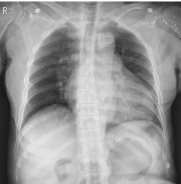
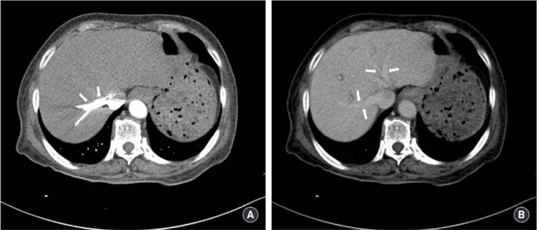
Acupuncture is regarded as a relatively safe procedure, but it may have various complications. Cardiac tamponade after acupuncture is an extremely rare compli- cation but can have fatal outcomes. We present a 73-year-old female patient with decreased consciousness and dyspnea after acupuncture at an oriental medical clinic. Initial vital signs were a blood pressure of 60/40 mmHg and a heart rate of 110 beats/min. Echocardiography and chest computed tomography were per- formed in the emergency department to determine the cause of the shock, and the result came out as hemopericardium. The patient underwent through an emer- gency cardiac surgery under the diagnosis of cardiac tamponade following acu- puncture and was fully recovered after surgery.

Keywords: Acupuncture, Cardiac tamponade, Complication

Cardiac Tamponade Associated with Acupuncture

Joo Hwan Lee

1

, Sang Chan Jin

2

, Woo Ik Choi

2

, Dong Kyu Lee

1

1

Department of Emergency Medicine, Daegu Fatima Hospital, Daegu, Korea

2

Department of Emergency Medicine, Keimyung University School of Medicine, Daegu, Korea

Introduction

Acupuncture is used to relieve or treat a certain disease by inserting one or more needles in a specific site of the body [1]. Acupuncture has been widely practiced in the East as a therapeutic intervention, and acknowledgment of its utility and safety lead to gradual spread to the West [2].

Acupuncture is regarded as relatively safe, as the risk of a serious adverse event after acupuncture is estimated to be only 0.05 per 10,000 treatments, and 0.55 per 10,000 individual patients. However, severe and fatal adverse events are being constantly reported [3]. We report a case of cardiac tamponade after acupuncture with decreased consciousness and dyspnea, with a review of relat- ed literature.

Case report

A 73-year-old female patient with no known previous illness developed de- creased consciousness and dyspnea after visiting an oriental clinic had acu- puncture applied at epigastric and scapular area to treat intermittent epigastric pain and dyspepsia which started a month earlier. She was transferred to our hospital’s emergency department (ED). At the time arrived at the ED, mental status was drowsy, and the initial vital signs were a blood pressure of 60/40 mmHg, a pulse rate of 110 beats/min, a respiratory rate of 23 breaths/min, and a body temperature of 35.0°C. On physical examination, jugular venous disten- sion was observed, with a distant heart sound on auscultation, and the abdo- men was flat without tenderness. Laboratory findings were as follows: white blood cell count, 6,570/mm

3

(neutrophils 30%); hemoglobin, 13.2 g/dL; hema- Received: March 8, 2020

Revised: May 5, 2020 Accepted: May 6, 2020 Corresponding Author:

Joo Hwan Lee, M.D.

Department of Emergency Medicine, Daegu Fatima Hospital, 99, Ayangro, Dong-gu, Daegu 41199, Korea Tel: +82-53-940-7139 E-mail: [email protected] pISSN 2092-8335 · eISSN 2733-5380 Keimyung Med J 2020 39(1):48-51 https://doi.org/10.46308/kmj.2020.00038

Case Report

This is an open-access article distributed under the

terms of the Creative Commons Attribution license

(http://creativecommons.org/licenses/by-nc/4.0/),

which permits unrestricted use, distribution, and

reproduction in any medium, provided the original

work is properly cited.

(2)

49 계명의대학술지 제39권 1호 2020

http://www.e-kmj.org

Keimyung Med J

tocrit, 39.9%; plateletcount, 167,000/mm

3

; aspartatetransam- inase, 76 IU/L; alaninetransaminase, 24 IU/L; CK-MB, 1.0 ng/mL; and troponin-I, 0.072ng/mL. Electrocardiography revealed arterial fibrillation without change of the ST seg- ment. The chest X-ray showed enlarged, globular-shaped heart (Fig. 1); the echocardiography, pericardial effusion; and chest computed tomography (CT) scan, a small amount of pneumothorax in left hemithorax along with a large volume of pericardial effusion (Fig. 2,3). Any specific finding was not shown except hepatic congestion in abdominal CT (Fig. 4).

Based on the aforementioned test findings and the patient’s symptoms immediately after acupuncture, she was diagnosed as cardiac tamponade caused by acupuncture. A pericardial window operation was done, concerning the risk of heart in- jury with pericardiocentesis via bedside transthoracic echo- cardiography. 300cc of bloody pericardial effusion was drained through pericardial window creation, and membra- nous tissues of the blood clot were found. The patient fully recovered after surgery and was discharged without compli- cations.

Discussion

Acupuncture is performed not only to control pain but also for a variety of reasons, such as obesity control, cessation of smoking, relief of tinnitus, post-stroke rehabilitation, treat-

ment of rheumatic diseases, etc [4]. It is generally regarded as a safe procedure, if performed by a well-trained and experi- enced practitioner, but it can lead to various complications in practice [5]. Most of adverse events of acupuncture are mild, such as bleeding, hematoma, local infection, headache, and myalgia, but there can be serious adverse events such as neu- ral damage, pneumothorax, hepatitis, perforation of internal organs, spinal cord damage, and cardiac tamponade [6].

Fig. 1. Chest X-ray performed after the patient arrived at the ED, showing an enlarged cardiac silhouette with a clear lung field.

Fig. 2. Chest CT scan showing a small amount of pneumothorax in the left hemithorax (arrows).

Fig. 3. Chest CT scan showing pericardial fluid collection (arrows).

(3)

http://www.e-kmj.org Cardiac Tamponade Following Acupuncture

50

Keimyung Med J

Generally, 2cm- to 5cm-long, 26- to 32-gauge needles are used in acupuncture, which can damage the nervous system and blood vessels if they penetrate the thoracic and abdomi- nal viscera [7,8]. Therefore, cardiac tamponade can occur, though rarely, when acupuncture is performed in the thoracic cage, and this may be fatal [9]. According to the review of lit- erature of Ernst and Zhang [9], 26 patients were diagnosed with cardiac tamponade after acupuncture in 17 studies re- ported up to 2010, in which eleven of the patients fully recov- ered after surgery or other treatment, and 14 died.

Cardiac tamponade generally occurs after the heart is com- pressed through the accumulation of fluid, pus, blood, clots, or gas in the pericardium [10]. Common causes include hem- orrhage from chest trauma, metastatic malignancy, pericardi- tis, uremia, etc [11]. The mechanism of cardiac tamponade caused by acupuncture includes cardiac tissue or vessel injury by the needle, and damage of the right ventricular wall due to the inflow of the needle through the bloodstream [12,13]. Di- rect cardiac damage is also associated with penetration of the sternum through congenital sternal foramen, a malformation found in 5-8% of the population [14]. It is usually hidden by

tendon fibers, connective tissue, bone, so it cannot be easily detected through palpation. Lee et al. [15] reported a case of a patient who presented chest pain after acupuncture in his up- per abdomen. The patient was diagnosed with cardiac tam- ponade caused by a broken acupuncture needle that moved to the right ventricle and was detected in the fluoroscopy, com- puted tomography, and trans-esophageal echocardiography, and the needle was successfully removed through cardiac sur- gery. A recent case of cardiac injury due to acupuncture was in four cases (Table 1).

In this case, a patient without any previous illness was transferred to the ED in shock state, just after undergoing acupuncture in her scapula and upper abdomen. Echocardi- ography confirmed pericardial effusion and pneumothorax was presented on chest CT scan, but no foreign body was de- tected. A pericardial window operation was performed, acu- puncture needles were not found and the biopsy results after the surgery showed no specific finding other than blood clots, which ruled out secondary causes such as metastatic malig- nancy. The patient was diagnosed with acute cardiac tampon- ade due to direct cardiac injury by acupuncture needle. In

B Fig. 4. Abdominal CT scan showing contrast regurgitation to the hepatic vein (A) and dilatation of the hepatic vein (B).

A B

Table 1. Acupuncture related heart injuries

Reference Patient information Punctured site Adverse event Follow up

Kirchgatterer et al. (2000) [16] 83/F Sternum Cardiac tamponade Recovered

Park et al. (2005) [17] 49/F Shoulders and upper back Cardiac tamponade Recovered

Song and Park (2010) [18] 69/F Shoulders and neck Myocardium injury Unknown

Kim et al. (2011) [19] 54/F Chest, abdomen Hemopericardium Recovered

(4)

51 계명의대학술지 제39권 1호 2020

http://www.e-kmj.org

Keimyung Med J

chest CT scan, pneumothorax occurs in the anterior part of the left lung, so it is estimated that the pericardiac tamponade is caused by upper abdomen acupuncture rather than the scapula.

Complications related to acupuncture result from the lack of anatomical knowledge and hygiene of the practitioners [20]. Practitioner education on the human anatomy and hy- giene should be strengthened, and patient education on acu- puncture complications and precautions are needed to pre- vent severe complications. Also, practitioners should be aware of the possible adverse events following acupuncture and should be able to treat emergency cases. Emergency physi- cians should be aware of the occurrence of cardiac tamponade after acupuncture through a detailed medical examination to improve the patient prognosis through early diagnosis and treatment.

Conflict of interest

All authors declare no conflicts-of-interest related to this article.

References

1. Eo EK, Shin JS, Choi OK. Pneumothorax following by acupunc- ture. J Korean Soc Emerg Med. 1996;7:429-33.

2. NIH Consensus Conference. Acupuncture. JAMA. 1998;

280:1518-24.

3. White A. A cumulative review of the range and incidence of sig- nificant adverse events associated with acupuncture. Acupunct Med. 2004;22:122-33.

4. Vickers A, Wilson P, Kleijnen J. Acupuncture. Qual Saf Health Care. 2002;11:92-7.

5. Carron H, Epstein BS, Grand B. Complications of acupuncture.

JAMA. 1974;228:1552-4.

6. Macpherson H, Scullion A, Thomas KJ, Walters S. Patient re- ports of adverse events associated with acupuncture treatment: a prospective national survey. Qual Saf Health Care. 2004;13:349-

55.

7. Vilke GM, Wulfert EA. Case reports of two patients with pneu- mothorax following acupuncture. J Emerg Med. 1997;15:155-7.

8. Peuker ET, White A, Ernst E, Pera F, Filler TJ. Traumatic compli- cations of acupuncture. Therapists need to know human anato- my. Arch Fam Med. 1999;8:553-8.

9. Ernst E, Zhang J. Cardiac tamponade caused by acupuncture: a review of the literature. Int J Cardiol. 2011;149:287-9.

10. Spodick DH. Acute cardiac tamponade. N Engl J Med. 2003;

349:684-90.

11. Strobbe A, Adriaenssens T, Bennett J, Dubois C, Desmet W, Mc- Cutcheon K, et al. Etiology and long-term outcome of patients undergoing pericardiocentesis. J Am Heart Assoc. 2017;6. DOI:

10.1161/jaha.117.007598.

12. Schechter DC, Gilbert L. Injuries of the heart and great vessels due to pins and needles. Thorax. 1969;24:246-53.

13. Kataoka H. Cardiac tamponade caused by penetration of an acu- puncture needle into the right ventricle. J Thorac Cardiovasc Surg. 1997;114:674-6.

14. Peuker E, Grönemeyer D. Rare but serious complications of acu- puncture: traumatic lesions. Acupunct Med. 2001;19:103-8.

15. Lee BK, Lee SH, Ko JY, Yoo BS, Yoon J, Choe KH, et al. A case of cardiac tamponade due to penetration of the right ventricule by an acupunture needle. Korean Circ J. 1999;29:1129-32.

16. Kirchgatterer A, Schwarz CD, Höller E, Punzengruber C, Hartl P, Eber B. Cardiac tamponade following acupuncture. Chest.

2000;117:1510-1.

17. Park JH, Shin HJ, Choo SJ, Song JK, Kim JJ. Successful removal of migrated acupuncture needles in a patient with cardiac tam- ponade by means of intraoperative transesophageal echocardio- graphic assistance. J Thorac Cardiovasc Surg. 2005;130:210-2.

18. Song BG, Park JR. Acupuncture needles over the myocardium.

Heart Lung Circ. 2010;19:480.

19. Kim JH, Kim SH, Lee YJ, Hong JS, Ahn R, Hong ES. Hemoperi- cardium following acupuncture. Yonsei Med J. 2011;52:207-9.

20. Leung PC, Zhang L, Cheng KF. Acupuncture: complications are

preventable not adverse events. Chin J Integr Med. 2009;15:229-

32.

수치

Fig. 2. Chest CT scan showing a small amount of pneumothorax  in the left hemithorax (arrows).
Table 1. Acupuncture related heart injuries

참조

관련 문서

In our study, a set of the terms extracted from the RSNA reporting templates of CT Head, DX Chest, MR Spine, NM Bone Scan, and US Abdomen exams was compared with its matching

Chest CT scan obtained at admission shows extensive patchy areas of ground-glass opacities with upper and middle lung zone predominance and small nodular

Chest CT scan(after 6 cycles of

Subclavian vein occlusion occurs from 3.6 to 9.0% after implantation of a cardiac rhythm device. 1)2) Engorged veins over the anterior chest wall can be caused by stenosis or

Preoperative photograph of the left eye (A) and B-scan ultrasonography of the left eye on day 41 (B) : Corneal opacity and shallow anterior chamber were