176 This is an Open-Access article distributed under the terms of the Creative Commons Attribution Non-Commercial License (http://
creativecommons.org/licenses/by-nc/3.0) which permits unrestricted non-commercial use, distribution, and reproduction in any medium, provided the original work is properly cited.
J. Mushrooms 2021 September, 19(3):176-183 http://dx.doi.org/10.14480/JM.2021.19.3.176 Print ISSN 1738-0294, Online ISSN 2288-8853
© The Korean Society of Mushroom Science
Minkyeong Kim(Researcher), Hyehyun Hong(Graduate student), Jung-Hwan Kim(Graduate student), Seung-Young Kim(Professor), Changmu Kim(Senior Reseaercher)
*Corresponding author E-mail : [email protected]
Tel : +82-32-590-7110,Fax : +8232-590-7472 Received August 27, 2021
Revised September 6, 2021 Accepted September 6, 2021
자생 Tuber himalayense 자실체 추출물의 항염증 활성
김민경
1· 홍혜현
2· 김정환
2· 김승영
2· 김창무
3*
1국립생물자원관 미생물자원과
2선문대학교 제약생명공학과
3국립생물자원관 유용자원분석과
Anti-inflammatory activity of indigenous Tuber himalayense in Korea
Minkyeong Kim
1, Hyehyun Hong
2, Jung-Hwan Kim
2, and Seung-Young Kim
2, and Changmu Kim
3*
1Microorganism Resources Division, National Institute of Biological Resources, Incheon 22689, Korea
2Department of Pharmaceutical Engineering & Biotechnology, Sunmoon University, Asan 31460, Korea
3Biological and Genetic Resources Assessment Division, National Institute of Biological Resources, Incheon, 22689, Korea
ABSTRACT:In this study, the anti-inflammatory activity of an extract of the fruiting body of the Tuber himalayense (TH) truffle collected from oak growing areas in Korea was investigated. The extract was not cytotoxic at concentrations below 100µg/mL in an experiment evaluating inflammation inhibitory effect in lipopolysaccharide (LPS)-stimulated RAW 264.7 macrophages. LPS- induced nitric oxide (NO) and prostaglandin E2 (PGE2) production was inhibited by the extract in a concentration-dependent manner. Western blot assay results indicated that the anti-inflammatory activity of TH extract was likely caused by the reduced production of NO and PGE2 via suppression of induced NO synthase and cyclooxygenase-2 gene expression. In addition, TH extract effectively inhibited the production of interleukin (IL)-1β and IL-6 by macrophages. Thus, TH extract effectively inhibits the overexpression of various inflammatory mediators and could be valuable in formulating anti-inflammatory foods and medicines that target these components.
KEYWORDS:Anti-inflammation, Fruiting body extract, Truffle, Tuber himalayense
서 론
Tuber(덩이버섯)속은 세계 3대 진미중의 하나인 Truffle(송
로버섯)로 더 알려져 있는 자낭균문(Ascomycota), 주발버섯 목(Pezizales), 덩이버섯과(Tuberaceae)에 속하는 종들이다.
세계적으로 가장 유명한 외생균근형성 균류 중 하나이며 (Trappe, 1979), 전세계적으로 약 220여종이 알려져 있고 주로 참나무 뿌리와 공생관계를 이루며 생육한다(Bonito et al., 2013; Benucci et al., 2012). Tuber는 자실체 (fruiting body)의 색깔, 모양 그리고 크기에 따라 다양한 종으로 분류되며 독특한 향과 풍미가 특징이다(Bonito et al., 2016). Tuber는 초기 자실체 형성시 흰색을 띠며, 점 차 크기가 커지면서 어두워진다. 대부분의 성숙한 Tuber 의 자실체는 갈색에서 어두운 갈색을 띠고, 구형으로 자 라며 표면에 돌출된 다각형의 돌기로 덮여 있다(Xiao et al., 2015; Patel et al., 2017). 국내에서는 Tuber aestivum subsp. uncinatum 만이 유일하게 보고되었다가(Shin et al., 1995), 최근 T. huidongense와 T. himalayense가 각각 포항과 단양에서 발견되어 새롭게 보고되었다(Park et al., 2020; Park et al., 2021).
Tuber의 생리활성에 대한 연구는 식재료로 잘 알려진
종들 중심으로 이루어져 왔다. 주로 유럽에서 발견되며 고가의 송로버섯으로 알려진 T. melanosporum(Périgord black truffle)은 항종양활성(Zhao et al., 2014), white truffle 이라고 불리는 T. magnatum는 항산화활성 및 항염 활성이 보고되었다(Beara et al., 2014). T. himalayense와 함께 Asian black truffle 로 분류되는 T. indicum 역시 항 산화활성에 대한 연구결과가 있다(Guo et al., 2011).
염증 반응은 세포의 손상을 초기 단계에서 억제하고 상 처부분의 치유에 필수적인 생체내 방어체계에 해당되지만, 과도한 염증 혹은 염증의 결과로 발생하는 조직의 손상, 질병 등이 패혈증, 관절염 등을 유발하여 다양한 질병의 위험요소로 작용한다. 다양한 염증관련 물질이나 메커니 즘이 알려져 있어, 특정 물질이 어떠한 부분에 영향을 미 치는지 확인하는 실험을 주로 진행한다. 염증 조절 유전자 인 inducible nitric oxide synthase(iNOS)와 cyclooxygenase- 2(COX-2)는 활성화된 대식세포에서 발현되어 NO, PGE2
와같은 다량의 염증 매개체를 합성하는 것으로 알려져 있다(Wynn and Vannella, 2016). 이들의 발현은 인체 내 에서 병리적으로 중요한 작용을 하는데, iNOS로부터 과 량 합성된 NO는 다양한 염증성 질환과 밀접한 관련이 있 는 것으로 알려져 있다. 또한 COX-2는 통증과 염증을 유 발하는 prostaglandins의 분비를 자극하는 효소로서 통증 과 염증을 유발하고 세포증식에 관여하는 등 여러 염증 관련 질병의 원인으로도 알려져 있다(Kim et al., 2013;
Kong, 2014; Laskin and Pendino, 1998). Prostaglandin E2(PGE2)는인체의 면역반응에 중요한 역할을 하는 염증 반응의 매개체로 phospholipase A2(PLA2)의 효소 작용에 의해 arachidonic acid로부터 합성 된다(Lee et al., 2014).
본 연구에서는 우리나라에서 채집된 자생 T. himalayense 의 항염증 활성을 검증하고자 LPS로 유도된 RAW 264.7 대식세포에 자실체 열수 추출물을 이용하여, 염증매개체 로 알려진 NO, PGE2, iNOS, COX-2, 그리고 pro- inflammatory cytokine의 억제를 측정하는 방법을 이용하 였다. T. himalayense는 가장 최근에 국내에 서식이 확인 된 종이며 같은 속의 종에서 다양한 생리활성에 대한 보 고가 있어, 항염증 활성을 보일 가능성이 높을 것으로 판 단되었다.
재료 및 방법
채집 표본의 분류 및 동정
본 연구에 사용된 Tuber himalayense NIBRFG0000505337 는 2019년 9월 27일에 인천광역시 국립생물자원관 내 참 나무 인공 생육지대에서 채집되었다(Fig. 1A-B). 표본의 동정을 위해 AccuPrep Genomic DNA 추출 키트 (Bioneer, Daejeon, Korea)를 사용하여 DNA를 추출하였 다. 추출한 DNA는 International transcribed spacer(ITS), Large-subunit rRNA(LSU), β-tubulin(TUB), 그리고
translation elongation factor 1-alpha(TEF-1α)의 구간을 ITS1F–ITS4(Gardes and Bruns, 1993; White et al., 1990), LR0R–LR7(Rehner and Samuels, 1994; Vilgalys and Hester, 1990) BT2a–BT2b(Glass and Donaldson, 1995) 그리고 EF1α Tuber_f–EF1α Tuber_r(Bonito et al., 2013) primer를 이용하여 증폭하였으며 Macrogen(Seoul, Korea)사에 의뢰하여 염기서열을 분석하였다. Geneious 9.0.5(Kearse et al, 2012) 를 이용해 분석된 염기서열을 NCBI의 blast tool을 사용하여 근연종을 확인하였다. 계통 분석은 MEGA 7software(Kumar et al., 2016)를 사용하 여 Maximum Likelihood방법으로 GenBank의 레퍼런스 서열과 비교하여 계통수를 작성하였으며, bootstrap 1,000 반복하여 분석하였다(Kimura, 1980). 형태 동정을 위해 광학현미경(NIKON 80i , Nikon, Japan)으로 미세구조를 관찰하였으며, 전자현미경(JSM -6390LV, JEOL, Japan) 을 통해 포자를 추가 관찰하였다(Fig. 1C-J).
자실체 열수 추출을 통한 분석용 시료 확보
채집한 시료는 열풍건조 후 분쇄하여 시료 1 g에 10배 의 증류수를 가하여 24시간동안 열수 추출을 2회 진행하 였으며, 회수한 1차 추출물과 2차 추출물을 혼합하여 5.0μm의 공극 크기를 갖는 필터(TOYO, Japan)로 여과하 였다. 여과된 추출물은 동결건조하여 분말화하였으며, 분 말화된 추출물을 Ethyl acetate로 분획한 뒤, 진공 농축하 여 실험에 사용하였다.
분석용 대식세포의 준비
본 연구에서 사용된 LPS는 Sigma-Aldrich(St. Louis, MO, USA)에서 구입하였으며, 마우스 대식세포인 RAW 264.7은 한국세포주은행에서 분양 받아 10% fetal bovine serum(FBS)과 100 U/mL penicillin, 100 μg/mL streptomycin 이 포함 된 Dulbecco's Modified Eagle Medium(DMEM, Gibco, NY, USA) 배양액을 사용하여 37oC, 5% CO2 incubator에서 배양하였다. 이후 2일을 주기로 계대 배양 하여 세포가 안정된 상태에서 실험을 진행하였다.
세포 생존율 측정
세포의 생존율은 3-(4,5-dimethylthiazole-2-yl)-2,5- diphenyl-tetrazolium bromide(MTT) 환원 방법을 이용하 여 측정하였다(Denizot and Lang, 1986). RAW 264.7 세 포를 24-well plates에 8.0 × 10⁴ cells/well이 되도록 분주 한 뒤, 37oC, 5% CO2조건의 incubator에서 24시간 동안 전 배양하였으며, 농도별로 희석한 시료와 LPS(1 μg/mL) 를 동시 처리하여 동일한 조건하에 24시간 동안 배양하였 다. 이후 MTT 시약을 첨가하여 37oC에서 3시간 동안 반 응하여 생성된 보라색 formazan을 DMSO로 용해시킨 뒤, 96-well plates에 옮겨 담아 Microplate reader(Thermo Scientific, MA, USA)를 이용해 570 nm에서 흡광도를 측
정하였다. 각 시료에 대한 평균 흡광도를 측정하였으며, 대조군의 흡광도 측정값과 비교하여 세포 생존율을 평가 하였다
Nitric oxide(NO) 생성 억제 활성 측정
LPS로 유도된 RAW 264.7 세포에서 Nitric oxide(NO) 생성 억제 활성은 Griess 반응을 이용하여 측정하였다.
24-well plates에 세포를 8.0 × 10⁴ cells/well이 되도록 분 주한 뒤, 37oC, 5% CO2 조건의 incubator에서 24시간 전 배양 하였으며, 농도별로 희석 된 시료와 LPS(1 μg/mL)를 동시 처리하여 동일한 조건하에 24시간 배양하였다. 이후 상등액과 Griess 시약 [1% (w/v) sulfanilamide, 0.1% (w/v) naphylethylenediamine in 2.5%(v/v) phosphoric acid]
100 μL를 동량 혼합하여 실온에서 10분간 암반응 후 Microplate reader(Thermo Scientific, MA, USA)를 이용 해 540 nm에서 흡광도를 측정하였다.
Prostaglandin E2(PGE2) 생성 억제 활성 측정
RAW 264.7 세포를 24-well plates에 8.0 × 10⁴ cells/
well만큼 분주하여 24시간 전 배양한 뒤, 농도별로 희석된 시료와 LPS(1 μg/mL)를 동시 처리하여 동일한 조건하에 24시간 동안 배양하였다. 24시간 후 배양 배지를 회수한 뒤, 원심분리(10,000 rpm, 3 min)하여 침전물을 제거한 상등액에서 PGE2의 생성량을 측정하였으며, 상등액에 존재 하는 PGE2의 함량은 mouse enzyme-linked immunosorbent assay(ELISA) kit (R&D Systems Inc., Minneapolis, MN, USA)를 이용하여 측정하였다.
Western blot analysis
RAW 264.7 세포를 4.0 × 10⁵ cells/well로 분주하여 37oC, 5% CO2 조건의 incubator에서 24시간 동안 전 배 양하였으며, 이후 배양 배지로 희석된 시료와 LPS(1 μg/
mL)를 동시 처리하여 동일 조건으로 24시간 배양하였다.
24시간 후 각 well의 세포를 PBS로 3회 세척하였으며, lysis buffer[1 × RIPA(Upstate Cell Signaling Solution, NY, USA), 1 mM phenylmethylsulfonyl fluoride(PMSF), Fig. 1. Morphological features of Tuber himalayense (NIBRFG0000505337), Ascocarp (A), peridium and gleba (B), asci (C–E), ascospores (F–G), SEM images of ascospores(H–J). Scale bars B = 1 cm, C = 100 μm, D–E = 50 μm, F–G = 10 μm, H–J = 10 μm.
1 mM Na3VO4, 1 mM NaF, 1 μg/mL aprotinin, 1 μg/mL pepstatin, and 1 μg/mL leupeptin]를 이용하여 단백질을 1 시간 동안 lysis하였다. 이후 13,000 rpm, 20 min의 조건으 로 원심분리하여 상등액을 회수 하였으며, BCA ki(Bio- Rad, USA)를 사용하여 정량한 단백질을 10%의 polyacrylamide gel에 전기영동하고 poly-vinylidene difluoride(PVDF) membrane(Milipore, Burlington, MA, USA)에 200 mA, 2시간 동안 전이시켰다. 5% skim milk 를 포함한 0.05% Tween 20/Tris-buffered saline(0.05%
T/TBS)에 단백질이 전이된 membrane을 넣고 상온에서 1 시간 30분간 blocking 시킨 후, 4oC에서 18시간동안 1차 항체와 반응시켰다. 1차 항체는 iNOS antibody(1 : 5,000, Bio-Rad, USA), COX-2 antibody(1 : 1,000, Rockland Immunochemicals, Inc. USA), β-actin antibody clone AC-74(1 : 10,000, Sigma, USA), phospho-ERK1/2 antibody, phospho-SAPK/JNK(Thr180/Tyr185) antibody, phospho-p38 antibody, IκB-α antibody, phospho-NF-κB antibody(1 : 1,000, Cell signaling Tech, Danvers, MA, USA)를 이용하였다. 반응이 끝난 후 0.05% T/TBS 용액 으로 4회 세척 후 1 : 10,000으로 희석한 2차 항체 (Jackson ImmunoResearch, USA)에 담근 뒤, 상온에서 1 시간 30분간 반응하였다. 반응 후 0.05% T/TBS 용액으 로 4회 세척한 membrane은 ECL kit(Bio-rad, USA)와 imaging densitometer(model GS-700, Bio-rad, USA)을 이용하여 단백질 발현을 측정하였으며, 그 결과는 ImageJ program(NIH, Bethesda, MD, USA)을 통해 β-actin 대비 iNOS와 COX-2 단백질 발현 면적을 수치화한 뒤 그래프 로 나타내었다.
Pro-inflammatory cytokine(TNF-α, IL-6, IL-1β) 생성 억제 활성 측정
RAW 264.7 세포를 24-well plates에 8.0 × 10⁴ cells/
well로 분주하여 37oC, 5% CO2 조건의 incubator에서 24 시간 동안 전 배양하였으며, 농도 별로 희석된 시료와 LPS(1 μg/mL)를 동시에 처리하여 동일한 조건하에 24시 간 동안 배양하였다. 이후 배양 배지를 회수하여 원심분 리(10,000 rpm, 3 min)하여 침전물을 제거한 상등액을 pro-inflammatory cytokine의 생성량 측정에 사용하였으 며, 상등액에 존재하는 cytokine의 함량은 Mouse TNF-α ELISA Kit(Invitrogen, California, USA), Mouse IL-6 ELISA Kit(BD Biosciences, California, USA), Mouse IL-1β ELISA Kit(R&D Systems Inc., Minneapolis, MN, USA)를 이용하여 측정하였다.
통계처리
모든 실험은 3회 반복하여 측정하였으며, 그 결과는 평 균값 ± 표준편차로 나타내었다. 통계적 분석은 각 처리 구 간의 유의성(*p < 0.05; **p < 0.01) 검증을 위해 분산분석 (analysis of variance, ANOVA)후 student’s t-test로 다중 비교를 실시하였다.
결과 및 고찰
채집 표본의 분류 및 동정
채집된 표본(NIBRFG0000505337)이 대한 형태적 관찰 결과(Fig. 1)와 분자계통학적 분석을 통해 Tuber himalayense 인 것으로 확인되었다. ITS와 LSU의 염기서열로 작성한
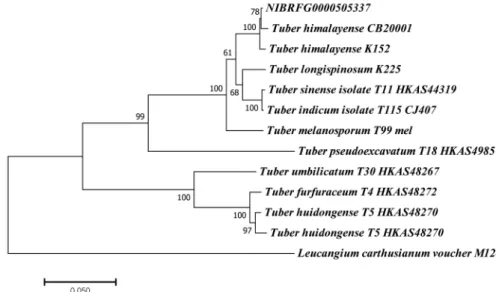
Fig. 2. Phylogenetic tree of Tuber himalayense(NIBRFG0000505337). The tree inferred from concatenated alignment of internal transcribed spacer(ITS) and Large-subunit rRNA(LSU) sequences. The topological structure was determined using a Maximum Likelihood method based on the Kimura 2-parameter model. The sample from our study is indicated in bold. Scale bar indicates the number of substitutions per site for each branch.
계통수에서 T. himalayense(CB20001 및 K152)와 단계통 임을 보여주었다(Fig. 2). Park 등(2021)에 의해 보고된 T.
himalayense와 형태 및 분자계통학적 분석결과 같은 종으 로 확인되었다. 또한 TUB 유전자 및 TEF-1α 유전자는 각각 T. himalayense LC312253(100%), T. himalayense LC312271(100%)와 높은 염기서열 유사성을 보였다.
세포 생존율 측정
T. himalayense(시료명: TH) 열수 추출물의 세포 생존 률을 조사하기 위하여 25, 50, 100 µg/ml 농도로 희석한 시료와 LPS(1 μg/mL)를 동시 처리하여 24시간동안 배양 한 후 MTT 분석을 진행한 결과, 가장 고농도인 100 µg/
ml 에서 85.9±1.2%로 시료를 처리한 모든 농도에서 80%
이상의 세포 생존율을 보여 RAW264.7 세포에 대한 독성 이 나타나지 않음을 확인하였다(Fig. 3). 따라서 추후 진 행될 실험에서는 세포 독성이 나타나지 않은 25, 50, 100 µg/ml 농도를 사용하였다.
NO 생성 억제 활성 측정
T. himalayense가 LPS로 증가된 NO의 생성에 미치는 영향을 조사하기 위하여 RAW 264.7 세포를 LPS(1 μg/
mL)로 자극을 유발한 뒤, 시료를 25, 50, 100 µg/ml 농도 로 동시 처리하여 24시간 후 NO 생성량을 측정하였다.
그 결과, TH를 처리한 RAW 264.7 세포는 LPS 단독 처 리군에 비하여 NO생성을 39.4 ± 0.6%, 60.6 ± 1.0%, 79.1 ± 0.9% 억제하였으며, 가장 고농도인 100 µg/ml에서 는 무처리군과 유사한 수준의 저해 활성을 확인하였다 (Fig. 4). 이러한 결과는 TH추출물이 염증인자 중 하나인 NO를 효과적으로 억제하며, NO의 다량 생성으로 인해 유발되는 염증을 표적으로 하는 항염증 소재로 적용 할 수 있음을 시사한다. 염증반응에서 iNOS로부터 생성된
다량의 NO는 염증반응을 심화하고 패혈성 쇼크, 류마티 스 관절염과 같은 여러 염증성 질환을 유발하는 것으로 알려져 있다(Laskin and Pendino, 1998).
Prostaglandin E2 (PGE2) 생성 억제 활성 측정
T. himalayense 처리에 의한 인체의 면역반응에 중요한 역할을 Prostaglandin E2(PGE2)의 생성 억제활성을 조사 하기 위하여 RAW 264.7 세포에 LPS(1 μg/mL)로 자극을 유발한 뒤, 시료를 25, 50, 100 μg/mL를 동시 처리하여 24시간 배양한 후 PGE2의 생성량을 측정하였다. 그 결과, TH를 처리한 RAW 264.7 세포는 LPS 단독 처리군에 비하 여 PGE2 생성을 11.8 ± 0.4%, 45.4 ± 3.7%, 67.9 ± 0.8% 억 제하는 것을 확인하였다(Fig. 5). PGE2의 합성은 COX-2 에 의해 촉진되는 것으로 보고되어 있으며, 염증자극 발
Fig. 3. Inhibitory effects of TH on cell viability in RAW 264.7 cells. The production of nitric oxide was assayed in the culture medium of cells stimulated with LPS (1 μg/mL) for 24 h in the presence of TH (25, 50 and 100 μg/mL).
Cytotoxicity was determined by MTT assay. Result is expressed as percentages compared to the respective values obtained for the control.
Fig. 4. Inhibitory effects of TH on nitric oxide production in RAW 264.7 cells. The production of nitric oxide was assayed in the culture medium of cells stimulated with LPS (1 μg/
mL) for 24 h in the presence of TH (25, 50 and 100 μg/mL).
Result is expressed as percentages compared to the respective values obtained for the control. Data represent the means ± SD with three separate experiments. *p<0.05; **p<0.01.
Fig. 5. Inhibitory effects of TH on PGE2 production in RAW 264.7 cells. The production of PGE2 was assayed in the culture medium of cells stimulated with LPS (1 μg/mL) for 24 h in the presence of TH extract (25, 50, and 100 μg/mL).
Result is expressed as percentages compared to the respective values obtained for the control. Data represent the means ± SD with three separate experiments. *p<0.05; **p<0.01.
생시 발현되는 COX-2로부터 생성된 과량의 PGE는 통증 과 염증을 유발하고 세포증식에 관여하는 등 여러 염증관 련 질병의 원인으로도 알려져 있다(Kong, 2014).
iNOS 및 COX-2 발현 억제 활성 측정
앞서 진행된 실험에서 NO, PGE2생성 억제 활성을 측 정한 결과, TH를 처리한 두 염증인자의 생성이 농도 의존 적으로 억제되는 것을 확인하였으므로 본 실험에서는 TH 에 의한 NO, PGE2의 생성 억제가 inducible nitric oxide synthase(iNOS), cyclooxygenase-2(COX-2)의 발현에 기 인한 것인지 조사하기 위하여 Western blot을 실시하였다.
염증조절 유전자인 iNOS, COX-2에 의해 활성화된 대식 세포에서 NO, PGE2와 같은 다량의 염증매개체를 발현하 는 것으로 알려져 있다(Wynn and Vannella, 2016). RAW 264.7 세포에 LPS(1 μg/mL)로 자극을 유발한 뒤, 시료를 25, 50, 100μg/ml 농도로 동시 처리하여 24시간 후 iNOS와 COX-2의 발현을 조사한 결과, iNOS의 발현이 농도 의존적으로 감소하는 경향을 보였으며, 가장 고 농 도인 100 μg/ml에서는 무처리군과 유사한 저해 수준이 확 인되었다(Fig. 6a). COX-2의 발현 또한 농도 의존적으로 감소하였으며(Fig. 6b), 이들의 발현 감소가 NO, PGE2의 생성 억제와 유사한 경향을 보이는 것을 볼 때, TH처리에 의한 억제 활성은 염증 관련 유전자의 발현 조절로부터 기인한 것으로 판단된다. 염증인자의 생성을 조절하는 iNOS와 COX-2는 항염증에서 중요한 표적으로 작용하기 때문에 염증조절 유전자를 억제하는 TH의 활성은 iNOS, COX-2의 억제 기전을 통한 항염증 소재로의 가능성을 시 사한다.
Pro-inflammatory cytokine(TNF-α, IL-6, IL-1β) 생성 억제 활성 측정
LPS 및 염증유발 자극으로 인해 활성화된 대식세포에서 분비되는 염증성 cytokine은 발열 및 조직을 손상시키고, 다른 염증인자의 합성을 자극함으로써 염증반응을 촉진하 는 것으로 알려져 있으며, 대표적인 pro-inflammatory cytokine에는 tumor necrosis factor-α(TNF-α), interleukin- 1β(IL-1β)와 interleukin- 6(IL-6)가 있다(Jung et al., 2013). 이들은 염증 자극 발생 시 대량으로 생산되어 인체 질환을 악화시키는 것으로 알려져 있는데. 그 중 TNF-α 는 endotoxin shock에 중요한 인자로 인체에 치명적인 독 성을 유발하여 패혈증의 원인이 되는 것으로도 보고되었 다(Han et al., 2014; Bae et al., 2009). 따라서 본 연구는 TH의 pro-inflammatory cytokine 생성 억제 활성을 조사 하기 위하여 RAW 264.7 세포에 LPS(1 μg/mL)로 자극을 유발한 뒤, 시료를 25, 50, 100 μg/mL를 동시 처리하여 24시간 배양한 후 TNF-α, IL-6, IL-1β 의 생성량을 측정 하였다. 그 결과, TH는 25, 50, 100 μg/mL 농도에서 LPS 단독 처리군 대비 TNF-α의 생성을 14.0 ± 3.7%, 27.5 ±
3.6%, 36.9 ± 2.5% 억제 하였으며, IL-6를 14.4 ± 2.0%, 43.1 ± 5.1%, 79.1 ± 1.0%, IL-1β를 12.9 ± 0.9%, 38.0 ± 3.5%, 63.5 ± 3.4% 억제하였다(Fig. 7). 이러한 결과는 TH가 TNF- α, IL-6, IL-1β를 억제함으로써 염증반응의 심화를 방지하 며, TNF-α보다 IL-1β, IL-6를 더욱 효과적으로 억제함을 확인하였다.
적 요
본 연구는 국내 참나무 생육지에서 채집된 송로버섯의 일종인 T. himalayense 자실체 추출물(TH)의 항염증 활성 을 확인하였다. LPS로 유도된 RAW 264.7 대식세포를 대 상으로 한 염증 억제 실험에서 TH는 100 μg/ml 이하의 농도에서 세포독성을 보이지 않았으며 LPS에 의해 증가 된 NO와 PGE2의 생성을 농도 의존적으로 억제하였다.
Western blot 분석 결과로 볼 때, TH의 항염증 활성은 Fig. 6. Inhibitory effects of TH on the protein level of iNOS and COX-2 expression in LPS-stimulated RAW 264.7 cells.
Cells 4.0 × 10⁵ cells/well were pre-incubated for 24 h, and then treated with LPS (1 μg/mL) and Sample for 24 h. The protein levels of a) iNOS, b) COX-2 were analyzed by western blot. β-actin was used as the control.
iNOS와 COX-2 유전자의 발현억제에 의해 NO와 PGE2
의 생성이 감소된 것으로부터 유발된 것임을 입증한다.
또한, TH가 대식세포에 의해 생산 및 분비되는 cytokine IL-1β와 IL-6의 생성을 효과적으로 저해함을 확인하였다.
T. himalayense는 다양한 염증 매개체들의 과발현을 효과 적으로 억제하며, 이를 표적으로 하는 항염증 관련 식품 및 의약품에 다양하게 활용 될 수 있을 것으로 기대된다.
추후 염증 예방 물질로 적용 될 수 있는 가능성을 높이기 위하여 염증성 작용 기전에 대한 자세한 연구가 필요할 것으로 사료된다.
감사의 글
본 연구는 환경부 국립생물자원관의 지원을 받은 기관 연구과제(NIBR202102107)에 의하여 수행된 연구결과의 일부로 이에 감사드립니다.
REFERENCES
Bae GS, Jo BY, Kim MS, Park KC, Koo BS, Seo SW, Kim SG, Yun SW, Jung WS, Ham YW, Song HJ, et al. 2009. Anti- inflammatory effects of Sophora japonica aqueous extract.
Korean J Orient Physiol Pathol 23: 1392-1398.
Beara IN, Lesjak MM, Četojević-Simin DD, Marjanović ZS, Ristić JD, Mrkonjić ZO, Mimica-Dukić NM. 2014. Phenolic profile, antioxidant, anti-inflammatory and cytotoxic activities of black (Tuber aestivum Vittad.) and white (Tuber magnatum Pico) truffles. Food Chem 165: 460-466.
Benucci GM, Gógán Csorbai A, Baciarelli Falini L, Bencivenga M, Di Massimo G, Donnini D. 2012. Mycorrhization of Quercus robur L., Quercus cerris L. and Corylus avellana L.
seedlings with Tuber macrosporum Vittad. Mycorrhiza 22(8):
639-646.
Bonito G, Smith ME, Nowak M, Healy RA, Guevara G, Cázares E, et al. 2013. Historical biogeography and diversification of truffles in the Tuberaceae and their newly identified southern hemisphere sister lineage. PLoS One 8(1): e52765.
Bonito GM, Smith ME. 2016. General systematic position of the truffles: evolutionary theories. In: Zambonelli A, Iotti M, Murat C (eds). True Truffle (Tuber spp.) in the World—Soil Ecology, Systematics and Biochemistry. Soil Biology. Springer, Cham. Vol 47: 3-18.
Denizot F, Lang R. 1986. Rapid colorimetric assay for cell growth and survival. Modifications to the tetrazolium dye procedure giving improved sensitivity and reliability. J Immunol Methods 89(2): 271-277.
Gardes M, Bruns TD. 1993. ITS primers with enhanced specificity for basidiomycetes-‐application to the identification of mycorrhizae and rusts. Mol Ecol 2(2): 113-118.
Glass NL, Donaldson GC. 1995. Development of primer sets designed for use with the PCR to amplify conserved genes from filamentous ascomycetes. Appl Environ Microbiol 61(4):
1323-1330.
Guo T, Wei L, Sun J, Hou CL, Fan L. 2011. Antioxidant activities of extract and fractions from Tuber indicum Cooke &
Massee. Food Chem 127(4): 1634-1640.
Han MH, Lee MH, Hong SH, Choi YH, Moon JS, Song MK, Kim MJ, Shin SJ, Hwang HJ. 2014. Comparison of anti- inflammatory activities among ethanol extracts of Sophora flavescens, Glycyrrhiza uralensis and Dictamnus dasycarpus, and their mixtures in RAW 264.7 murine macrophages.
Korean J Life Sci 24(3): 329-335.
Jung SH, Kim SJ, Jun BG, Lee KT, Hong SP, Oh MS, Jang DS, Choi JH. 2013. α-Cyperone, isolated from the rhizomes of Fig. 7. Inhibitory effects of TH on production of pro-
inflammatory cytokines in RAW 264.7 cells. The production of a) TNF-α b) IL-6, and c) IL-1β was assayed in the culture medium of cells stimulated with LPS (1 μg/mL) for 24 h in the presence of TH (25, 50, and 100 μg/mL). Results are expressed as a percentage of the control. Data represent the means ± SD with three separate experiments. *p<0.05;
**p<0.0.
Cyperus rotundus, inhibits LPS-induced COX-2 expression and PGE2 production through the negative regulation of NFκB signalling in RAW 264.7 cells. J Ethnopharmacol 147(1): 208-214.
Kearse M, Moir R, Wilson A, Stones-Havas S, Cheung M, Sturrock S, Buxton S, Cooper A, Markowitz S, Duran C, et al.
2012. Geneious Basic: an integrated and extendable desktop software platform for the organization and analysis of sequence data. Bioinformatics 28(12): 1647-1649.
Kim DH, Hwang EY, Son JH. 2013. Anti-inflammatory activity of Carthamus tinctorious seed extracts in Raw 264.7 cells. J Life Sci 23(1): 55-62.
Kimura M. 1980. A simple method for estimating evolutionary rates of base substitutions through comparative studies of nucleotide sequences. J Mol Evol 16(2): 111-120.
Kong CS. 2014. Anti-inflammatory activity of the solvent- partitioned fractions from Spergularia marina in LPS- stimulated RAW 264.7 cells. Prev Nutr Food Sci 19(4): 261- 267.
Kumar S, Stecher G, Tamura K. 2016. MEGA7: molecular evolutionary genetics analysis version 7.0 for bigger datasets.
Mol Biol Evol 33(7): 1870-1874.
Laskin DL, Pendino KJ. 1995. Macrophages and inflammatory mediators in tissue injury. Annu Rev Pharmacol Toxicol 35:
655-677.
Lee SY, Hyun JM, Kim SS, Park SM, Park KJ, Choi YH, Kim SH, Yu SN, Ahn SC. 2014. Anti-inflammatory effect of Citrus unshiu peels fermented with Aspergillus niger. J Life Sci 24(7):
750-756.
Park H, Gwon JH, Lee JC, Kim HS, Oh DS, Eom AH. 2020.
Report on Tuber huidongense, a truffle species previously unrecorded in Korea. Korean J Mycol 48(4): 505-510.
Park H, Gwon JH, Lee JC, Kim HS, Oh DS, Eom AH. 2021.
Morphological and phylogenetic characteristics of Tuber himalayense collected from rhizosphere of Quercus dentata in Korea. Korean J Mycol 49(1): 101-108.
Patel S, Rauf A, Khan H, Khalid S, Mubarak MS. 2017. Potential health benefits of natural products derived from truffles: a review. Trends Food Sci Technol 70: 1-8.
Rehner SA, Samuels GJ. 1994. Taxonomy and phylogeny of Gliocladium analysed from nuclear large subunit ribosomal DNA sequences. Mycol Res 98(6): 625-634.
Shin KS, Park JS, Yoshimi S. 1995. Note on Tuber aestivum subsp.
uncinatum newly recorded in Korea. Kor J Mycol 23(1): 10- 13.
Trappe JM. 1979. The orders, families, and genera of hypogeous Ascomycotina (truffles and their relatives). Mycotaxon 9:
297-340.
Vilgalys R, Hester M. 1990. Rapid genetic identification and mapping of enzymatically amplified ribosomal DNA from several Cryptococcus species. J Bacteriol 172(8): 4238-4246.
White TJ, Bruns T, Lee S, Taylor JL. 1990. Amplification and direct sequencing of fungal ribosomal RNA genes for phylogenetics.
PCR protocols: a guide to methods and applications. PCR Protocols. Academic Press: 315-322.
Wynn TA, Vannella KM. 2016. Macrophages in tissue repair, regeneration, and fibrosis. Immunity 44(3): 450-462.
Xiao DR, Liu RS, He L, Li HM, Tang YL, Liang XH, Chen T, Tang YJ. 2015. Aroma improvement by repeated freeze–thaw treatment during Tuber melanosporum fermentation. Sci Rep 5: 17120.
Zhao W, Wang XH, Li HM, Wang SH, Chen T, Yuan ZP, Tang YJ.
2014. Isolation and characterization of polysaccharides with the antitumor activity from Tuber fruiting bodies and fermentation system. Appl Microbiol Biotechnol 98(5): 1991- 2002.