The importance of the metabolic syndrome (MetS) in the pathogenesis of cardio- vascular disease has increasingly been addressed. Recently, accumulating epidemiological and clinical evidence has shown that MetS is associated with increased risks for cardiovascular disease
1,2and type 2 diabetes mellitus.
3,4More recently, MetS was found to be an independent predictor for cardiovascular disease in hypertensive subjects.
5Furthermore, several subsequent studies reported that MetS was associated with the hypertension- related target organ damage
6-8such as left ventricular hypertrophy and impaired arterial distensibility, suggesting MetS with subclinical organ damage as a mediator of enhanced cardiovascular risks.
The mechanisms underlying the association of MetS in hypertension with the
Increased Inflammation, Reduced Plasma Phospholipid Eicosapentaenoic Acid and Reduced Antioxidant
Potential of Treated Hypertensive Patients with Metabolic Syndrome
Min-Jeong Shin,
1Eugene Shim,
2Borum Kang,
1Sungha Park,
3Sang-Hak Lee,
3Chi Young Shim,
3Eunju Park,
4and Namsik Chung
31Department of Food and Nutrition, Korea University, Seoul; 2National Hypertension Center, Yonsei University Health System, Seoul;
3Cardiology Division, Department of Internal Medicine, Yonsei University College of Medicine, Seoul;
4Department of Food and Nutrition, Kyungnam University, Masan, Korea.
Purpose: In the present study, we tested whether the presence of metabolic syndrome (MetS) would worsen the features of inflammation, plasma omega 3 fatty acid levels and antioxidant potential in treated hypertensive patients. Materials and Methods: Two groups were classified by the components of MetS: a reference group of treated hypertensive subjects: hypertension (HTN) group (n = 39) and with more than two additional MetS components: HTN with Mets group (n = 40). We further compared the parameters between HTN group and HTN with MetS group. Results: The results showed that age (p < 0.001) and body mass index (BMI) (p < 0.001) were significantly different between HTN group and HTN with MetS group. Age- and BMI-adjusted total radical trapping antioxidant potential (TRAP) (p < 0.01) was significantly lower, whereas age- and BMI-adjusted CD (p <
0.05) and interleukin (IL) 6 (p < 0.05) were significantly higher in HTN with MetS group than in HTN group.
Moreover, HTN with MetS group had significantly lower levels of age- and BMI-adjusted plasma phospholipid eicosapentaenoic acid (EPA) than HTN group (p < 0.05). On the other hand, the levels of age- and BMI-adjusted intracellular cell adhesion molecule-1 (ICAM-1), adiponectin and high molecular weight (HMW)-adiponectin were not significantly different between the groups. Conclusion: In conclusion, our results showed increased inflammatory marker, reduced antioxidant potential and EPA levels in treated hypertensive patients in the presence of MetS, suggesting the importance of changes of therapeutic lifestyle to modify the features of MetS.
Key Words: Metabolic syndrome X, hypertension, oxidative stress, eicosapentaenoic acid, antioxidants, cytokines
Received: December 31, 2008 Revised: March 12, 2009 Accepted: March 20, 2009
Corresponding author: Dr. Namsik Chung, Cardiology Division, Department of Internal Medicine, Yonsei University College of Medicine, 250 Seongsan-ro, Seodaemun-gu, Seoul 120-752, Korea.
Tel: 82-2-2228-8444, Fax: 82-2-312-1568 E-mail: [email protected]
∙The authors have no financial conflicts of interest.
© Copyright:
Yonsei University College of Medicine 2009
INTRODUCTION
development of cardiovascular disease remain uncertain.
However, several pathways which are mediated by endo- thelial dysfunction,
9arterial stiffness
5,10and inflammation may be involved.
11Given that hypertension, as a primary part of MetS, is usually accompanied by other cardiome- tabolic risk factors,
12it is possible that the cardiovascular risk profiles are greatly exaggerated in the presence of underlying MetS in hypertension, thus possibly leading to increased risks for cardiovascular morbidity and mortality.
In the present study, we hypothesized that the presence of MetS would result in unfavorable patterns for cardio- vascular risk profiles in treated hypertensive patients. To test this possibility, we evaluated the inflammatory markers and antioxidant potential together with blood omega 3 fatty acid compositions which have emerged as a risk factor for cardiovascular disease in hypertension with- or without MetS.
Subjects
One hundred thirty-five hypertensive patients who had been diagnosed and treated at Yonsei Cardiovascular Center participated in the present study. Subjects with any of the following conditions were excluded from participation:
valvular heart disease, peripheral vascular disease, signifi- cant systemic disease, history of inflammatory disease and/or on anti-inflammatory medications, patient taking aldosterone antagonists at the time of study enrollment, clinically significant atrioventricular conduction distur- bance, history of atrial fibrillation or other serious arrhy- thmia, severe hypertension (> 210/130 mmHg) or serum creatinine greater than 1.4 mg/dL. They underwent a brief physical examination for measurement of seated blood pressure using a sphygmomanometer with an appropriate cuff. Two measurements were taken at least five minutes apart and the mean value was used for analysis. Weight was measured using a standard balance beam scale or an elec- tronic scale and height was measured using a height rod of a standard beam scale, or a wall-mounted stadiometer.
Body Mass Index (BMI) was calculated as weight in kg divided by height in meters squared. Waist circumference was measured twice to the nearest 0.1 cm with a flexible tape measure at the level of the minimum circumference, usually at the level of the navel. All patients gave written informed consent, and the Institutional Review Board at the Yonsei University Medical Center approved the study protocol.
Classification of MetS
MetS was defined as three or more of the following abnor- malities according to modified NCEP ATP III definition
(ATP III criteria and the WHO Western Pacific Region obesity criteria)
13,14: 1) abdominal obesity, waist circum- ference ≥ 90 cm in men, ≥ 80 cm in women; 2) hypertrigly- ceridemia, ≥ 150 mg/dL; 3) high-density lipoprotein (HDL)- cholesterol, < 40 mg/dL in men and < 50 mg/dL in women;
4) hypertension (systolic blood pressure ≥ 130 mmHg or diastolic pressure ≥ 85 mmHg) or on anti-hypertensive medication; 5) high fasting glucose, ≥ 110 mg/dL or under treatment for diabetes.
Serum lipid profiles, glucose, insulin and HOMA Fasting blood samples were taken, and serum cholesterol, low-density lipoprotein (LDL)-cholesterol and HDL-choles- terol were measured by enzymatic methods with commer- cially available kits (Choongwae, Seoul, Korea). Serum triglyceride levels were analyzed using a total glycerol test kit (Roche, Basel, Switzerland). All determinants were done on a Hitachi 747 auto-analyzer (Hitachi Ltd., Tokyo, Japan). Fasting serum glucose concentrations were mea- sured by the glucose oxidase method using a Beckman Glucose Analyzer (Beckman Instruments, Irvine, CA, USA).
The fasting serum insulin level was measured with an immu- noradiometric assay and a gamma counter (Hewlett Packard, Meriden, CT, USA). We calculated homeostasis model of insulin resistance (HOMA-IR) using the equation HOMA- IR = fasting insulin (µU/mL) × glucose (mmol/L)/22.5.
15Conjugated dienes in LDL
Baseline LDL conjugated dienes (CD) were determined according to Ahotupa, et al.
16with little modification. One hundred µL of plasma was added to 700 µL of heparin citrate buffer (0.064 M trisodium citrate, 50,000 IU/L heparin, pH 5.05), and the suspension was allowed to stand for 10 min at room temperature. The insoluble lipoproteins were then sedimented by centrifugation at 1,000 g for 10 min. The pellet was resuspended in 100 µl of 0.1 M Na- phosphate buffer containing 0.9% NaCl (pH 7.4). Lipids were extracted from 100 µL of LDL suspension by chloro- form-methanol (2 : 1), dried under nitrogen, then redissolved in cyclohexane and analyzed spectrophotometrically at 234 nm. Oxidation during the sample preparation was pre- vented by adding ethylenediaminetetraacetic acid (EDTA).
Plasma total radical trapping antioxidant potential (TRAP)
TRAP was measured by a modification of the photometric method according to Rice-Evans and Miller.
17The method for measuring antioxidant activity is based on the antioxi- dants’ inhibition of absorbance of the radical cation, 2,2’- azinobis (3-ethylbenzothiazoline 6-sulfonate) (ABTS
+).
The ABTS
+radical cation was formed by the interaction of ABTS
+(150 µM) with ferrylmyoglobin radical species,
MATERIALS AND METHODS
generated by the activation of metmyoglobin (2.5 µM) with H
2O
2(75 µM). Ten µL of sample/buffer/Trolox-standard were added to tubes containing 400 µL of phosphate buffered saline (PBS) buffer, 20 µL of metmyoglobin and 400 µL of ABTS, and the contents were mixed by vortex- ing. The reaction was initiated by addition of 170 µL of H
2O
2. Absorbance was measured with a spectrophotometer at 734 nm after 6 min of incubation. Values were expressed as trolox equivalent antioxidant capacity (TEAC), which is defined as mM concentration of Trolox antioxidant capacity, determined by using a calibration curve.
Plasma phospholipid eicosapentaenoic acid and docosahexaenoic acid contents
Total lipids were extracted according to the method of Folch, et al., and the phospholipid fraction was isolated by using thin-layer chromatography with hexane : diethyl ether : acetic acid (80 : 20 : 2) development solvent. The phospho- lipid fractions were then directly transesterified to prepare fatty acid methyl esters (FAMEs) by the method of Lepage and Roy.
18The FAME of individual fatty acids of phos- pholipids was separated on a gas chromatograph (model 6890, Agilent Technologies Inc, Palo Alto, CA, USA) equipped with a capillary column (SP-2560; 100m, Supelco, Bellefonte, PA, USA) as previously described.
19Peak retention times were identified by comparison with known standard (37 component FAME mix, Supelco, Bellefonte, PA; GLC37, NuCheck Prep, Elysian, MN, USA) and ana- lyzed with the ChemStation software (Agilent Technol-
ogies). Plasma phospholipid levels of EPA and DHA were expressed as the percentage of total fatty acids.
Assay of plasma IL-6, ICAM-1, adiponectin, and HMW-adiponectin
Plasma interleukin-6 (IL-6, R&D Systems, Minneapolis, MN, USA), intercellular cell adhesion molecule-1 (ICAM- 1, R&D Systems, Minneapolis, MN, USA), adiponectin (Linco Research, St. Charles, MO, USA) and high molecular weight adiponectin (HMW-adiponetin, Linco Research, St.
Charles, MO, USA) were measured using an enzyme-linked immunoassay according to manufacturer’s instructions.
Statistical analysis
The Statistical Package for Social Science (SPSS; SPSS Inc, Chicago, IL, USA) 12.0 software package was used for statistical analysis. Data are presented as mean ± S.E. Each variable was examined for normal distribution, and abnor- mally distributed variables were log-transformed. Frequency distributions were tested by χ
2test among the groups.
General Linear Model (GLM) was used to test the differ- ences of parameters between the groups after adjusting age and BMI. p values < 0.05 were considered statistically significant.
Of the 135 treated hypertensive subjects, 124 subjects were
RESULTS
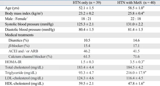
Table 1. Baseline Characteristics and Serum Lipids and HOMA-IR of Subjects according to the Presence of MetS HTN only (n = 39) HTN with MetS (n = 40)
Age (yrs) 52.1 ± 1.5 58.5 ± 1.0
*Body mass index (kg/m
2) 23.2 ± 0.2 25.8 ± 0.4
*Male : Female
�18 : 21 22 : 18
Systolic blood pressure (mmHg) 125.3 ± 2.1 131.0 ± 2.2
Diastolic blood pressure (mmHg) 80.4 ± 1.5 81.4 ± 1.5
Medical treatments
Diuretics (%) 10.5 14.6
β-blocker (%) 15.4 17.1
ACEI and / or ARB 46.2 41.5
Calcium channel blocker (%) 61.5 71.6
HOMA-IR 1.5 ± 0.3 3.5 ± 0.3
*Total cholesterol (mg/dL) 183.4 ± 4.4 184.5 ± 4.2
Triglyceride (mg/dL) 93.3 ± 4.7 216.0 ± 17.9
*LDL-cholesterol (mg/dL) 124.3 ± 4.6 116.4 ± 4.5
HDL-cholesterol (mg/dL) 59.5 ± 2.1 47.8 ± 1.6
*HOMA-IR, homeostasis model of insulin resistance; MetS, metabolic syndrome; HTN, hypertension; ACEI, angiotensin converting enzyme inhibitors; ARB, angiotensin II receptor blocker; LDL, low-density lipoprotein; HDL, high-density lipoprotein.
Values are mean ± SE.
*p < 0.001.
�χ2test.
available for the classification of MetS and further analyzed for this study. Based on the criteria of MetS, two groups were classified by the components of MetS: a reference group of treated hypertensive subjects: HTN group (n = 39) and with more than two additional MetS components:
HTN with Mets group (n = 40), and we further compared the parameters between HTN group and HTN with MetS group. The proportions of patients taking these medica- tions were similar between the groups (Table 1).
Table 1 describes baseline characteristics and clinical parameters between HTN group and HTN with MetS group. Age (p < 0.001) and BMI (p < 0.001) were signi- ficantly different between the groups. Systolic and diastolic blood pressure and gender distribution were not signifi- cantly different. As expected, serum concentrations of TG (p < 0.001) and HDL-cholesterol (p < 0.001) and the levels of HOMA-IR (p < 0.001) were significantly different bet- ween the groups. On the other hand, serum concentrations of total cholesterol and LDL-cholesterol were similar between the two groups.
Table 2 compares LDL-CD, TRAP, phospholipid DHA and inflammatory markers between the two groups.
Because age and BMI were significantly different between the groups, we used the age- and BMI-adjusted values for all analyses. The results showed that age- and BMI-adjusted TRAP (p < 0.01) was significantly lower, whereas age- and BMI-adjusted CD (p < 0.05) was significantly higher in HTN with MetS group than in HTN group. On the other hand, the levels of age- and BMI-adjusted ICAM-1, adipo- nectin and HMW-adiponectin were not significantly different between the groups.
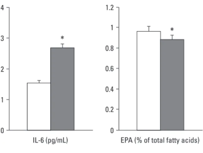
Moreover, HTN with MetS group had significantly lower levels of age- and BMI-adjusted plasma phospho- lipid EPA (p < 0.05) and significantly higher levels of age- and BMI-adjusted IL-6 (p < 0.05) than in HTN group, whereas no differences were found in DHA between the two groups (Fig. 1).
To examine the synergistic effects of MetS in hypertension on cardiovascular risk profiles, we evaluated plasma LDL- CD as a marker for lipid peroxidation, TRAP as a reflection of antioxidant potential, inflammation (IL-6, ICAM-1, total- and HMW- adiponectin) and plasma phospholipid omega 3 fatty acid contents. Our results showed that the levels of LDL-CD and IL-6 were increased whereas the levels of TRAP and plasma phospholipid EPA were decreased in hypertensive patients with MetS compared to those in hypertensive patients.
It is well demonstrated that increased oxidative stress underlies the pathophysiology of hypertension by directly influencing vascular wall cells
20and oxidative stress has been associated with the onset of cardiovascular complica- tions in subjects with the metabolic syndrome.
21Several observational and experimental studies showed impaired antioxidant systems with decreases in antioxidant capacity and increases in lipid peroxidation in MetS,
22which is
DISCUSSION
Table 2. Age- and BMI-adjusted Lipid Peroxidation, Inflammatory Markers and Plasma DHA Contents of Subjects according to the Presence of MetS
HTN only (n = 39) HTN with MetS (n = 40)
LDL-CD (µM) 3.0 ± 0.9 5.8 ± 0.8*
TRAP (mM) 1.38 ± 0.02 1.32 ± 0.01
�Adiponectin (µg/mL) 8.99 ± 0.81 7.48 ± 0.78
HMW-Adiponectin (µg/mL) 4.43 ± 0.80 3.50 ± 0.78
DHA (% of total fatty acids) 3.18 ± 0.22 3.32 ± 0.21
BMI, body mass index; DHA, docosahexaenoic acid; MetS, metabolic syndrome; HTN, hypertension; LDL-CD, conjugated dienes in low- density lipoprotein; TRAP, total radical trapping antioxidant potential; HMW, high molecular weight adiponectin.
Values are Mean ± S.E. All data presented are adjusted for age and BMI.
*p < 0.05.
�p < 0.01.
Fig. 1. Comparison of age- and BMI-adjusted levels of IL-6 and EPA between hypertension group and hypertension with MetS group (*p < 0.05). BMI, body mass index; IL-6, interleukin-6; EPA, eicosapentaenoic acid; MetS, metabolic syndrome.
consistent with our results. Hyperglycemia, a key compo- nent of the MetS, might trigger oxidative stress by several mechanisms either independently or associated with other conditions.
23It includes glucose auto-oxidation, advanced glycated end product (AGE) formation, abnormal arachi- donic acid metabolism and its coupling to cyclooxygenase catalysis.
23The enhanced lipid peroxidation and reduced antioxidant potential in hypertension with MetS, shown in the present study, can be explained, in part, through the mechanisms mentioned above. Considering the athero- genic effect of oxidative alterations in the atherosclerotic process,
24our results provide a possible mechanism which links MetS in hypertension to increased cardiovascular risks.
Inflammation has been established to be a major media- tor of increased cardiovascular risks.
25As evidenced by elevations of circulating levels of C-reactive protein (CRP) and several inflammatory cytokines including IL-6 and tumor necrosis factor- α (TNF-α), chronic inflammation has been associated with hypertension and MetS.
26,27Con- sistent with the earlier findings,
26,27we observed in the present study that plasma levels of IL-6 were significantly higher in the hypertensive patients with MetS. Adiponec- tin, a most abundant adipokine, is primarily released from adipose tissue, and adipose tissue is believed to be an im- portant mediator of oxidative stress and inflammation.
28The profound metabolic effects of adiponectin on glucose and lipid metabolism provide an evidence to link obesity-related chronic inflammation and atherosclerosis.
28,29HMW-adi- ponectin is a multimeric form of adipoenctin and has been suggested as a more sensitive biomarker for insulin resistance and abdominal adiposity than total adiponectin.
29In con- trast to the previous studies,
28-31there were no significant differences in total- and HMW-adiponectin levels between the groups. This may be attributed, in part, to the study population with narrow range in BMI (mean 24.6 kg/m
2, 20.0-29.8) in the present study. However, we found gender- specific differences in total- and HMW adiponectin between hypertension group and hypertension with MetS group. In females, plasma levels of total- and HMW adiponectin were significantly lower in hypertension with MetS group than hypertension group, but not in males (data not shown).
Also, the effect of MetS in hypertension on the levels of TRAP was mainly derived from females (data not shown).
Our results partly support that MetS may have different relative importance for cardiovascular disease according to gender, and that the prognostic impact of MetS is better in females than in males.
2,6,32Emerging evidence showed that omega-3 fatty acids (EPA and DHA) have a variety of beneficial effects on blood pressure, platelet aggregation, inflammatory responses and vasodilation as well as lipid and lipoprotein metabo- lism.
33Furthermore, omega 3 fatty acids were found to
protect cardiovascular morbidity and mortality,
34,35suggest- ing as an emerging risk factor for cardiovascular disease.
The mechanisms by which omega 3 fatty acids exhibit cardioprotective effects include that they alter membrane physical characteristics and the activity of membrane- bound proteins.
36Furthermore, they can interact with ion channels and also act as ligands for several nuclear tran- scription factors.
36The major finding in this study was that plasma phospholipid EPA content was significantly lower in hypertensive patients with MetS than that in hypertensive patients, whereas no difference was observed in DHA.
Given that EPA is known to inhibit the production of proin- flammatory eicosanoids more efficiently than DHA,
37it may modulate inflammatory conditions, oxidative stress accompanied by reduced antioxidant potential, which was not experimentally proved in this study. However, only plasma phosholipid EPA, but not DHA, was slightly reduced, therefore, it is also possible to consider potential metabolic alterations. EPA may in part originate from retro- conversion of DHA in the peroxisome which requires partial β-oxidation.
38Thus, diminished levels of EPA and concomitantly enhanced DHA may occur under conditions of reduced peroxisomal β-oxidation in the MetS. Further experimental studies are needed to elucidate the mechanism for these relationships described in this study.
The main limitations of this study are small sample size and cross-sectional setting, thus making it hard to draw causality. Also, we could not exclude the possibility that lack of nutritional information on fatty acids and fish con- sumption might confound the results. Nevertheless, our results extended several previous studies by showing synergistic effects of MetS in hypertension on inflammation and oxidative stress. Furthermore, we found decreased plasma phospholipid EPA in hypertensive patients with MetS, suggesting possible impact of MetS on inflam- matory response in hypertension.
To conclude, our results showed increased inflammatory marker, reduced plasma phospholipid EPA content and reduced antioxidant potential in treated hypertensive patients in the presence of MetS. Considering that blood and tissue omega 3 fatty acids are the reflections of dietary intakes,
36our results suggest the importance of changes of thera- peutic lifestyle to modify the features of MetS. Intensive nutritional program needs to be encouraged in these hyper- tensive patients with MetS in clinical settings.
This work was supported by the Korea Science and Engi- neering Foundation (KOSEF) grant funded by the Korea government (MOST) (R01-2008-000-20879-0).
ACKNOWLEDGEMENTS
1. Isomaa B, Almgren P, Tuomi T, Forsén B, Lahti K, Nissén M, et al. Cardiovascular morbidity and mortality associated with the metabolic syndrome. Diabetes Care 2001;24:683-9.
2. McNeill AM, Rosamond WD, Girman CJ, Golden SH, Schmidt MI, East HE, et al. The metabolic syndrome and 11-year risk of incident cardiovascular disease in the atherosclerosis risk in communities study. Diabetes Care 2005;28:385-90.
3. Ford ES, Li C, Sattar N. Metabolic syndrome and incident diabetes:
current state of the evidence. Diabetes Care 2008;31:1898-904.
4. Grundy SM, Cleeman JI, Daniels SR, Donato KA, Eckel RH, Franklin BA, et al. Diagnosis and management of the metabolic syndrome: an American Heart Association/National Heart, Lung, and Blood Institute Scientific Statement. Circulation 2005;112:
2735-52.
5. Schillaci G, Pirro M, Vaudo G, Gemelli F, Marchesi S, Porcellati C, et al. Prognostic value of the metabolic syndrome in essential hypertension. J Am Coll Cardiol 2004;43:1817-22.
6. Schillaci G, Pirro M, Pucci G, Mannarino MR, Gemelli F, Siepi D, et al. Different impact of the metabolic syndrome on left ventricular structure and function in hypertensive men and Women. Hypertension 2006;47:881-6.
7. Cuspidi C, Meani S, Fusi V, Severgnini B, Valerio C, Catini E, et al. Metabolic syndrome and target organ damage in untreated essential hypertensives. J Hypertens 2004;22:1991-8.
8. Leoncini G, Ratto E, Viazzi F, Vaccaro V, Parodi D, Parodi A, et al. Metabolic syndrome is associated with early signs of organ damage in nondiabetic, hypertensive patients. J Intern Med 2005;
257:454-60.
9. Lteif AA, Han K, Mather KJ. Obesity, insulin resistance, and the metabolic syndrome: determinants of endothelial dysfunction in whites and blacks. Circulation 2005;112:32-8.
10. Schiffrin EL. Vascular stiffening and arterial compliance. Impli- cations for systolic blood pressure. Am J Hypertens 2004;17:39S- 48S.
11. Rutter MK, Meigs JB, Sullivan LM, D’Agostino RB Sr, Wilson PW. C-reactive protein, the metabolic syndrome, and prediction of cardiovascular events in the Framingham Offspring Study.
Circulation 2004;110:380-5.
12. Aizawa Y, Watanabe H, Ramadan MM, Usuda Y, Watanabe T, Sasaki S. Clustering trend of components of metabolic syndrome.
Int J Cardiol 2007;121:117-8.
13. Expert Panel on Detection Evaluation, and Treatment of High Blood Cholesterol in Adults. Executive Summary of The Third Report of The National Cholesterol Education Program (NCEP) Expert Panel on Detection, Evaluation, and Treatment of High Blood Cholesterol in Adults (Adult Treatment Panel III). JAMA 2001;285:2486-97.
14. Steering Committee of the WHO Western Pacific Region, IASO
& IOTF. The Asia-Pacific perspective: redefining obesity and its treatment. Australia 2000.
15. Mattews DR, Hosker JP, Rudenski AS, Naylor BA, Treacher DF, Turner RC. Homeostasis model assessment: insulin resistance and beta-cell function from fasting plasma glucose and insulin con- centrations in man. Diabetologia 1985;28:412-9.
16. Ahotupa M, Marniemi J, Lehtimäki T, Talvinen K, Raitakari OT, Vasankari T, et al. Baseline diene conjugation in LDL lipids as a direct measure of in vivo LDL oxidation. Clin Biochem 1998;31:
257-61.
17. Rice-Evans C, Miller NJ. Total antioxidant status in plasma and body fluids; in Methods in Enzymology. New York: Academic Press; 1994. p.279-93.
18. Lepage G, Roy CC. Direct transesterification of all classes of lipids in a one-step reaction. J Lipid Res 1986;27:114-20.
19. Lemaitre RN, King IB, Mozaffarian D, Sotoodehnia N, Rea TD, Kuller LH, et al. Plasma phospholipid trans fatty acids, fatal ischemic heart disease, and sudden cardiac death in older adults:
the cardiovascular health study. Circulation 2006;114:209-15.
20. Nakazono K, Watanabe N, Matsuno K, Sasaki J, Sato T, Inoue M. Does superoxide underlie the pathogenesis of hypertension?
Proc Natl Acad Sci U S A 1991;88:10045-8.
21. Furukawa S, Fujita T, Shimabukuro M, Iwaki M, Yamada Y, Nakajima Y, et al. Increased oxidative stress in obesity and its impact on metabolic syndrome. J Clin Invest 2004;114:1752-61.
22. Van Guilder GP, Hoetzer GL, Greiner JJ, Stauffer BL, Desouza CA. Influence of metabolic syndrome on biomarkers of oxida- tive stress and inflammation in obese adults. Obesity (Silver Spring) 2006;14:2127-31.
23. Grattagliano I, Palmieri VO, Portincasa P, Moschetta A, Palasciano G. Oxidative stress-induced risk factors associated with the metabolic syndrome: a unifying hypothesis. J Nutr Biochem 2008;
19:491-504.
24. Toshima S, Hasegawa A, Kurabayashi M, Itabe H, Takano T, Sugano J, et al. Circulating oxidized low density lipoprotein levels. A biochemical risk marker for coronary heart disease.
Arterioscler Thromb Vasc Biol 2000;20:2243-7.
25. Hansson GK. Inflammation, atherosclerosis, and coronary artery disease. N Engl J Med 2005;352:1685-95.
26. Hotamisligil GS, Arner P, Caro JF, Atkinson RL, Spiegelman BM. Increased adipose tissue expression of tumor necrosis factor- alpha in human obesity and insulin resistance. J Clin Invest 1995;
95:2409-15.
27. Kressel G, Trunz B, Bub A, Hülsmann O, Wolters M, Lichting- hagen R, et al. Systemic and vascular markers of inflammation in relation to metabolic syndrome and insulin resistance in adults with elevated atherosclerosis risk. Atherosclerosis 2009;202:263- 71.
28. Hotta K, Funahashi T, Bodkin NL, Ortmeyer HK, Arita Y, Hansen BC, et al. Circulating concentrations of the adipocyte protein adiponectin are decreased in parallel with reduced insulin sensitivity during the progression to type 2 diabetes in rhesus monkeys. Diabetes 2001;50:1126-33.
29. Lara-Castro C, Luo N, Wallace P, Klein RL, Garvey WT. Adipo- nectin multimeric complexes and the metabolic syndrome trait cluster. Diabetes 2006;55:249-59.
30. Aso Y, Yamamoto R, Wakabayashi S, Uchida T, Takayanagi K, Takebayashi K, et al. Comparison of serum high-molecular weight (HMW) adiponectin with total adiponectin concentrations in type 2 diabetic patients with coronary artery disease using a novel enzyme-linked immunosorbent assay to detect HMW adiponectin. Diabetes 2006;55:1954-60.
31. Seino Y, Hirose H, Saito I, Itoh H. High molecular weight multi- mer form of adiponectin as a useful marker to evaluate insulin resistance and metabolic syndrome in Japanese men. Metabolism 2007;56:1493-9.
32. Hunt KJ, Resendez RG, Williams K, Haffner SM, Stern MP: San Antonio Heart Study. National Cholesterol Education Program versus World Health Organization metabolic syndrome in
REFERENCES
relation to all-cause and cardiovascular mortality in the San Antonio Heart Study. Circulation 2004;110:1251-7.
33. Jacobson TA. Secondary prevention of coronary artery disease with omega-3 fatty acids. Am J Cardiol 2006;98:61i-70i.
34. Lemaitre RN, King IB, Mozaffarian D, Kuller LH, Tracy RP, Siscovick DS. n-3 Polyunsaturated fatty acids, fatal ischemic heart disease, and nonfatal myocardial infarction in older adults:
the Cardiovascular Health Study. Am J Clin Nutr 2003;77:319-25.
35. Hu FB, Bronner L, Willett WC, Stampfer MJ, Rexrode KM, Albert CM, et al. Fish and omega-3 fatty acid intake and risk of coronary heart disease in women. JAMA 2002;287:1815-21.
36. Harris WS. The omega-3 index as a risk factor for coronary heart disease. Am J Clin Nutr 2008;87:1997S-2002S.
37. Harris WS, Sands SA, Windsor SL, Ali HA, Stevens TL, Magalski A, et al. Omega-3 fatty acids in cardiac biopsies from heart transplantation patients: correlation with erythrocytes and response to supplementation. Circulation 2004;110:1645-9.
38. Su HM, Moser AB, Moser HW, Watkins PA. Peroxisomal straight-chain Acyl-CoA oxidase and D-bifunctional protein are essential for the retroconversion step in docosahexaenoic acid synthesis. J Biol Chem 2001;276:38115-20.