Veterinary Science
*Corresponding author
Tel: +82-31-467-1807; Fax: +82-31-467-1814 E-mail: [email protected]
An inactivated vaccine to control the current H9N2 low pathogenic avian influenza in Korea
Jun Gu Choi
1, Youn Jeong Lee
1,*, Yong Joo Kim
1, Eun Kyoung Lee
1, Ok Mi Jeong
1, Haan Woo Sung
2, Jae Hong Kim
3, Jun Hun Kwon
11
National Veterinary Research and Quarantine Service, Anyang 430-824, Korea
2
Department of Veterinary Medicine, Kangwon National University, Chunchon 200-701, Korea
3
Laboratory of Avian Diseases, College of Veterinary Medicine, Seoul National University, Seoul 151-742, Korea
The H9N2 subtype low pathogenic avian influenza is one of the most prevalent avian diseases worldwide, and was first documented in 1996 in Korea. This disease caused serious economic loss in Korea's poultry industry.
In order to develop an oil-based inactivated vaccine, a virus that had been isolated in 2001 (A/chicken/Korea/01310/
2001) was selected based on its pathogenic, antigenic, and genetic properties. However, in animal experiments, the efficacy of the vaccine was found to be very low without concentration of the antigen (2
7to 2
10hemagglutinin unit).
In order to overcome the low productivity, we passaged the vaccine candidate virus to chicken eggs. After the 20th passage, the virus was approximately ten times more productive compared with the parent virus. For the most part, the passaged virus maintained the hemagglutinin cleavage site amino acid motif (PATSGR/GLF) and had only three amino acid changes (T133N, V216G, E439D, H3 numbering) in the hemagglutinin molecule, as well as 18 amino acid deletions (55-72) and one amino acid change (E54D) in the NA stalk region. The amino acid changes did not significantly affect the antigenicity of the vaccine virus when tested by hemagglutination inhibition assay. Though not complete, the vaccine produced after the 20th passage of the virus (01310 CE20) showed good protection against a homologous and recent Korean isolate (A/chicken/Korea/Q30/2004) in specific pathogen- free chickens.
The vaccine developed in this study would be helpful for controlling the H9N2 LPAI in Korea.
Keywords: AI, avian influenza, H9N2, inactivated vaccine, LPAI
Introduction
Avian influenza virus (AIV) is an enveloped virus that belongs to the Orthomyxoviridae family and has an eight segmented, single stranded, negative sense RNA genome.
Among the proteins encoded by the genome, there are two surface glycoproteins, hemagglutinin (HA) and neuram- inidase (NA). AIV is classified into subtypes according to the combination of 16 HA and 9 NA molecules [10,26].
Among the many subtypes, the H9N2 AIV is thought to have originated from shorebirds and gulls [30], and rapidly spread to become one of the most prevalent diseases in domestic poultry worldwide. It has also caused serious economic loss in the poultry industry [5,16,17]. In Korea, the first H9N2 low pathogenic avian influenza (LPAI) outbreak occurred in 1996 [15]. Since 2000, and it has become endemic (especially in layer farms) [13,16]. Many studies have demonstrated that there are several distinct H9N2 AIV lineages, and indicated the Korean H9N2 viruses formed a unique antigenic and phylogenetic cluster [13,15-18].
Although immunization with this vaccine is not complete, it is one of the most promising control measures for the H9N2 LPAI to date. Some countries have used vaccines for H9N2 LPAI [5,17,22,29]; however, vast antigenic varia- tions exist even within the same subtype, and it is very difficult to select a vaccine strain that is effective on the virus in current circulation. In addition, some isolates do not grow to a high enough titer in the embryonated chicken eggs (ECEs) to achieve efficient vaccine productivity [31].
Korean animal health authorities took stamping-out and
compensation control policies with regards to H9N2 LPAI
when it occurred between 1996 and 1999. At that time,
vaccines for subtypes of AIV, including H9N2 LPAI, were
prohibited in Korea because they interfered in the
discrimination of naturally infected birds from vaccinated
birds. However, H9N2 LPAI became endemic, and the
policy was not reliable enough to cover each outbreak.
According to the 2004 Avian influenza standard operating procedures, Korean animal health authorities permitted the use of the vaccine for LPAI (especially the H9N2 subtype), and the Committee on the National AI Vaccine Campaign determined that using a single vaccine strain was the most effective strategy with which to simplify the H9N2 AI situation in Korea [7,20].
In this study, we present the characterization of the Korean H9N2 LPAI vaccine strain, and evaluated the efficacy of the pilot vaccine in specific pathogen-free (SPF) chickens.
Materials and Methods Viruses used in this study
All of the viruses were isolated by the National Veterinary Research and Quarantine Service using routine diagnostic practices. The infectious tissue homogenates were inoculated in the allantoic cavity of 9-11 day old SPF ECEs (Lohmann Valo SPF Cuxhaven, Germany) according to standard procedures [27]. The first H9N2 isolate in Korea, A/
Chicken/Korea/MS96/1996 (MS96) [15] was used, and A/Chicken/Korea/99029/1999 (99029) and A/Chicken /Korea/01310/2001 (01310) were used as representative isolates of the 1999 and 2001 strain, respectively. The 2001 strain was eventually chosen as the vaccine candidate. In order to test the antigenicity and the efficacy of the vaccine, a recent isolate, A/Chicken/Korea/Q30/2004 (04Q30), was used [16].
Pathogenicity test of Korean isolates in SPF and commercial broiler chickens
In order to determine the pathogenicity of the selected viruses in SPF and commercial broiler chickens, MS96, 99029, and 01310 were inoculated via the intra-tracheal route in eight 7-week-old SPF chickens (10
6.5EID
50/0.1 ml, 10
5.6EID
50/0.1 ml, and 10
7.1EID
50/0.1 ml, respectively) and fifteen 12-week-old commercial broiler chickens (10
5.2EID
50/0.1 ml, 10
5.7EID
50/0.1 ml, and 10
5.0EID
50/0.1 ml, respectively), which were confirmed to be free of antibodies against H9N2 AIV. In the experiment with SPF chickens, tracheal and cloacal swab samples were taken at 3, 5, 7, and 9 days post-inoculation (dpi). The swab samples were suspended in 3 ml of gentamicin-PBS (1%
gentamicin in PBS, pH 7.2), and were inoculated with 0.2 ml of samples into three 9-11 day old SPF ECEsvia the allantoic cavity route. The inoculated broiler chickens were reared for 2 weeks, and the mortality was recorded.
Antigenic relationship between Korean H9N2 AIVs After being propagated in ECEs, the viruses (MS96, 99029, and 01310) were inactivated by incubating them with 0.1% formalin at 20
oC for 10 h. The inactivation was confirmed by injecting formalin-treated virus into the allantoic cavities of 10-day-old ECEs, two times serially.
Virus inactivation was determined by hemagglutination negativity, using 1% chicken red blood cells. The inac- tivated virus was emulsified with oil adjuvant (Montanide ISA 70 SEPPIC, France) at a ratio of 3:7, and wasinjected into eight 6-week-old SPF chickens. The antisera were obtained at 3 weeks after injection. In order to determine the antigenic relationship between the selected viruses, we performed a cross hemagglutination inhibition (HI) test with each of the antisera and virus antigens (four HA unit), and the r-value was subsequently calculated [2].
HA and NA gene sequencing for comparison with recent Korean isolates
Viral RNA was extracted from infectious allantoic fluid using the Viral Gene-Spin Viral DNA/RNA extraction kit (iNtRON Biotechnology, Korea) and amplified using gene-specific primer sets by RT-PCR with a Qiagen one- step RT-PCR kit (Qiagen, USA) according to the method described by Hoffmann et al. [12]. The amplified product was excised from agarose gel and eluted using the GENECLEAN SPIN kit (Qbiogene, USA). The nucleotide sequences were analyzed by direct sequencing of the PCR products using ABI PRISM BigDye Terminator Cycle Sequencing Kits (Applied Biosystems, USA). The HA and NA gene nucleotide sequences of 01310 CE3 have been deposited in GenBank under accession number EU253561 and EU253562, respectively.
The nucleotide and deduced amino acid sequences of the HA and NA molecules were aligned by the Clustal W method with the MegAlign software (Lasergene 7.0;
DNASTAR, USA). The similarity of the HA and NA amino acid sequences were compared with recently studied Korean H9N2 AIVs [16].
Efficacy testing of vaccine candidate virus accord-
ing to the antigen contents and virus passage in ECEs
The selected vaccine candidate (01310 CE3, 2
7HA unit)
virus based on the above pathogenic, serologic, and molecular
data (refer to the Results section) was used to prepare the
vaccine according to the previously mentioned proced-
ures. The preparation of the high HA content vaccine (2
10HA unit) involved the concentration of the virus by
centrifugation (18,000 rpm, 4 h; Beckman, USA). The
vaccines were injected into ten 6-week-old SPF chickens
via the intramuscular (IM) route. The serum samples were
taken three weeks post-vaccination (wpv), and were
followed by the performance of the HI test. The HI test
results were analyzed using Student’s t-test. Statistical
significance was set a priori at α = 0.05. The chickens were
challenged with 01310 CE3 virus (10
6.0EID
50/0.1 ml) at 3
wpv via the oral route. Next, oropharyngeal and cloacal
swab samples were taken at 5 dpi for virus isolation. The
Fisher’s exact test was performed to compare the virus
isolation rate (α= 0.05).
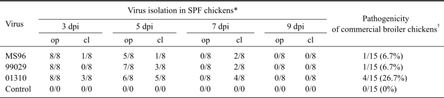
Table 1. Comparison of virus isolation and mortality of recent Korean H9N2 LPAIV
Virus
Virus isolation in SPF chickens*
Pathogenicity of commercial broiler chickens
†3 dpi 5 dpi 7 dpi 9 dpi
op cl op cl op cl op cl
MS96 8/8 1/8 5/8 1/8 0/8 2/8 0/8 0/8 1/15 (6.7%)
99029 8/8 0/8 7/8 3/8 0/8 2/8 0/8 0/8 1/15 (6.7%)
01310 8/8 3/8 6/8 5/8 0/8 4/8 0/8 0/8 4/15 (26.7%)
Control 0/0 0/0 0/0 0/0 0/0 0/0 0/0 0/0 0/15 (0%)
*7-week-old SPF chickens were inoculated via the intra-tracheal route with MS96 (106.5EID50), 99029 (105.6EID50), and 01310 (107.1EID50): virus isolation/total inoculated. †12-week-old AIV antibody-free commercial broiler chickens were inoculated via the intra-tracheal route with MS96 (105.2 EID50), 99029 (105.7EID50), and 01310 (105.0EID50): number of dead/total inoculated (% mortality). op: oropharyngeal, cl: cloacal.
In order to recover the highly growing vaccine virus in ECEs, the vaccine candidate virus was serially passaged through 9- to 11-day-old SPF ECEs and selected with high HA titer using chicken red blood cells. The passage number in ECEs was indicated as CE X (X stands for passage number) after the virus name.
Pathogenicity test of the selected vaccine virus in SPF ECEs and chickens
In order to test the changes in the pathogenicity of the passaged vaccine viruses in chicken eggs, the viruses (CE5, CE10, CE15, CE20, and CE40) were diluted in PBS and inoculated in the allantoic cavities of 10-day-old SPF ECEs. In addition, the mortality was checked after 48 h of incubation.
For the pathogenicity test of the selected vaccine virus in chickens, eight 6-week-old SPF chickens were intra- venously inoculated with 0.2 ml of 1/10 diluted virus, and we noted the mortality for 10 days according to standard procedures [31].
Antigenicity testing and the optimum growth condition of the vaccine virus
The vaccines of the antigenicity test were prepared with 01310 CE6, CE20, and CE40 viruses. In addition, the HA titers of the viruses were adjusted to 2
9HA units. Eight 6-week-old SPF chickens were then vaccinated at 0.5 ml/chickenvia the IM route. After vaccination, serum samples were taken at 3 wpv, and the HI assay was then performed with the MS96 CE3, 01310 CE20, and 04Q30 CE3 viruses. A one-way ANOVA test was subsequently performed.
In order to determine the optimum growth conditions of the vaccine strain in ECEs, the virus was serially diluted 10-fold with 1% gentamicin-PBS (10
-3-10
-7), and was inoculated into 60 SPF ECEs. At 8, 16, 24, 32, 40, and 48 hours after incubation, 10 eggs were harvested during each dilution and the HA titer was determined with 1% chicken
red blood cells [27].
Vaccine efficacy test
The vaccine was prepared with the 01310 CE20 (2
10HA unit) strain as per the procedures mentioned in the Materials and Methods section, and was injected at a concentration of 0.5 ml/chicken via the intramuscular route (thigh muscle) into each of eight 6-week-old SPF chickens. Three weeks after vaccination, the parent virus (01310 CE3, 10
5.5EID
50/0.1 ml) and recent Korean isolate (04Q30 CE3, 10
5.5EID
50/0.1 ml) were challenged intranasally.
To isolate the challenged virus, oropharyngeal and cloacal swab samples were taken at 1, 3, and 5 dpi from each chicken and suspended in 3 ml of gentamicin-PBS. The samples were subsequently inoculated into three 9- to 11-day-old SPF ECEs respectively, followed by assessment of the virus growth from the allantoic fluids. At 5 dpi, the chickens were sacrificed and the various tissues were taken with different scissors in order to prevent contamination. Next, the 10% (w/v) tissue homogenates were tested for viral growth in three 9- to 11-day-old SPF ECEs. In order to titrate the virus from various tissues, tissue homogenates were pooled in a group of equal volume, and were then titrated using SPF ECEs.
Results
Vaccine candidate selection
In order to select the vaccine candidate, we chose the
MS96, 99029, and 01310 viruses as the representative
viruses of the year. In addition, we compared the virus
replication potency and pathogenicity for SPF and
commercial broiler chickens. For the SPF chickens, all of
the viruses had a zero mortality rate; however, the viruses
were isolated at 3 to 5 dpi from oropharyngeal swab
samples, and 3 to 7 dpi from cloacal swab samples. At 5
dpi, the MS96, 99029, and 01310 viruses were isolated
from 5/8, 7/8, and 6/8 oropharyngeal swab samples and
Table 3. Comparison of vaccine efficacy according to the antigen content
Antigen content (HA unit)
HI titer at 3 weeks post
vaccination
Virus isolation at 5 dpi
†Oropharyngeal
swab
Cloacal swab 01310 CE3 (2
7) 8.2 ± 0.6* 7/10
‡9/10
‡01310 CE3 (2
10) 8.8 ± 0.4* 1/10
‡3/10
‡Control − 10/10 10/10
*Mean ± SD of the titer analyzed with the Student’s t-test, α = 0.05, p
= 0.022. †The chickens were challenged intranasally with the homolo- gous virus (01310 CE3, 106.0EID50/0.1 ml): virus isolation / total tested. ‡Fisher’s exact test, p = 0.020, respectively.
Table 4. Comparison of virus titer and mortality in ECEs and SPF chickens of passaged vaccine candidate
Passage No.
HA titer (log
2)
Virus titer (log
10EID
50/0.1 ml)
Mortality in ECEs*
Mortality in SPF chicken
†CE5 7 7.5 3/10 nt
CE7 8 nt
‡nt nt
CE10 9 8.5 2/10 nt
CE15 10 8.4 2/10 nt
CE20 10 8.7 6/10 0/8
CE30 10 nt nt nt
CE40 10 nt 5/10 nt
*Mortality at 2 dpi incubation time. The viruses were inoculated at 104.5EID50/0.1 ml each. †The 8 SPF chickens were intravenously inoculated with 0.2 ml of 1/10 diluted (107.7EID50/0.1 ml) infectious allantoic fluid and observed for 10 days. ‡nt: not tested.
Table 2. Antigenic relationship between Korean isolates
Antiserum Virus
MS96 99029 01310
MS96 169* (1.00
†) 60 (0.73) 274 (0.90)
99029 239 158 (1.00) 181 (0.61)
01310 274 181 549 (1.00)
*Mean HI titer from 8 chickens. †r-value, r = (r1 × r2)1/2. r1 = heterolo- gous titer with virus 2 / homologous titer with virus 1. r2 = heterolo- gous titer with virus 1 / homologous titer with virus 2.
1/8, 3/8, and 5/8 cloacal swab samples, respectively.
Furthermore, in commercial broiler chickens, the 01310 strain showed a 26.7% mortality rate, whereas a 6.7%
mortality rate was recorded for each of the other two viruses (Table 1).
To elucidate the antigenic relationship between the selected Korean isolates, the r-value was calculated with a cross HI titer between the viruses. The values ranged from 0.61 to 1.00, with no significant differences between the viruses (Table 2).
Based on our pathogenic and serologicdata, we chose the 01310 virus as a vaccine candidate. Moreover, when we compared the HA and NA amino acid sequences deduced by nucleotide sequences, the 01310 CE3 virus was found to have 95.5-98.6% and 94.6-98.5% similarity with the recent Korean H9N2 AIVs isolated from 2002 to 2004, respectively (data not shown).
Efficacy of the selected vaccine candidate virus according to the antigen contents
In order to elucidate the efficacy of the vaccine candidate virus, ten SPF chickens were immunized with different antigen contents. Chickens immunized with a high antigen vaccine (2
10HA unit) showed a similar but statistically significant (p < 0.05) antibody titer compared to the low antigen content (2
7HA unit) vaccine (Table 3). In addition, in the low antigen content vaccine group, the challenged viruses were recovered from 7/10 and 9/10 chickens from oropharyngeal and cloacal swab samples at 5 dpi, respectively. It was believed that the vaccine was not effective to protect against viral shedding. However, in high antigen content groups, the challenged virus was isolated from 1/10 or 3/10 oropharyngeal/cloacal swab samples, respectively. Although neither of the vaccines were able to completely protect against viral shedding, the high antigen content vaccine was more effective than the low antigen content vaccine upon comparison of the virus recovery rate in the oropharyngeal/cloacal swab samples (p = 0.02, respectively) (Table 3).
Biological characteristics of the vaccine virus The vaccine candidate virus (01310 CE3) grew in ECEs at around 2
7HA units and about 10
7.0EID
50/0.1 ml, as did most of the field isolates (personal observation, data not presented), and we were not able to determine the expected vaccine efficacy with unconcentrated antigen (Table 3).
Therefore, we attempted to get highly growing phenotyped viruses through egg passage. When the 01310 strain was passaged in SPF ECEs as a vaccine candidate, the virus titer increased with each ECE passage (Table 4). After the 15th passage in ECEs, the virus titer showed 2
10HA units stably;
this was approximately 10 times greater than that of the parent virus. Moreover, the 20th passaged virus showed the highest titer, 10
8.7EID
50/0.1 ml (Table 4). As shown in Fig. 1, the 01310 CE20 showed the highest HA titer when inoculated with 10
4.7EID
50/0.1 ml and incubated for 32 hours. The mean HA titer at that point was 9.7 ± 0.5 (log
2).
However, as the 01310 virus was passaged in SPF ECEs,
the mortality of the chicken eggs also increased. During 48
Table 5. Immunogenicity and comparison of serological relationship with Korean isolates
Antisera
HI titer (mean ±SD, log
2) with homologous / heterologous Ag MS96 01310, CE20 04Q30, CE3
01310, CE6 −
‡7.8 ± 0.5* −
01310, CE20 8.8 ± 0.7
†8.3 ± 0.7*
†9.1 ± 1.0
†01310, CE40 − 8.4 ± 0.7* −
Control 0.0 0.0 0.0
The values were analyzed with a one-way ANOVA test. α = 0.05, *p
= 0.12, †p = 0.15. ‡ not tested.
Table 6. Vaccine efficacy test of the 01310 CE20 strain against the parent virus (01310 CE3) and the recent Korean isolate (04Q30 CE3) Challenge
virus Vaccination*
Oropharyngeal (op) /
cloacal (cl) swab sample
†Tissue homogenate (10% w/v)
‡1dpi 3 dpi 5 dpi
Brain Trachea Lung Spleen Kidney Cecal Tonsil
op cl op cl op cl
01310
CE3 + 0/7 0/7 0/7 0/7 0/7 0/7 0/7 (nt) 0/7 (0) 0/7 (0) 0/7 (0) 0/7 (0) 0/7 (0)
− 2/8 0/8 7/8 0/8 7/8 6/8 0/8 (nt) 5/8 (10
1.4) 0/8 (0) 1/8 (0) 0/8 (0) 4/8 (10
5.4) 04Q30
CE3
+ 2/8 0/8 2/8 0/8 0/8 0/8 0/8 (nt) 0/8 (0) 0/8 (0) 0/8 (0) 0/8 (0) 0/8 (0)
− 6/8 0/8 8/8 3/8 8/8 5/8 0/8 (nt) 8/8 (10
4.4) 6/8 (10
2.0) 2/8 (0) 2/8 (10
1.6) 5/8 (10
6.0)
*Vaccine prepared as described in Materials and Methods with 01310 CE20 (210 HA unit) and vaccinated intramuscularly at 0.5 ml/chicken.
†Virus isolation / total tested. ‡Tissues were taken at 5 dpi, virus isolation / total tested (virus titers of the pooled samples, EID50/0.1 ml), nt:
not tested.