서 론
원발성 충수돌기선암종은 매우 드문 질환이며 전체 소화 기암의 0.5% 미만을 차지하고 있다. 원발성 충수돌기암은 크게 4가지로 분류되며, 그 중 85%는 유암종(carcinoid)이며, 점액성낭종선암종(micinous cyst adenocarcinoma)과 대장형 선암종(colonic type adenocarcinoma), 유암선종(adenocarcinoid) 이 8%, 4%, 2%로 보고되고 있다.(1-3)
1882년 Berger가 최초로 원발성 충수돌기선암종을 발표 한 이래 현재까지 500여 예가 보고되어 있으며, 국내 보고 는 수예에 불과할 뿐이다.(1)
원발성 충수돌기암은 임상적으로 진단이 매우 어려우며, 급성 충수돌기염 진단하에 응급수술 시행 후 병리조직 검 사에서 충수돌기암으로 확진되는 경우가 대부분이다. 원발 성 충수돌기암이 가장 흔히 전이되는 장소는 복강이고, 그 외 림프절, 간, 난소, 복벽, 폐의 순서로 보고되고 있다.(2,4) 저자들은 수술 전 말단 회장 종양의 간전이로 진단 후 수 술 도중 시행한 동결조직병리검사에서 원발성 충수돌기선 암종의 간 전이가 발견되어 우결장반절제술 및 간우엽절제 술을 시행한 예를 경험하여 원발성 충수돌기선암종의 간전 이로 수술적 절제를 시행한 최초 예라 생각되어 문헌고찰 과 함께 보고하고자 한다.
증 례
과거력상 특이소견이 없는 53세 된 남자 환자가 내원 1주
간전이를 동반한 충수돌기선암종의 수술적 절제
영남대학교 의과대학 외과학교실 황형철·이동식·윤성수·김홍진
course was uneventful; the patient was discharged at POD
#21 after the application of systemic chemotherapy. We de- tected no evidence of recurrence five months after surgery.
(J Korean Surg Soc 2007;72:75-79)
Key Words: Primary adenocarcinoma, Appendix, Liver me- tastasis
중심 단어: 원발성 선암종, 충수돌기, 간전이 Department of Surgery, College of Medicine, Yeungnam Uni- versity, Daegu, Korea
Surgical Resection of Liver Metastasis from Adenocarcinoma of the Appendix
Hyung Chul Hwang, M.D., Dong Shik Lee, M.D., Sung Su Yun, M.D. and Hong Jin Kim, M.D.
Primary appendiceal adenocarcinoma is a rare neoplasm, which constitutes less than 0.5% of all gastrointestinal neoplasms. Very few cases of primary tumors of the appen- dix have been reported in the literature; and no prior case reports of surgical resection of appendiceal adenocarcinoma with liver metastasis have been published. In this study, we report for the first time a case of the surgical resection of liver metastasis from a case of adenocarcinoma of the ap- pendix. (Case) A 53-year-old man was admitted complaining of intermittent abdominal pain. The patient’s CEA level was mildly elevated, at a level of 30.8 ng/ml; the AFP was 1.93 ng/ml, and all additional blood tests were normal. Abdominal CT revealed a 9 cm sized malignant-appearing peripheral enhancing mass in the right lobe of the liver. In an effort to rule out metastatic cancer originating from the GI tract, we conducted gastroendoscopy and colonoscopy. However, we were unable to detect any malignant lesions. Therefore, we conducted a whole body fusion PET scan, which revealed a hot uptake of FDG at the right lobe of the liver, as well as a terminal ileum. We concluded with a diagnosis of primary small bowel malignancy with liver metastasis; the patient underwent surgery. During surgery, we detected a very large malignant-appearing tumor in the right lobe of the liver, and an appendix which appeared suspicious for malig- nancy, and measured 8.0 cm in length and 1.5 cm in dia- meter. The frozen biopsy of the appendix at surgery con- firmed a malignant adenocarcinoma of the appendix, coupled with vascular invasion. Thus, we conducted a right hemi- hepatectomy and a right hemicolectomy. The post-operative
책임저자:김홍진, 대구광역시 남구 대명5동 317-1번지 ꂕ 705-717, 영남대학교 의과대학 외과학교실 Tel: 053-620-3585, Fax: 053-624-1213 E-mail: [email protected]
접수일:2006년 4월 18일, 게재승인일:2006년 9월 1일
75
성, HBsAb 양성, HBcAb는 양성이었으며, HBeAg, HBeAb, Anti-HCV는 음성이었다.
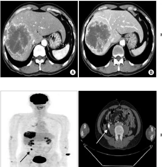
간초음파 검사 및 복부전산화단층촬영(Fig. 1)에서 간우엽 에 저혈관성의 변연부가 증대된 악성 양상을 보이는 종괴 가 관찰되었다. 비장비대나 간경화, 복수 등의 소견은 관찰
를 위한 수술을 시행하였다. 간우엽 절제술 후 동결조직병 리검사에서 전이성 선암종이 발견되었으며, 충수돌기의 비 대소견이 있어 시행한 충수돌기절제술 후 동결조직병리 검 사에서 원발성 선암종이 발견되어 우결장반절제술을 시행 하였다.
Fig. 1. (A) The abdominal CT re- vealed a 9 cm sized malig- nant appearing peripheral enhancing mass in the rig- ht lobe of the liver. (B) In delayed phase, mass was not rapidly washing out, then gradually enhancing inward mass.
Fig. 2. Fusion PET/CT scan was shown hypermetabolic le- sion (SUV max: 13, size:
91 mm) in the right lobe of the liver. Both arrow showed focal FDG uptake in a terminal ileum.
Fig. 5. (A) Microscopic appearance of the appendix showing mucus secreting glands (left), and infiltrative pattern of neoplastic glands and some desmoplastic stromal response (right) (H&E, ×100), (B) Microscopic appearance of the liver showing metastatic adenocarcinoma with central necrossis (H&E, ×100).
Fig. 4. Gross findings of liver. (A) The cut surface shows well defined yelloish solid mass lesion, measuring 9.5×8.0
×9.0 cm in size. (B) On multiple serial section, gray yellowish solid mass is pre- sented. The mass is exten- ded to resection margin but not involvement of rese- ction margin. There is no cirrhotic change.
Fig. 3. Gross findings of appendix. (A) The appendicial external serosal surface is smooth. This appendix specimen measuring 8.0 cm in length and 1.5 cm in diameter. (B) Macroscopic appearance of the resected appendix, revealing a 3.0 cm sized lesion in the base of the appendix. Lumen is filled with mucous material.
중이며 5-FU (Fluorouracil) 및 Irinotecan (CamptoⓇ)에 의하여 항암약물요법 치료 중이다.
고 찰
충수돌기에 발생하는 원발성 선암종은 전체 소화기계에 발생하는 악성종양의 0.5% 미만을 차지할 정도로 매우 드 물다.(1-3) 이러한 희귀한 빈도 때문에 수술 전이나 수술 중 에 진단되기 어렵고, 급성충수돌기염이나 충수돌기주위 농 양 의심 하에 개복술 시행 후 병리조직검사상 확진이 되는 경우가 대부분이다.(2,5,6) 남자에 약간 많고 대장 선암종과 비슷하게 40대 이상에서 호발하며, 유암선종의 경우 여자 에서 호발하는 경향이 있다고 보고되고 있다.(2,3,5,6)
증상은 우하복부 동통, 종괴 촉지 등 충수돌기염이나 충 수돌기주위 농양의 소견을 보이거나 장기간의 간헐적인 복 통을 호소하기도 하며 장폐쇄, 체중감소, 장출혈 등이 동반 될 수 있으며, 여자인 경우에는 난소 종괴로 오인되기도 한 다.(1,5,6)
충수돌기선암종의 진단방법으로는 임상증상, 복부초음 파검사, 복부전산화단층촬영 및 대장바리움조영술 등을 이 용할 수 있으나 수술 전 진단하기는 어렵다. 저자들의 예에 서도 수술 전 검사에서 복부초음파검사, 복부전산화단층촬 영을 시행하였으나 간우엽에 악성 양상을 보이는 종괴 외 에 특이소견은 보이지 않았다. 혈청암태아성항원치가 증가 되어 있어 미발견된 소화기계 기원의 선암종을 의심하여 위내시경, 대장내시경을 시행하였으나, 특이소견이 관찰되 지 않았다. 양전자방출전산화단층촬영에서 말단 회장부위 에 국소적인 FDG 섭취증가를 보이는 병변이 관찰되어 원 발병소로 의심하였다. 말단 회장 종양의 간전이로 진단 후 수술 시행 도중 동결조직병리검사에서 원발성 충수돌기선 암종의 간전이로 진단되었다. 환자는 충수돌기선암종에서 흔히 관찰되는 종괴가 촉지되거나, 충수돌기염 혹은 충수 돌기주위 농양의 소견이 관찰되지 않았다.
원발성 충수돌기선암종의 우결장반절제술의 필요성에
있다는 이론적 근거하에 적극적인 간절제를 시행하는 이유 가 된다.(10,11) 충수돌기선암종의 경우 주로 림프절을 통하 여 전이되나 혈행성으로 간이나 폐로 전이되기도 하며, 천 공으로 인하여 복벽에 전이되기도 한다.(2,4) 충수돌기의 림 프절은 말단 회장 및 맹장주위의 림프절로 배출되므로, 충 수돌기선암종의 경우 십이지장 제3부위의 전방부와 회결 장동맥 기시부의 림프절 전이 유무를 확인하여야 한다.(5,6) 저자들의 경우에도 술 중 동결조직검사로 충수돌기선암종 의 간전이를 진단 후 원발병소인 충수돌기를 포함한 우결 장반절제술과 주위 림프절 곽청술 및 적극적인 간우엽절제 술을 시행하였으며 향후 추적관찰이 필요할 것으로 보인 다.
충수돌기선암종의 예후는 비교적 불량한 것으로 보고되 고 있으며, Andersson 등(4)은 5년 생존율을 18∼30%로 보 고하였고 Nitecki 등(2)은 Duke's stage에 따라 원격전이가 있을 경우 5년 생존율을 6%로 보고하였다. 충수돌기암의 예후에 영향을 미치는 요소로는 조직학적 분류가 가장 중 요하며, 조직학적 분류 중에서도 선암종이 예후가 가장 나 쁘다. 다른 요소로는 침윤 정도와 원격전이 여부, 수술 술식 등이 있다.(1,5,6)
충수돌기암의 원격전이의 경우 소수의 예에서 보조적 항 암요법의 효과가 제한적으로 보고되었다.(3,12,13) 저자들 은 5개월째 5-FU, Irinotecan (CamptoⓇ)을 이용한 항암요법 중으로 술 후 5개월 동안의 추적관찰에서 재발이나 전이의 소견은 관찰되지 않았다.
REFERENCES
1) Park IJ, Yu CS, Kim HC, Kim JC. Clinical features and pro- gnostic factors in primary adenocarcinoma of the appendix. J Korean Gastroenterol 2004;43:29-34.
2) Nitecki SS, Wolff BG, Schlinkert R, Sarr MG. The natural history of surgically treated primary adenocarcinoma of the appendix. Ann Surg 1994;219:51-7.
3) Rutledge RH, Alexander JW. Primary appendiceal malignan- cies: rare but important. Surgery 1992;111:244-50.
4) Andersson A, Bergdahl L, Boquist L. Primary carcinoma of the appendix. Ann Surg 1976;183:53-7.
5) Jang KC, Lee YB, Kim SC, Park YH. A case of primary adenocarcinoma of the appendix. J Korean Gastroenterol 1992;24:866-9.
6) Song TJ, Moon HY, Koo BH. Clinical review of the appen- diceal tumor. J Korean Surg Soc 1992;43:719-24.
7) Kabbani W, Houlihan PS, Luthra R, Hamilton SR, Rashid A.
Mucinous and nonmucinous appendiceal adnocarcinomas: dif- ferent clinicopathological features but similar genetic altera- tions. Mod Pathol 2002;15:599-605.
8) Ozakyol AH, Saricam T, Kabukcuoglu S, Cage T, Erenoglu E. Primary appendiceal adenocarcinoma. Am J Clin Oncol 1999;22:458-9.
9) Mohamed F, Chang D, Sugrabaker PH. Third look surgery and
beyond for appendiceal malignancy with peritoneal dissemina- tion. J Surg Oncol 2003;83:5-12.
10) Blumgart LH, Fong Y. Surgical options in the treatment of hepatic metastasis from colorectal cancer. Curr Probl Surg 1995;32:335-421.
11) Park IJ, Kim HJ, Kim HC, Yu CS, Chang HM, Ryu MH, et al. Comparative analysis of colorectal cancer with liver metastasis identified preoperatively vs. intraoperatively. J Korean Soc Coloproctol 2004; 20:378-83.
12) Chang AE, Schneider PD, Sugarbaker PH, Simpson C, Culnane M, Steinberg SM. A prospective randomized trial of regional vs. systemic continuous 5-FU chemotherapy in the treatment of colorectal metastases. Ann Surg 1987;206:685- 93.
13) Moertel CG, Weiland LH, Nagorney DM, Dockerty MB. Car- cinoid tumors of the appendix: treatment and prognosis. N Engl J Med 1987;317:1699-701.