www.krspine.org
Effect of the Corrective Osteotomy in Ankylosing Spondylitis to Quality of Life(QOL)
Ye-Soo Park, M.D., Chang-Hun Lee, M.D., Jae-Hoon Kim, M.D., Tae-Hwan Kim, M.D.
J Korean Soc Spine Surg 2011 Mar;18(1):13-18.
Originally published online March 31, 2011;
doi: 10.4184/jkss.2011.18.1.13
Korean Society of Spine Surgery
Department of Orthopaedic Surgery, Ewha Womans University Collge of Medicine
#911-1 Mok-dong, Yangcheon-gu, Seoul, 158-710, Korea Tel: 82-2-2646-6808 Fax: 82-2-2646-6804
©Copyright 2011 Korean Society of Spine Surgery pISSN 2093-4378 eISSN 2093-4386
The online version of this article, along with updated information and services, is located on the World Wide Web at:
http://www.krspine.org/DOIx.php?id=10.4184/jkss.2011.18.1.13
This is an Open Access article distributed under the terms of the Creative Commons Attribution Non-Commercial License (http://
creativecommons.org/licenses/by-nc/3.0) which permits unrestricted non-commercial use, distribution, and reproduction in any medium, provided the original work is properly cited.
Journal of Korean Society of
Spine Surgery
Effect of the Corrective Osteotomy in Ankylosing Spondylitis to Quality of Life(QOL)
Ye-Soo Park, M.D., Chang-Hun Lee, M.D., Jae-Hoon Kim, M.D., Tae-Hwan Kim, M.D.*
Department of Orthopaedic Surgery, Guri Hospital, Hanyang University College of Medicine, Hospital for Rheumatic Disease, Hanyang University College of Medicine, Guri, Korea*
Study Design: This is retrospective study.
Objective: We evaluated the radiologic changes and quality of life after corrective osteotomy in kyphotic deformity of ankylosing spondylitis.
Summary of Literature Review: There is few study about that relationship between corrective osteotomy and quality of life.
Materials and Methods: Retrospective study about 16 patients who underwent corrective osteotomy from 2005 September to 2007 December was done. Radiologic assessments of sagittal balance were performed on the criteria of thoracic kyphosis, lumbar lordosis, distance between the vertical line on midpoint of C7 and posterosuperior point of S1 pre and postoperatively. Disease specific instruments: the Bath ankylosing spondylitis disease activity index (BASDAI) and the Bath ankylosing spondylitis functional index (BASFI) were applied. Clinical assessments were performed with short form-36 through interview and telephone.
Results: The mean thoracic kyphosis was changed from 46.1 degrees to 39.3 degrees. The mean lumbar lordosis was corrected from - 7.4 degrees to - 38.4 degrees, and the mean distance between vertical lines of C7 and S1 was improved from 127.1mm to 30mm. There were significant changes in the subgroup of Physical function, Role physical, Vitality, Social function, Role emotional, Mental health. (p<0.05) The changes of BASDAI, BASFI, Bodily pain and General health were not significant. And similar improvements in the radiological results and SF-36 scores were in the 6 patients with Andersson lesion.
Conclusion: The parameters of radiographic assessment were improved after corrective osteotomy in the fixed kyphotic deformity of ankylosing spondylitis. General function, social function and mental health were also improved.
Key Words: Ankylosing spondylitis, Corrective osteotomy, Quality of life
Received: August 23, 2010 Revised: March 4, 2011 Accepted: March 4, 2011 Published Online: March 31, 2011 Corresponding author: Ye-Soo Park, M.D.
Department of Orthopaedic Surgery, Guri Hospital, Hanyang University College of Medicine, 249-1, Kyomoon-dong, Guri 471-701, Korea TEL: 82-31-560-2316, FAX: 82-31-557-8781
E-mail: [email protected]
“This is an Open Access article distributed under the terms of the Creative Commons Attribution Non-Commercial License (http://
creativecommons.org/licenses/by-nc/3.0/) which permits unrestricted non-commercial use, distribution, and reproduction in any medium, provided the original work is properly cited.”
INTRODUCTION
Ankylosing spondylitis is a chronic inflammatory disease, and thus for its treatment, clinicians consider more the improvement of quality of life together with reduction of the disease activity than complete cure. To assess the response to treatments, nu- merous studies on the measurement of the disease activity and the improvement of quality of life have been conducted.1,2) In the field of orthopedic surgery, studies on corrective osteotomy techniques for kyphotic deformity of ankylosing spondylitis, complications, and radiological results have been conducted primarily.3-6) Kim et al. have reported clinical evaluation after the correction of secondary kyphosis of ankylosing spondylitis by the application of the modified Arthritis Impact Measurement Scales (AIMS) questionnaire, and performed comprehensive evaluation by dividing to function, activity, pain, and satisfaction level.7)
However, the AIMS is an evaluation method focusing on
rheumatoid arthritis.8) It thus was thought that the survey ap- plying tools to evaluate general quality of life would be mean- ingful. Therefore, together with the evaluation of radiographs taken prior to and after corrective osteotomy, we evaluated the improved level of quality of life in ankylosing spondylitis by the application of the Short form-36 (SF-36) that is a tool to
Ye-Soo Park et al Volume 18 • Number 1 • March 2011
www.krspine.org 14
measure quality of life pertinent to general health. In addition, the effect of Andersson lesion on radiological as well as clinical results were assessed.
RESEARCH SUBJECTS AND METHODS
A retrospective study was conducted on 16 ankylosing spondylitis patients performed corrective osteotomy for fixed kyphotic deformity from September 2005 to December 2007 and available for longer than 24 months follow-up observation. As surgical methods, kyphotic deformity was corrected by pedicle subtraction osteotomy, and for cases associated with Andersson lesion, by considering the location of lesion and the kyphotic level, at the first surgery, together with pedicle subtraction osteotomy, Smith-Petersen osteotomy was performed. At the second osteotomy, the anterior pyramid with Andersson lesion was removed by anterior approach, and using meshes as well as autogenous ribs, interbody fusion was performed additionally.
As radiological evaluation, on the lateral images of the spine taken in the standing position prior to surgery and after surgery, the thoracic kyphotic angle formed by the upper border of 1st thoracic vertebra and the lower border of 12th thoracic vertebra, the lumbar kyphotic angle formed by the upper border of 1st lumbar vertebra and the lower border of 5th lumbar vertebra, and the measurement of sagittal imbalance applied C7 plumb line that measures the distance between the posterosuperior corner of 1st sacral vertebra and the line transversing the center of 7th cervical vertebra and vertical to the ground.) Kyphosis was presented as positive values, and lordosis was presented as negative values.
As clinical evaluation, the disease activity was measured by the presurgical as well as postsurgical Bath Ankylosing Spondylitis
Disease Activity Index (BASDAI),9) the level of function was evaluated by the Bath Ankylosing Spondylitis Functional Index (BASFI),10) and the improved level of quality of life was assessed by the Short Form-36 questionnaire at the time of patients visit- ing or telephone interviews.11)
Based on evaluation results, the results of patients with Ander- sson lesion performed anterior interbody fusion were compared with the results of ankylosing spondilytis patient performed only posterior corrective osteotomy by the nonparametric test Mann- Whitney test.
RESULTS
Among 16 patients, the male was 13 patients and the female was 3 patients. Their mean age was 36.5 years (30 - 67 years).
Pedicle subtraction osteotomy was performed on all 16 cases.
Patients associated with Andersson lesion were 6 cases (5 males and 1 female), and at the time of the second surgery, anterior corpectomy and interbody fusion were performed additionally.
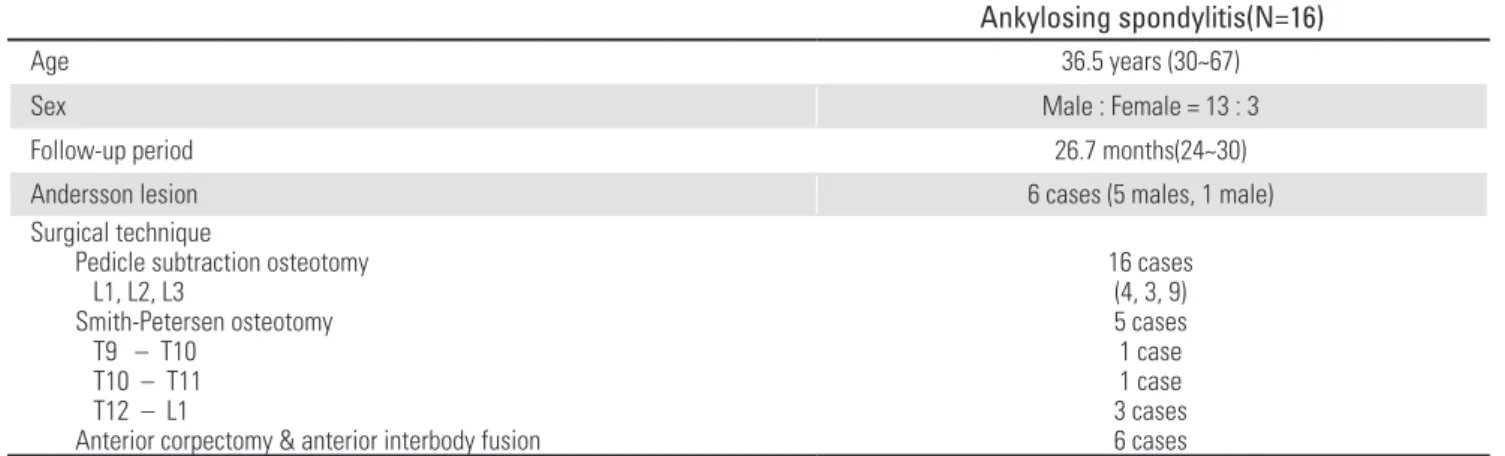
Among them, for 5 cases with kyphotic deformity in the vicin- ity of the lesion, together with pedicle subtraction osteotomy, Smith-Petersen osteotomy was performed at the time of first surgery. Pedicle subtraction osteotomy was performed on the 1st lumbar vertebra in 4 cases, on the 2nd lumbar vertebra in 3 cases, and on the 3rd lumbar vertebra in 9 cases. Smith-Petersen osteotomy was performed between the 9th thoracic vertebra and the 10th thoracic vertebra in 1 case, the 10th thoracic bertebra and the 11th vertebra in 1 case, and the 12th thoracic vertebra and the 1st lumbar vertebra in 3 cases. Postsurgical complica- tions were not observed (Table1).
Table 1. Demographic data
Ankylosing spondylitis(N=16)
Age 36.5 years (30~67)
Sex Male : Female = 13 : 3
Follow-up period 26.7 months(24~30)
Andersson lesion 6 cases (5 males, 1 male)
Surgical technique
Pedicle subtraction osteotomy L1, L2, L3
Smith-Petersen osteotomy T9 – T10
T10 – T11 T12 – L1
Anterior corpectomy & anterior interbody fusion
16 cases (4, 3, 9) 5 cases 1 case 1 case 3 cases 6 cases
1. Radiological results
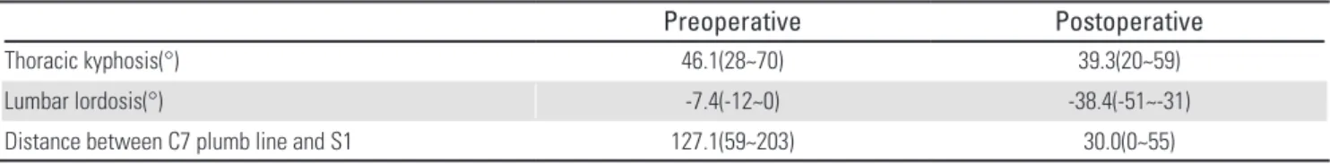
The kyphosis of the thoracic vertebras prior to surgery was corrected from 46.1 degrees (28 - 70 degrees) prior to surgery to average 39.3 degrees (20 - 59 degrees) after surgery. The lumbar lordosis was improved from -7.4 degrees (-12 - 0) prior to surgery to -38.4 degrees (-51 -31 degrees) after surgery. The distance from C7 plumb line to the posterosuperior corner of S1 was improved from 127.1 mm (59 - 203 mm) to 30 mm (0 - 55 mm)(Table2).
2. Clinical results
The BASDAI prior to surgery was measured to be 5.9 points and after surgery was 4.8 points. The BASFI prior to surgery was 6.0 points and after surgery was 5.0 points (Table3). The values did not show statistically significant differences.
Among the SF-36 scores, physical function was increased from average 45.1 points prior to surgery to average 52.8 points after surgery (p=0.018). role limitation due to physical health was increased from average 43.1 points prior to surgery to average 56.9 points after surgery (p=0.012). Vitality was improved from average 44.5 points prior to surgery to average 67.7 points after surgery(p=0.008). Social function was improved from average 50.3 points to average 56.8 points after surgery (p=0.026), role limitation due to emotional problems was improved from average 46.6 points prior to surgery to 52.0
after surgery(p=0.017), and mental health was improved from average 50.9 points prior to surgery to average 64.1 after surgery (p=0.033). bodily pain was improved from average 49.8 points prior to surgery to average 51.1 points after surgery (p=0.263).
General health was improved from average 52.6 points prior to surgery to average 55.3 points after surgery (p=0.314), nonetheless, statistically significant differences were not shown (Fig. 1). physical component summary was improved presurgical 45.2 points to 52.3 points after surgery, and mental component summary was improved from presurgical 51.9 points to 62.0 points after surgery(Fig. 2).
3.Comparison of results according to Andersson lesion Patients associated with Andersson lesion were 6 cases. In radiological evaluation, the thoracic kyphosis was improved from presurgical 49.1 degrees (30 - 69 degrees) and after surgery was 42.3 degrees (25 - 57 degrees), lumbar lordosis was improved from presurgical -8.8 degrees (-12-5 degrees) to -39.0 degrees after surgery (-51-32 degrees), and C7 plumb line was improved from 120.8 mm (54 - 184 mm) prior to surgery to 29.6 mm (0 - 41 mm) after surgery. In the group without Andersson lesion, after pedicle subtraction osteotomy, thoracic kyphosis prior to surgery was 45.2 degrees (28 - 70 degrees) and after surgery was 38.9 degrees (20 - 59 degrees), the lumbar lordosis prior to surgery was -7.1 degrees (-11 - 0 degrees) and
Fig 1. Significant improvements were seen 6 categories (physical function, role physical, vitality, social function, role emotional, mental health) after corrective osteotomy(p-value<0.05). There were also improvements in bodily pain and general
Fig 2. Improvement in mental component summary was better than that in physical component summary (p-value<0.05).
Table 2. Radiologic results after corrective osteotomy
Preoperative Postoperative
Thoracic kyphosis(°) 46.1(28~70) 39.3(20~59)
Lumbar lordosis(°) -7.4(-12~0) -38.4(-51~-31)
Distance between C7 plumb line and S1 127.1(59~203) 30.0(0~55)
Ye-Soo Park et al Volume 18 • Number 1 • March 2011
www.krspine.org 16
after surgery was -39.2 degrees (-44-31 degrees), C7 plumb line was improved from 134.2 mm (81 - 203 mm) to 34.2 mm(15 - 85 mm), and significant differences between the two groups were not shown (Table 4). Similarly, the difference of the improvement of SF-36 score was not also significantly different.
DISCUSSION
Ankylosing spondylitis is a chronic inflammatory disease. Not only it restricts the routine life of patient greatly but also due to mental withdrawal, it interferes with social life. In addition, if fixed kyphotic deformity is developed, forward looking becomes impossible, the sagittal imbalance is developed, and thus the center of gravity moves forwad, and pride is hurt due to problems in appearance, which mediates great effects on the quality of life of ankylosing spondylitis patients.
In 1945, Smith-Petersen et al.12) performed osteotomy on kyphotic deformity in rheumatoid arthritis patients. Afterward, several investigators reported surgical techniques and postsurgical complications. Nevertheless, the literatures reporting osteotomy for kyphotic deformity in ankylosing spondylitis are mostly on methodological approach for correction and focused on the re- duction of complications, and studies reporting clinical outcomes after kyphotic deformity correction are very rare.
In 1995, Halm et al.13) analyzed clinical outcomes after cor- rection by the survey using the modified AIMS in ankylosing spondylitis patients. In 2000, Kim et al.7) modified the question- naire survey suitable to the characteristic of Koreans such as sit- ting on the floor and cultural background, and reported clinical results. After the correction of kyphosis deformity, functional changes, indoor activity, outdoor activity, psychosocial activity
and pain showed significant improvement in comparison with prior to surgery. The overall subjective satisfaction level on sur- gery also showed statistically significant satisfaction. Nonethe- less, patients underwent osteotomy on the 4th lumbar vertebra reported that it was not comfort to sit on the floor.
The AIMS is a method developed by Meenan et al.14) in 1980 to evaluate the quality of life of arthritis patients, however, its re- liability has not been validated.15) In our study, clinical outcomes of corrective osteotomy were assessed by the application of the SF-36 that has been used widely among the generic health- related quality of life (HR-QOL) instruments, and its high reli- ability and adequacy were validated.16,17) In comparison with prior to surgery, physical function, restriction of the role of body, vitality, social function, the restriction of emotion, and psycho- logical health were significantly improved after surgery. This is in agreement with previous research results that significant improvement was shown in most categories. It is thought that forward looking as well as upward looking became possible, and the balance of sagittal plane was achieved, which was of help to not only functional improvement, but also psychologi- cal stability and psychiatric health. Actually, patients showed subjective satisfaction on experiencing abundant changes of life as upper looking became possible. In the summary of the results of the above categories, the level of physical health and the level of psychological health were all improved.
Nonetheless, it was shown that osteotomy did not exert great effects on pain and general health. This is different from previous studies reporting that after corrective osteotomy, not only back- ache, but also general pain as well as pain in the joint of other upper and lower extremities were reduced. We thought that the disease activity and the level of function exerted effects. Indeed, Table 3. The Bath ankylosing spinal functional index† and the Bath ankylosing spondylitis disease activity index†† were not changed
significantly after corrective osteotomy.
Preoperative Postoperative P-value
BASFI† 6.0±1.9 5.0±1.0 0.45
BASDAI†† 5.9±2.0 4.8±2.1 0.53
Table 4. There were not significant change between the groups of patients with thoracic kyphotic deformity and Andersson lesion and the groups of patients with thoracic kyphotic deformity in radiologic parameters (*p-value=0.36, **p-value=0.14, ***p-value=0.62).
Thoracic kyphotic deformity
with Andersson lesion (N=6) Thoracic kyphotic deformity without Andersson lesion (N=10)
Thoracic kyphosis(°)* 49.1(30~69) 42.3(25~57) 45.2(28~70) 38.9(20~59)
Lumbar lordosis(°)** -8.8(-12~-5) -39(-51~-32) -7.1(-11~0) -39.2(-44~-31)
Distance between C7 plumb line and
S1(mm)*** 120.8(54~184) 29.6(0~41) 134.2(81~203) 34.2(15~85)
in our patients, the BASDAI and the BASFI that represent the activity of disease were not significantly different from prior to surgery. Particularly, it was understood that in cases with the BASDAI higher than 4 points, the control of the disease activity was not appropriate, and thus they could be considered as cases requiring the consideration of the change of therapeutics. It was average 5.9 points prior to surgery and 4.8 points after surgery, and thus the level of pain according to the disease activity medi- ated effects on clinical results could not be ruled out. In regard to the BASFI, it was improved in comparison with prior to surgery, nonetheless, different from the physical domain of SF-36, sig- nificant differences were not shown. It could be thought to be due to that first, the BASFI is a visual analogue scale measure- ment instrument, the SF-36 is a tool to evaluate each category only by 3 stages or 5 stages, and the SF-36 focuses on the overall function and the satisfaction level of patient. On the other hand, the BASFI is an evaluation that assesses whether specific activity could be performed in ankylosing spondylitis patients.
Together with this, the effect of osteotomy on the improvement of overall health condition was limited, and it was thought that besides spinal kyphosis deformity, pain and overall health con- dition were closely associated with the disease activity of chronic inflammatory diseases.
Concerning surgical treatments for Andersson lesion, good re- sults of interbody fusion in the area with the lesion have been re- ported.18,19) In our study, similarly, in addition to posterior trans- pedicular corrective osteotomy for kyphotic deformity, after the removal of the anterior vertebra, interbody fusion was performed additionally, and radiologically, good results were shown. In clinical results, similarly, it was confirmed that although surgery was unavoidable because of Andersson lesion, it was confirmed if appropriate correction could be performed, the improvement of quality of life was experienced.
CONCLUSION
Our study was conducted on a small number of patients, and some results were different from previous research results.
Nonetheless, it was confirmed that in ankylosing spondylitis patients, corrective osteotomy for fixed kyphotic deformity could correct the sagittal imbalance, and improve physical as well as psychological quality of life. Such ankylosing spondylitis
is a chronic inflammatory disease, and thus the disease activity could not be controlled only by surgical treatments. Hence, it is thought that the satisfaction level of surgery could be elevated when aggressive treatments are administered additionally.
REFERENCES
1. Turan Y, Duruöz MT, Cerrahoglu L. Quality of life in patients with ankylosing spondylitis: a pilot study.
Rheumatol Int. 2007;27:895-9.
2. Ozdemir O. Quality of life in patients with ankylosing spondylitis: relationships with spinal mobility, disease activity and functional status. Rheumatol Int. 2010 Jan 5.
[Epub ahead of print]
3. Bradford DS, Schumacher WL, Lonstein JE, Winter RB.
Ankylosing spondylitis: experience in surgical management of 21 patients. Spine. 1987;12:238-43.
4. Van Royen BJ, De Gast A. Lumbar osteotomy for correction of thoracolumbar kyphotic deformity in ankylosing spondylitis. A structured review of three methods of treatment. Ann Rheum Dis. 1999;58:399-406.
5. Niemeyer T, Hackenberg L, Bullmann V, Liljenqvist U, Halm H. Technique and results of monosegmental transpedicular subtraction osteotomy in patients with ankylosing spondylitis and fixed kyphotic deformity of the spine. Z Orthop Ihre Grenzgeb. 2002;140:176-81.
6. Yoo MC, Lee SE, Kim KT, Lee HK, Cho JH. Clinical and Radiological Features of Ankylosing Spondylitis . J Korean Soc Spine Surg. 1995;2:72-80.
7. Kim KT, Cho YJ, Hong GP, Park BJ, Lee JH. Clinical Results after Corrective Osteotomy of the Kyphotic Deformity in Ankylosing Spondylitis. J Korean Soc Spine Surg. 2000;7:61-9.
8. Lillegraven S, Kvien TK. Measuring disability and quality of life in established rheumatoid arthritis. Best Pract Res Clin Rheumatol. 2007;21:827-40.
9. Garrett S, Jenkinson T, Kennedy LG, Whitelock H, Gaisford P, Calin A. A new approach to defining disease status in ankylosing spondylitis: the Bath Ankylosing Spondylitis Disease Activity Index. J Rheumatol. 1994;21:2286-91.
10. Calin A, Garrett S, Whitelock H, et al. A new approach to defining functional ability in ankylosing spondylitis: the development of the Bath Ankylosing Spondylitis Functional Index. J Rheumatol. 1994;21:2281-5.
11. Ware JE Jr. SF-36 health survey update. Spine.
Ye-Soo Park et al Volume 18 • Number 1 • March 2011
www.krspine.org 18
2000;25:3130-9.
12. Smith-Petersen MN, Larson CB, Aufranc OE. Osteotomy of the spine for correction of flexion deformity in rheumatoid arthritis. Clin Orthop Relat Res. 1969;66:6-9.
13. Halm H, Metz-Stavenhagen P, Zielke K. Results of surgical correction of kyphotic deformities of the spine in ankylosing spondylitis on the basis of the modified arthritis impact measurement scales. Spine. 1995;20:1612-9.
14. Meenan RF, Gertman PM, Mason JH. Measuring health status in arthritis. The arthritis impact measurement scales.
Arthritis Rheum. 1980;23:146-52.
15. Haywood KL, Garratt AM, Dawes PT. Patient-assessed health in ankylosing spondylitis: a structured review.
Rheumatology. 2005;44:577-86
16. Hays RD, Sherbourne CD, Mazel RM. The RAND 36- Item Health Survey 1.0. Health Econ. 1993;2:217-27.
17. Essink-Bot ML, Krabbe PF, Bonsel GJ, Aaronson NK. An empirical comparison of four generic health status measures.
The Nottingham Health Profile, the Medical Outcomes Study 36-item Short-Form Health Survey, the COOP/
WONCA charts, and the EuroQol instrument. Med Care.
1997; 35:522-37
18. Fang D, Leong JC, Ho EK, Chan FL, Chow SP. Spinal pseudarthrosis in ankylosing spondylitis. Clinicopathological correlation and the results of anterior spinal fusion J Bone Joint Surg Br. 1988;70:443-7.
19. Kim KT, Suk KS, Lee SH, Bae SC. The treatment of spinal pseudoarthrosis in ankylosing spondylitis. J Korean Orthop Assoc. 2005;40:312-20.
강직성 척추염에서 교정 절골술이 삶의 질에 미치는 영향
박예수 • 이창훈 • 김재훈 • 김태환*
한양대학교 의과대학 정형외과학교실, 류마티스병원*
연구 계획: 교정 절골술을 시행한 강직성 척추염 환자를 대상으로 삶의 질에 대한 후향적인 연구
목적: 강직성 척추염에 의한 후만 변형에 대해 교정 절골술을 시행한 후 방사선학적 평가와 더불어 삶의 질 향상에 대한 임상적 평가를 시행하고자 하였 다.
선행 문헌의 요약: 기존의 연구들은 강직성 척추염의 수술 후 방사선학적 평가에 국한되어 있었고, 환자의 시각에서 교정 절골술이 삶의 질 향상에 기여 하는 정도를 확인하는 연구가 부족하였다.
대상 및 방법: 2005년 9월부터 2007년 12월까지 고정된 후만 변형에 대해 교정 절골술을 시행한 16명의 강직성 척추염 환자를 대상으로 후향적 연 구를 시행하였다. 수술적 방법으로는 척추경 후방 절골술을 시행하여 변형을 교정하였고, Andersson 병변을 동반한 경우에는 전방 추체 제거술, 추체 간 유합술을 추가적으로 시행하였다. 술 후 방사선학적 평가로는 흉추 후만각, 요추 전만각, 그리고 제 7 경추 수선까지의 거리를 이용하여 시상면 불 균형의 수술 전, 후의 변화를 조사하였다. 질병 특이적인 도구로 the Bath ankylosing spondylitis disease activity index (BASDAI) 와 the Bath ankylosing spondylitis functional index (BASFI)를 측정하여 질병 활성도를 파악하였다. 임상적 평가로는 전화 면담 및 설문지를 통해 SF-36을 이용하여 삶의 질 향 상 정도를 측정하였다.
결과: 흉추 후만각은 술 전 평균 46.1도에서 술 후 평균 39.3도로 교정되었고, 요추 전만각은 술 전 - 7.4도에서 술 후 - 38.4도, 제7 경추체에서 내린 수 선과 제1천추체 후상부와의 거리는 127.1mm에서 30mm로 호전되었다. BASDAI와 BASFI는 술 전과 비교하여 유의한 차이를 보이지 않았고, SF-36 score는 술 전에 비해 신체기능, 신체역할제한, 활력, 사회기능, 감정역할제한, 정신건강 부분에서 술 후 유의하게 증가하였고(p<0.05), 통증과 일반건강 부분은 유의한 변화를 보이지 않았다. Andersson 병변을 동반한 환자는 6명으로 방사선학적 평가 및 SF-36 score에서 후만 변형에 대해서만 척추경 제 거 절골술을 시행한 군과 비교하여 유의한 차이를 보이지 않았다.
결론: 고정된 척추 후만 변형을 보이는 강직성 척추염 환자들은 교정 절골술 이후 방사선학적으로 호전되었으며, 일상 생활과 사회 생활 및 정신 건강 정도가 향상되는 것을 확인할 수 있었다.
색인 단어: 강직성 척추염, 교정 절골술, 삶의 질 약칭 제목: 강직성 척추염 환자의 삶의 질