서론
만성 치주염은 치아를 지지하는 결체조직과 골조직의 파괴
로 이어지는 염증성 질환이다. 이러한 치주염은 치주병원균과 같은 환경인자와 숙주 방어간의 불균형에 의해 발생하는 다 인자성 질환으로 숙주방어기전은 유전적 요소, 호르몬, 영양 등에 의해 영향을 받을 수 있다1). 하지만 치주질환의 병리기 전을 조절하는데 있어서 영양의 역할은 명확하지 않다2).
치주질환의 치료는 비외과적 치주치료와 외과적 치주치 료로 구분되며, 여러 연구에서 비외과적, 외과적 치주치료 후 치주낭 깊이의 감소, 임상부착수준의 증진, 치은염 지수 의 감소가 보고되었다3-4). 치주치료에 부가적으로 doxycy-
비외과적․ 외과적 치주치료와 병행 투여된 비타민 C 특수영양 보충용 식품이 치주질환의 치유과정에 미치는 효과
백영란, 박진우, 이재목, 서조영
*경북대학교 치의학전문대학원 치주과학교실
The effect of vitamin-C containing neutraceutical on periodontal wound healing as an adjunct to non-surgical or surgical periodontal treatment
Young-Ran Baek, Jin-Woo Park, Jae-Mok Lee, Jo-Young Suh
*Department of Periodontology, School of Dentistry, Kyungpook National University
ABSTRACT
Purpose: There are numerous reports about the usefulness of antibiotics such as doxycycline or metronidazole in the conventional treatment for the patients with chronic periodontal diseases. However, seldom are the reports about effects of vitamins or nutraceuticals. The purpose of this study was to evaluate the effects of nutrient supplement including multiple vitamins and neutraceuticals with PRF-K2 from plants and seaweed in treatment of the patients with chronic periodontitis which is needed a nonsurgical or a surgical treatment by evaluating the clinical parameters and the gingival crevicular fluid volume.
Methods: The systemically healthy and nonsmoking patients diagnosed with chronic periodontitis were divided into a nonsurgical group and a surgical group. They were also divided into the test group with nutrient supplements and the control group without nutrient supplements. In the nonsurgical group, the clinical parameters (probing depth, clinical attachment level, sulcus bleeding index, and plaque index) and the gingival crevicular fluid volume were checked on baseline, at 1 week, at 3 week and at 9 week after a supplement treatment. In the surgical group, the clinical parameters and the gingival crevicular fluid volume were also checked at 15 week after a surgical treatment.
Results: In both nonsurgical and surgical groups, reduction of pocket depth and increment of clinical attachment level were revealed in the test group compared with the control group, but there was not statistically significant difference (p>0.05), and sulcus bleeding index was decreased with statistically significant difference (p<0.05). In addition, plaque index was decreased with statistically significant difference (p<0.05) in the nonsurgical group. Gingival crevicular fluid volume was decreased with statistically significant difference (p<0.05) at week 9 in both non-surgical and surgical groups.
Conclusions: In conclusion, our results demonstrate that providing nutrient supplement in both nonsurgical or surgical periodontal treatments may improve gingival inflammation and gingival crevicular fluid.
(J Korean Acad Periodontol 2009;39:157-166)
KEY WORDS: ascorbic acid; dietary supplements; periodontitis; gingvial crevicular fluid.
* Correspondence: Jo-Young Suh, D.D.S Ph.D
Department of Periodontology, School of Dentistry, Kyungpook National University, 188-1, Samduk-dong 2ga, Jung-gu, Daegu, 700-412, Korea
E-mail: [email protected], Tel: 82-53-600-7521, Fax: 82-53-427-3263
* 본 연구는 오스코텍(주)의 연구비 지원으로 수행됨.
Received: Apr. 7, 2009; Accepted: Jun. 2, 2009
cline이나 metronidazole 등 항생제에 대한 연구는 많이 보 고 되었지만5-6), 비타민이나 nutraceutical의 효과를 연구 한 것은 거의 없다.
특정 영양소의 결핍에 대하여 비교적 잘 알려진 반면 치 료와 병행하여 영양 공급의 효과에 대한 연구는 거의 없다.
영양과 관련하여 특히 비타민은 괴혈병, 급성 괴사성 궤양 성 치은염 등과 관련하여 치주영역에서 상당한 주목을 받아 왔다7-8).
정상적인 생리학적 기능에 필요한 영양 중에서 비타민 B 복합체는 치주 창상 치유에 중요한 역할을 할지도 모른다9). 비타민 B1으로 알려진 티아민은 당분을 에너지로 전환시켜 근육과 신경의 정상적인 기능에 필요하며10), 비타민 B2로 알려진 리보플라빈은 정상적인 성장, 근육의 발달, hair coat에 필수적이다11). 비타민 B6인 피리독신은 아미노산의 이용을 위해 체내에서 사용된다12). 또한 비타민 B 복합체는 다른 창상 치유과정에 긍정적인 효과를 미치는 것으로 보고 되었다13).
비타민 C가 치주조직에 영향을 미치는 기전을 살펴보면, 비타민 C 결핍은 프롤린의 hydroxylation에 영향을 주어 교원질 합성의 결핍을 야기하며14), 치은 점막의 내독소 투과 성을 증가시키고15) 중성구 기능을 감소시켜 숙주 면역 반응 을 약화시킨다16).
치주질환에 대한 음식물로 섭취된 비타민 C에 대한 효과 는 동물과 인간에 대한 연구에서 상반되는 효과가 보고되어 왔다. 초기 동물연구에서 Glickman17)은 비타민 C 결핍이 더 깊은 치주낭과 증가된 치주조직 파괴를 보인다고 보고한 반면 초기 역학 연구의 대부분에서 혈장 비타민 C 수준과 치주질환의 심도 사이에 연관성이 없는 것으로 나타났다18). 그러나, 최근의 역학 연구들에서는 비타민 C와 치주질환 사
이의 유의한 상관성이 보고되고 있다.
Nishida 등19)은 현재 또는 이전 흡연자에서 음식물로 섭 취된 비타민 C는 치주질환과 약하지만 유의한 상관성을 관 찰하였으며, Amarasena 등20)은 노인환자에서 혈청 비타민 C와 부착소실간의 반비례적 관계를 보고하였다. 또한 Munoz 등21)은 치주질환자에게 식물의 추출물과 coenzyme, 비타민이 포함된 neutraceutical을 투여했을 때, 치은지수, 출혈지수, 치주낭 깊이에 있어서 실험군에서 더 큰 개선을 보인다고 보고하였다. 또한 최근의 연구에서 Kim 등22)은 치 주유지관리기 환자에게 비타민이 포함된 neutraceutical을 공급하는 것이 치주건강을 증진시키는데 도움이 될 수 있음 을 시사하였다.
이번 연구는 만성 치주염환자의 비외과적, 외과적 치주치 료 과정에서 새송이 버섯, 가시오가피, 전칠삼 추출물을 원 료로 하는 혼합물(PRF-K2)과 소량의 비타민 B 복합체 및 다량의 비타민 C가 함유된 비타민 C 특수영양 보충용 식품 을 복용했을 때 일반 치주치료 환자에 비해 임상지수 변화 나 치은 열구액 수준의 변화에 어떤 영향을 보이는지 평가 하기 위해 시행되었다.
재료 및 방법
1. 대상
경북대학교 병원 치주과에 내원한 환자 중 만성 치주염으로 진단 받은 35~70세의 환자를 대상으로 하였다(Table 1, 2).
초진시 적어도 20개의 치아가 존재하며, 각각의 사분악에 탐침시 출혈을 보이는 치아가 있고, 임상 부착수준 및 탐침
Table 1. The Demographic Characteristics of Subjects in Non-surgical Group
Group Number Age(mean age±SD) Male/Female
Test 20 47.11±11.95 12/8
Control 21 41.43±9.35 13/8
Table 2. The Demographic Characteristics of Subjects in Surgical Group
Group Number Age(mean age±SD) Male/Female
Test 10 45.20±7.97 3/7
Control 10 43.30±5.12 5/5
치주낭 깊이가 5~9 mm 이상인 치아를 적어도 2개를 가지 는 환자를 연구 대상으로 선정하였다. 급성 감염이 발생한 환자, 만성질환의 의학적 기왕력을 가지는 환자, 임산부, 흡 연자와 또한 3개월 이내로 치면 세마를 실시하거나 치주치 료를 받은 환자는 제외하였다. 비외과적 치주치료가 완료된 환자는 비외과적 치료군으로, 사분악 중 한 부위라도 외과 적 치주치료가 시행된 환자는 외과적 치료군으로 분류되었 다. 2005년 10월부터 2006년 11월까지 이상의 조건을 만족 하며 연구에 참여하는데 동의 서명한 환자들이 비외과적 치 주치료 군에서 실험군에 20명(평균나이 47.1세), 대조군에 21명(평균나이 41.4세)이며 외과적 치주치료 군에서 실험군
에 10명(평균나이 45.2세), 대조군에 10명(평균나이 43.3세) 이었다. 위 연구는 XX대학교병원 임상시험심사위원회의 심 사를 통과하였다(의연 74005-27).
2. 방법
초진 시 서면 동의서가 제출되었으며 환자는 임의로 실 험군 및 대조군으로 분류되었다. 실험군에게는 비외과적, 외과적 치주치료와 병행하여 비타민 C 특수영양 보충식품 (PerioNutraⓇ Oscotec Inc. Cheonan, Korea)을 복용하게 하였다. 복용은 1회 2 캅셀씩, 1일 2회 시행되었다. 대조군은
Figure 1. Study design of non-surgical group (GCF, gingival crevicular fluid; PD, pocket depth; CAL, clinical attachment level; SBI, sulcus bleeding index; PI, plaque index; OHI, oral hygiene instruction).
Figure 2. Study design of surgical group (GCF, gingival crevicular fluid; PD, pocket depth; CAL, clinical attachment level; SBI, sulcus bleeding index; PI, plaque index; OHI, oral hygiene instruction; FO. flap operation).
보조제의 섭취없이 비외과적, 외과적 치주치료만 시행되었다.
비외과적 치료군과 외과적 치료군에서 공통적으로, 1주에 치석제거술이 시행되었으며, 3주에서 7주까지 국소마취하의 치은연하 치석제거술 및 치근활택술이 시행되었고(Fig. 1) 부가적으로 외과적 치료군에서는 9주에서 13주까지 치주판 막술이 시행되었다(Fig. 2). 매 내원시에는 실험군과 대조군 모두 구강 위생교육이 시행되었다.
1) 임상검사
숙달된 조사자가 비외과적 치료군에서는 실험시작, 1주, 3주, 9주에, 외과적 치료군에서는 실험시작, 1주, 3주, 9주 와 15주에 탐침 치주낭 깊이, 임상 부착수준, 치은 염증지 수, 치태지수를 측정하였으며, 모든 검사는 치아당 6부위에 서 측정하였다. 치주낭 깊이 및 임상 부착수준은 Willams probe(23W, Hu-Friedy, Chicago, USA)를 사용하여 0.5mm 단위로 측정하였다. 치은 염증도는 Muhlemann과 Son23)의 Sulcus bleeding index(SBI)를 사용하여 평가하였고 치태 지수는 Silness와 Loe24)의 Plaque Index(PI)에 따라 기록하 였다. 또한 비외과적 치료군과 외과적 치료군의 실험군에서 상, 하악 그리고 전치와 구치간의 비교가 시행되었다.
2) 치은 열구액 검사(Gingival Crevicular Fluid volume) 각 환자에서 전치부, 구치부, 상․ 하악에서 만성치주염을 대표적으로 나타낼 수 있는 치아를 선택하였다. 치주낭 깊 이가 4 mm 이상인 4개 부위를 선정(상악 우측 제1대구치, 하악 좌측 제1대구치, 상악 좌측 중절치, 하악 우측 중절치) 하여 비외과적 치료군에서는 실험시작, 1주, 3주, 9주에 치 은 열구액을 채취하고, 외과적 치료군에서는 실험시작, 1주, 3주, 9주와 15주에 치은 열구액을 채취하였다. 먼저 채취할 부위의 치태를 제거하고 건조 후, PeriopaperⓇ(Proflow Inc. NewYork, USA) 1개를 치주낭 내로 1 mm 정도 넣고 30초간 둔 후 제거하여 PeriotronⓇ 6,000(OraFlow Inc.
New York, USA)에서 치은 열구액 양을 측정하였다.
3) 통계학적 분석
모든 측정값은 평균과 표준오차로 정리하였다. 각 그룹 내에서 실험시작, 1주, 3주, 9주, 15주 사이의 임상 지수 및 치은 열구액의 양의 비교를 위해서 Friedman test가 시행 되었고, 시간에 따른 유의성을 분석하기 위하여 Bonferroni- corrected Wilcoxon signed-rank test가 시행되었다. p값
은 비외과적 치료군에서는 0.0167 미만, 외과적 치료군에서 는 0.0125 미만일 경우 통계적으로 유의하다고 간주되었다.
각 시점에서 실험군과 대조군 및 상, 하악 그리고 전치부와 구치부의 차이를 평가하기 위해 Mann-Whitney test를 시 행하였으며, p값이 0.05 미만일 경우 통계적으로 유의하다 고 간주되었다.
결과
1. 임상지수의 변화
1) 비외과적 치료 그룹
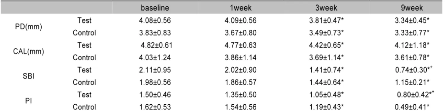
실험군과 대조군에 있어서 전악 임상지수는 Table 3에 제시되어 있다. 실험시작 시 두 군 사이의 임상지수는 통계 적으로 유의한 차이가 없었다. 실험군과 대조군 모두에서 실험시작과 비교하여 1주에는 큰 변화가 없었지만 3주, 9주 에 치주상태의 현저한 개선을 나타내었다(p<0.0167). 치주 낭 깊이의 감소는 모든 시점에서 두 군간 통계적으로 유의 한 차이를 보이지는 않았지만(p>0.05), 9주에서 대조군에 비해 실험군에서 더 감소되는 경향을 보였다. 임상부착수준 획득은 모든 시점에서 두 군간 유사한 경향을 보였으며 (p>0.05) 9주에서 대조군에 비해 실험군에서 크게 나타나 치주낭 깊이 감소와 유사한 경향을 보였다. 열구출혈 지수 (SBI)의 개선은 1, 3주에는 두 군간 유의한 차이를 보이지 않았지만, 9주에 대조군에 비해서 실험군에서 통계적으로 유의한 감소를 보였다(p=0.023). 치태지수의 개선은 1, 3주 에는 두 군간 유의한 차이가 없었으며 9주에 대조군에서 유 의하게 큰 감소를 보였다(p=0.016).
실험군 내 상 ․ 하악간의 비교에서 치주낭 깊이 감소와 임상부착증진이 유사하게 나타났으며 모든 시점에서 통계적 으로 유의한 차이를 나타내지 못했다(p>0.05)(Table 4). 실 험군 내 전치와 구치의 비교에서 전치부는 치주낭 깊이가 실험시작에 비하여 1주 0.09mm, 3주 0.29mm, 9주에 0.55 mm의 감소를 보인 반면 구치부는 1주 0.11 mm 증가, 3주 0.33mm 감소, 9주에 0.90 mm의 감소를 보였으며, 이 차이는 9주에 통계적으로 유의했다(p=0.001). 임상부착증진 은 전치부에서 실험시작에 비하여 1주 0.06mm, 3주 0.31 mm, 9주에 0.48mm 증가하였으며, 구치부에서 1주에 0.01 mm, 3주에 0.29mm, 9주에 0.71 mm 증가하였으며, 이 차
이는 모든 시점에서 통계적으로 유의하지 않았다 (p>0.05)(Table 5).
2) 외과적 치료 그룹
실험군과 대조군에 있어서 전악 임상지수는 Table 6에 제시되어 있다. 실험시작에서 모든 임상지수는 두 군 사이 에 통계적으로 유의한 차이가 없었다. 실험군과 대조군 모
두에서 실험시작과 비교하여 1주, 3주에는 큰 변화가 없었 으며 9주와 15주에 치주상태의 유의한 개선을 나타내었 다(p<0.0125). 치주낭 깊이의 감소는 모든 시점에서 두 군 간 통계적으로 유의한 차이를 보이지 않았다(p>0.05). 임상 부착수준 획득은 모든 시점에서 두 군간 유사한 경향을 보 였지만(p>0.05) 15주에 실험군에서 0.68mm, 대조군에서 0.49mm로 실험군에서 약간 크게 나타났다. 치은 염증지수
Table 3. Whole-Mouth Clinical Parameters for Test and Control Groups in Non-surgical group (Mean±SD)
baseline 1week 3week 9week
PD(mm) Test 4.08±0.56 4.09±0.56 3.81±0.47* 3.34±0.45*
Control 3.83±0.83 3.67±0.80 3.49±0.73* 3.33±0.77*
CAL(mm) Test 4.82±0.61 4.77±0.63 4.42±0.65* 4.12±1.18*
Control 4.03±1.24 3.86±1.14 3.69±1.14* 3.61±0.78*
SBI Test 2.11±0.95 2.02±0.90 1.41±0.74* 0.74±0.30*+
Control 1.98±0.56 1.86±0.57 1.44±0.64* 1.15±0.21*
PI Test 1.50±0.46 1.35±0.50 1.05±0.48* 0.80±0.42*+
Control 1.62±0.53 1.54±0.56 1.19±0.43* 0.49±0.41*
*significant difference compared to baseline (p<0.0167), +significant difference compared to control group (p<0.05)
Table 4. Clinical Parameters for Maxilla and Mandible in Test Group of Non-surgical Group (Mean±SD)
baseline 1week 3week 9week
PD(mm) Mx 4.11±0.55 4.07±0.52 3.81±0.48* 3.36±0.45*
Mn 4.06±0.75 4.12±0.78 3.81±0.60 3.32±0.50*
CAL(mm) Mx 4.74±0.68 4.70±0.82 4.49±074* 4.05±0.68*
Mn 4.88±0.77 4.80±0.66 4.53±0.60* 4.18±0.70*
*significant difference compared to baseline (p<0.0167), +significant difference compared to Mn (p<0.05)
Table 5. Clinical Parameters for Anterior and Posterior Segments in Test Group of Non-surgical Group (Mean±SD)
baseline 1weeks 3week 9week
PD(mm) Ant 3.69±0.46 3.60±0.45 3.40±0.35* 3.14±0.45*
Post 4.36±0.69 4.47±0.79 4.03±0.74* 3.46±0.78*+
CAL(mm) Ant 4.53±0.69 4.47±0.80 4.22±0.82* 4.05±0.77*
Post 5.03±0.72 5.02±0.65 4.74±0.57* 4.32±0.65*
*significant difference compared to baseline (p<0.0167), +significant difference compared to Ant. segment (p<0.05)
Table 6. Whole-Mouth Clinical Parameters for Test and Control groups in Surgical Group (Mean±SD)
baseline 1week 3week 9week 15week
PD(mm) Test 4.04±0.47 3.97±0.37 3.77±0.26 3.33±0.21* 3.06±0.26*
Control 4.13±0.86 4.09±0.94 3.89±0.89 3.46±0.80* 3.12±0.83*
CAL(mm) Test 4.19±0.46 4.17±0.37 4.05±0.40 3.55±0.38* 3.51±0.37*
Control 4.54±1.31 4.30±1.26 4.11±1.16 4.03±0.80 4.05±0.88
SBI Test 1.75±0.76 1.78±0.80 1.40±0.70 0.60±0.47*+ 0.40±0.21*+
Control 2.30±0.67 2.08±0.82 1.88±0.73* 1.25±0.50* 1.14±0.78
PI Test 1.28±0.89 1.28±0.85 0.95±0.68 0.48±0.15* 0.43±0.17*
Control 1.53±0.57 1.30±0.67 1.05±0.37 0.80±0.33* 0.56±0.21*
*significant difference compared to baseline (p<0.0125), +significant difference compared to control group (p<0.05)
의 개선은 1, 3주에는 두 군간 유의한 차이를 보이지 않았지만, 9주와 15주에 대조군에 비해서 실험군에서 통계적으로 유의한 감소를 보였다(p=0.007, 0.003). 치태지수의 개선은 모든 시 점에서 두 군간 유의한 차이를 보이지 않았다(p>0.05).
실험군 내 상․ 하악간의 비교에서 치주낭 깊이는 실험시 작에 비하여 상악에서 9주에 0.86mm, 15주에 1.22 mm 감 소하였고, 하악에서 9주에 0.53mm, 15주에 0.74 mm 감소 를 보여, 상악에서 약간 큰 경향을 나타내었지만 모든 시점 에서 통계적으로 유의한 차이를 보이지 않았다(p>0.05). 임 상부착증진은 실험시작에 비하여 상악에서 9주에 0.61 mm, 15주에 0.77mm의 증가를 보였고, 하악에서 9주에 0.59
mm, 15주에 0.66mm의 증가를 보였으며, 모든 시점에서 통계적으로 유의한 차이를 보이지 않았다(p>0.05) (Table 7).
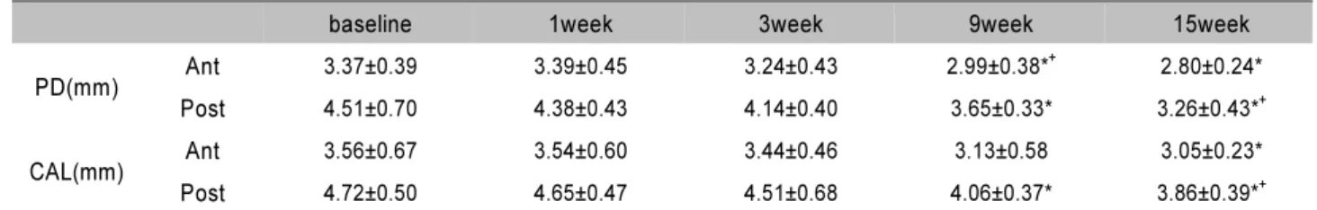
실험군 내 전치와 구치의 비교에서 전치부는 치주낭 깊이 가 실험시작에 비하여 1주 0.02 mm 증가, 3주 0.13mm 감 소, 9주에 0.38mm 감소, 15주에 0.57 mm 감소를 보인 반 면 구치부는 1주 0.13mm 감소, 3주 0.37 mm 감소 , 9주에 0.86mm의 감소, 15주에 1.25mm 감소를 보였으며, 이 차 이는 9주과 15주에 통계적으로 유의했다(p=0.004, 0.002).
임상부착증진은 전치부에서 실험시작에 비하여 1주 0.02 mm, 3주 0.12 mm, 9주에 0.43mm, 15주에 0.51 mm 증가
Table 7. Clinical Parameters for Maxilla and Mandible in Test Group of Surgical Group (Mean ± SD)
baseline 1week 3week 9week 15week
PD(mm) Mx 4.27±0.58 4.21±0.73 4.10±0.41 3.41±0.32* 3.05±0.30*
Mn 3.82±0.68 3.73±0.58 3.48±0.33 3.29±0.30* 3.08±0.37*
CAL(mm) Mx 4.34±0.81 4.40±0.68 4.24±0.56 3.73±0.51* 3.57±0.64*
Mn 4.04±0.65 3.95±0.54 3.67±0.46 3.45±0.24* 3.38±0.58*
*significant difference compared to baseline (p<0.0125), +significant difference compared to Mn (p<0.05)
Table 8. Clinical Parameters for Anterior and Posterior Segments in Test Group of Surgical Group (Mean ± SD)
baseline 1week 3week 9week 15week
PD(mm) Ant 3.37±0.39 3.39±0.45 3.24±0.43 2.99±0.38*+ 2.80±0.24*
Post 4.51±0.70 4.38±0.43 4.14±0.40 3.65±0.33* 3.26±0.43*+
CAL(mm) Ant 3.56±0.67 3.54±0.60 3.44±0.46 3.13±0.58 3.05±0.23*
Post 4.72±0.50 4.65±0.47 4.51±0.68 4.06±0.37* 3.86±0.39*+
*significant difference compared to baseline (p<0.0125), +significant difference compared to Ant. segment (p<0.05)
Table 9. Gingival Crevicular Fluid Volume of Study Sites in Test and Control Group of Non-surgical Group (Mean ± SD) Gingival crevicular fluid
baseline 1week 3week 9week
Test 83.40±30.60 78.35±33.05 70.51±31.17 44.98±14.72*+
Control 82.99±28.58 85.76±30.83 81.21±25.66 67.50±23.34
*significant difference compared to baseline (p<0.0167), +significant difference compared to control group (p<0.05)
Table 10. Gingival Crevicular Fluid Volume of Study Sites in Test and Control Group of Surgical Group (Mean ± SD) Gingival crevicular fluid
baseline 1week 3week 9week 15week
Test 85.83±27.27 78.23±32.10 64.30±16.24 52.28±17.70*+ 41.55±10.18*
Control 87.33±42.39 86.20±31.65 74.75±28.56 65.73±22.30 45.14±14.71*
*significant difference compared to baseline (p<0.0125), +significant difference compared to control group (p<0.05)
하였으며, 구치부에서 1주에 0.07mm, 3주에 0.21mm, 9주 에 0.66mm, 15주에 0.86mm 증가하였으며, 이 차이는 15 주에 통계적으로 유의하였다(p=0.007) (Table 8).
2. 치은 열구액 양의 변화
비외과적 치주치료 그룹과 외과적 치주치료 그룹의 치은 열구액 양의 변화는 Table 9, 10에 제시되어 있다. 비외과 적 치료그룹에서 치은 열구액(PeriotronⓇ 수치)은 실험시작 에 비하여 실험군에서 1주에 5.05, 3주에 12.89, 9주에 38.42의 감소를 보인 반면, 대조군에서는 1주에 2.77 증가, 3주에 1.78 감소, 9주에 15.49 감소를 보였다. 두 군간 차이 는 9주에 통계적으로 유의하였다(p=0.034). 외과적 치료그 룹에서 치은 열구액(PeriotronⓇ 수치)은 실험시작에 비하여 실험군에서 1주에 7.6, 3주에 21.53, 9주에 33.55, 15주에 44.28의 감소를 보였으며, 대조군에서는 1주에 1.13, 3주에 12.58, 9주에 21.6, 15주에 42.19의 감소를 보였다. 두 군간 의 차이는 9주에는 통계적으로 유의했으나(p=0.021), 15주 에는 유의하지 않았다.
고찰
이번 연구에서 실험군 대조군 모두에서 실험시작에 비하 여 치주상태의 현저한 개선을 나타내었으며, 이것은 여러 연구에서 비외과적, 외과적 치주치료 후 치주낭 깊이의 감 소, 임상부착수준의 증진, 열구출혈지수(SBI)의 감소가 보 고된 것과 일치한다3-4).
비타민 B, C가 창상 치유의 초기단계에서 작용하는 기전 은 비타민 B의 초기 세포증식의 촉진과 비타민 C에 의한 교 원질 합성의 증가로 설명될 수 있다25). 또한 티아민과 같은 비타민 B 복합체는 피부에서의 육아조직 형성 시 교원질에 영향을 미치는 것으로 알려져 있다26). 비타민 C는 인체에서 조직 손상을 일으키는 hydroxyl free radical을 청소하며27), 대식세포에 의한 superoxide anion의 분비를 억제하여28) 체내의 항산화 방어기전의 일부를 구성하고 있다. 치주 질 환 역시 병인성 세균과 숙주 면역 반응과의 상호 작용에 의 한 조직 손상을 야기하는 염증성 질환으로 free radical과 치주질환의 관계는 여러 연구에서 보고되었다29).
이번 연구는 만성 치주염 환자의 비외과적, 외과적 치주 치료 과정에서 여러 비타민 성분과 PRF-K2가 포함된 영양 보충용 식품을 복용했을 때 일반 치주치료 환자에 비해 임 상지수 변화나 치은 열구액 수준의 변화에 있어 유리한 효 과가 있는지를 알아보았다.
이 연구에서 사용된 영양 보충용 식품은 비타민 C (500mg/1g)가 성분 중 가장 많은 비율을 차지하고 있는데 치주질환에서 비타민 C 대체재의 효과에 대한 상반되는 의 견이 제시되어 왔다. Parfitt과 Hand30)은 1일 500mg 비타 민 C가 치은 건강에 아무런 효과를 가지지 못한다고 하였으 며, Vogel 등31)도 1일 1,500mg 비타민 C를 섭취했을 때 실 험적 치은염에 효과를 보이지 못한다고 하였다.
반면 실험적 비타민 C 결핍과 보조제에 관한 다른 연구 들은 치은 염증과 비타민 C 수준 사이의 직접적인 관계가 존재함을 보여주었다32). 좀 더 최근의 연구는1) 3년간의 관 찰에서 혈장 비타민 C 수준과 치주부착 상실이 음의 상관성 을 보인다고 하였으며, stepwise linear analysis에서 혈장 비타민 C 수준이 치주부착상실을 일으키는 변수의 3.9%를 설명할 수 있다고 하였다.
이번 연구에서 비외과적, 외과적 치주치료군 모두에서 치 주낭 깊이 감소와 임상 부착증진에서 영양 보충용 식품을 투여한 실험군과 대조군간 통계적으로 유의한 차이는 발견 되지 않았으며, 치은 염증지수는 실험군에서 유의한 감소를 보였다(P<0.05). 이 결과는 최근의 Staudte 등33)의 연구와 유사한 소견을 보이는데, 정상 범위 이하의 혈장 비타민 C 수준을 가지는 치주질환자에서 자몽(grapefruit)을 섭취시 켰을 때, 치은열구 출혈지수는 감소시켰으며, 치주낭 깊이 는 감소시키지 못했다.
치주치료와 병행하여 비타민을 포함한 보조제의 효과를 보고한 연구는 드물지만, 외과적 치주치료 후 비타민 B 복 합체를 투여한 다른 연구에서9) 180일 후 임상부착 증진에 유의한 차이를 보였다. 이번 연구의 결과와는 다른 양상으 로 이러한 차이는 대상 환자의 특성(나이, 샘플수, 흡연, 전 신질환 여부) 및 치주치료 유․ 무 및 방법의 차이와 영양 보 충용 식품 복용시기, 복용기간 및 추적관찰기간에 있어서의 차이에 기인할 것이다.
이번 연구에서 치은 염증의 감소와 같은 단기간의 결과는 대조군에 비하여 유의한 차이를 보였지만, 치주낭 깊이 감소 와 임상부착수준에서는 대조군과 차이를 보이지 않았다. 특 히 외과적 치주치료군의 경우 치주낭 깊이 감소에 있어서 보
조제의 효과가 나타나지 않는 것은 절제형 수술의 특성에 기 인하는 것으로 생각된다. 외과적 치주치료 후 임상부착수준 증진은 실험군에서 0.68mm, 대조군에서 0.49mm로 통계 적으로 유의하지는 않지만 실험군에서 약간 크게 나타났다.
실험군 내 상․ 하악 간의 비교에서는 치주낭 깊이 감소와 임상 부착증진에서 유의한 차이를 보이지 않았으나, 전치와 구치부의 비교에서는 구치부에서 치주낭 깊이 감소와 임상 부착증진이 더 크게 나타났다. 이것은 전치부에 비해 구치 부의 초기 치주낭 깊이가 컸기 때문으로 보이며, 초기 치주 낭 깊이가 클수록 변화량이 크게 나타난다는 이전의 연구들 과 일치하는 결과이다34).
치은 열구액을 이용한 분석은 표본의 수집이 특정 치아에 제한되어 부위 특이적이고 덜 침습적인 방법이다35). ab- sorbent filter paper를 이용하는 방법은 술자에 따라 삽입 깊이나 압력에 의해 차이를 야기할 수 있는 한계점은 있으 나, 이전의 여러 연구에서 치은 열구액의 양의 증가와 염증 심도 증가와의 연관성이 보고되었다36). 치은 열구액의 양은 PeriotronⓇ을 이용하여 측정되며, PeriotronⓇ 측정 수치가 20 이하인 경우 건강한 상태를 21~40인 경우 경도, 41~80은 중등도, 81 이상은 중증의 염증을 나타낸다고 알려져 있다37). 실험군과 대조군 모두 실험시작에서 81 이상의 중증의 염 증상태를 보였다. 비외과적 치료군과 외과적 치료군 모두에 서 비외과적 치료가 완료되고 2주의 치유기간이 허용된 9주 에 실험군과 대조군이 유의한 차이를 보였으며, 외과적 치 료가 완료된 15주에는 유의한 차이를 보이지 않았다. 치주 치료가 진행되면서 치은 열구액의 개선은 현저하였으며, 비 외과적 치주치료와 병행하여 투여하는 것이 외과적 치주치 료와 병행하여 투여하는 것에 비해 보조제의 효과가 더 큰 것으로 나타났다.
치은 열구액 수치의 변화는 실험군에서 대조군에 비해 치 은염증 지수가 유의하게 감소된 것과 일치하며 비타민과 PRF-K2를 포함한 영양 보충용 식품이 염증 감소에 효과가 있음을 시사한다. 특히 비외과적 치료군에서는 실험군에서 의 더 높은 치태지수와 낮은 치은염증 지수를 감안할 때 더 많은 염증성 자극인자에도 불구하고 더 낮은 치은염증 수준 을 유지했다라고 해석할 수 있다.
이번 연구에 사용된 보조제가 치주질환의 치유과정에서 보여준 염증 감소 효과는 이전 논문22)에서 나타난 치주 유지 관리기 환자에서 보여준 것와 유사한 결과이다. 보조제 복용 의 효과는 치은염증 반응의 억제 내지는 염증상태의 완화인
것으로 보이며, 이러한 치유촉진 효과는 치주염에 사용되는 다른 보조제에서 나타난 이전의 실험적인 연구38) 및 임상적 연구39)에서 나타난 효과와도 일치한다고 할 수 있다.
이번 연구는 다음과 같은 한계를 가지고 있다. 첫째, 이 영양 보충용 식품은 다양한 비타민과 neutraceutical의 복 합 제재이므로 각 구성성분의 역할 및 상호작용에 대한 분 석에 제한점을 지닌다. 둘째, 미생물학적 검사에 대한 정보 및 염증성 사이토카인 등에 대한 연구가 부족하다. 이를 극 복하기 위해 더 나은 연구방법의 모색과 장기간의 추적 관 찰 연구가 필요할 것이다.
결론적으로 이번 연구의 한계 내에서, 비외과적, 외과적 치주치료에 부가적으로 PRF-K2와 비타민을 함유한 영양 보충용 식품을 공급했을 때, 임상지수 중 치은염증 지수 개 선 및 치은 열구액 양에 있어 개선된 경향을 보이므로, 치주 치료에 부가적으로 영양 보충용 식품을 복용하였을 때 치주 질환의 치유에 도움을 줄 수 있을 것으로 사료된다.
참고문헌
1. Amaliya, Timmerman MF, Abbas F et al. Java project on periodontal diseases: the relationship between vitamin C and the severity of periodontitis. J Clin Periodontol 2007;34:299-304.
2. Alfano MC. Controversies, perspectives, and clinical im- plications of nutrition in periodontal disease. Dent Clin North Am 1976;20:519-548.
3. Pedrazzoli V, Kilian M, Karring T, Kirkegaard E. Effect of surgical and non-surgical periodontal treatment on perio- dontal status and subgingival microbiota. J Clin Periodontol 1991;18:598-604.
4, Becker W, Becker BE, Ochsenbein C et al. A longitudinal study comparing scaling, osseous surgery and modified Widman procedures. Results after one year. J Periodontol 1988;59:351-365.
5. Emingil G, Atilla G, Sorsa T et al. The effect of adjunctive low-dose doxycycline therapy on clinical parameters and gingival crevicular fluid matrix metalloproteinase-8 levels in chronic periodontitis. J Periodontol 2004;75:106-115.
6. Akincibay H, Orsal SO, Sengün D, Tözüm TF. Systemic administration of doxycycline versus metronidazole plus
amoxicillin in the treatment of localized aggressive perio- dontitis: a clinical and microbiologic study. Quintessence Int 2008;39:33-39.
7. Woolfe SN, Hume WR, Kenney EB. Ascorbic acid and pe- riodontal disease: a review of the literature. J West Soc Periodontol Periodontal Abstr. 1980;28:44-56.
8. Melnick SL, Alvarez JO, Navia JM, Cogen RB, Roseman JM. A case-control study of plasma ascorbate and acute ne- crotizing ulcerative gingivitis. J Dent Res 1988;67:855-860.
9. Neiva RF, Al-Shammari K, Nociti FH Jr, Soehren S, Wang HL. Effects of vitamin-B complex supplementation on pe- riodontal wound healing. J Periodontol 2005;76:1084-1091.
10. Muroyama K, Murosaki S, Yamamoto Y, Ishijima A, Toh Y. Effects of intake of a mixture of thiamin, arginine, caf- feine, and citric acid on adiposity in healthy subjects with high percent body fat. Biosci Biotechnol Biochem 2003;67:
2325-2333.
11. Kozik A, Korytowski W, Sarna T, Bloom AS. Interactions of flavins with melanin. Studies on equilibrium binding of riboflavin to dopa-melanin and some spectroscopic charac- teristics of flavin-melanin complex. Biophys Chem 1990;38:
39-48.
12. Morrow LE, Grimsley EW. Long-term diuretic therapy in hypertensive patients: effects on serum homocysteine, vita- min B6, vitamin B12, and red blood cell folate concentrations. South Med J 1999;92:866-870.
13. Aprahamian M, Dentinger A, Stock-Damgé C, Kouassi JC, Grenier JF. Effects of supplemental pantothenic acid on wound healing: experimental study in rabbit. Am J Clin Nutr 1985;41:578-589.
14. Berg RA, Steinmann B, Rennard SI, Crystal RG. Ascorbate deficiency results in decreased collagen production: un- der-hydroxylation of proline leads to increased intracellular degradation. Arch Biochem Biophys 1983;226:681-686.
15. Alvares O, Siegel I. Permeability of gingival sulcular epi- thelium in the development of scorbutic gingivitis. J Oral Pathol 1981;10:40-48.
16. Washko P, Rotrosen D, Levine M. Ascorbic acid in human neutrophils. Am J Clin Nutr 1991;54:1221-1227.
17. Glickman I. Acute vitamin C deficiency and the periodontal tissues; the effect of acute vitamin C deficiency upon the response of the periodontal tissues of the guinea pig to ar- tificially induced inflammation. J Dent Res 1948;27:
201-210.
18. Russell AL, Leatherwood EC, Consolazio CF, Vanreen R.
Periodontal disease and nutrition in South Vietnam. J Dent Res 1965;44:775-782.
19. Nishida M, Grossi SG, Dunford RG et al. Dietary vitamin C and the risk for periodontal disease. J Periodontol 2000;71:1215-1223.
20. Amarasena N, Ogawa H, Yoshihara A, Hanada N, Miyazaki H. Serum vitamin C-periodontal relationship in community-dwelling elderly Japanese. J Clin Periodontol 2005;32:93-97.
21. Muñoz CA, Kiger RD, Stephens JA, Kim J, Wilson AC.
Effects of a nutritional supplement on periodontal status.
Compend Contin Educ Dent 2001;22:425-428.
22. Kim YK, Chung HJ, Kim SW, Baek DH. The effect of neutraceutical containing PRF-K2 on periodontal condition during maintenance phase. J. Korean Aca Periodontol 2007;37:91-102.
23. Mühlemann HR, Son S. Gingival sulcus bleeding-a leading symptom in initial gingivitis. Helv Odontol Acta 1971;15:
107-113.
24. Silness J, Loe H. Periodontal disease in pregnancy. II.
Correlation between oral hygiene and periodontal condtion.
Acta Odontol Scand 1964;22:121-135.
25. Vaxman F, Olender S, Lambert A et al. Effect of pan- tothenic acid and ascorbic acid supplementation on human skin wound healing process. A double-blind, prospective and randomized trial. Eur Surg Res 1995;27:158-166.
26. Alvarez OM, Gilbreath RL. Thiamine influence on collagen during the granulation of skin wounds. J Surg Res 1982;32:24-31.
27. Halliwell B. Reactive oxygen species in living systems:
source, biochemistry, and role in human disease. Am J Med. 1991;91:14-22.
28. McGowan SE, Parenti CM, Hoidal JR, Niewoehner DE.
Ascorbic acid content and accumulation by alveolar macro- phages from cigarette smokers and nonsmokers. J Lab Clin Med 1984;104:127-134.
29. Kimura S, Yonemura T, Kaya H. Increased oxidative prod- uct formation by peripheral blood polymorphonuclear leu- kocytes in human periodontal diseases. J Periodontal Res 1993;28:197-203.
30. Parfitt GJ, Hand CD. Reduced plasma ascorbic acid levels and gingival health J Periodontol 1963;34:47-51.
31. Vogel RI, Lamster IB, Wechsler SA et al. The effects of
megadoses of ascorbic acid on PMN chemotaxis and ex- perimental gingivitis. J Periodontol 1986 ;57:472-479.
32. Leggott PJ, Robertson PB, Rothman DL, Murray PA, Jacob RA. The effect of controlled ascorbic acid depletion and supplementation on periodontal health. J Periodontol 1986;57:
480-485.
33. Staudte H, Sigusch BW, Glockmann E. Grapefruit con- sumption improves vitamin C status in periodontitis patients. Br Dent J 2005;199:213-217.
34. Pihlstrom BL, McHugh RB, Oliphant TH, Ortiz-Campos C.
Comparison of surgical and nonsurgical treatment of perio- dontal disease. A review of current studies and additional results after 61/2 years. J Clin Periodontol 1983;10:524-541.
35. Lamster IB. Evaluation of components of gingival crev- icular fluid as diagnostic tests. Ann Periodontol 1997;2:
123-137.
36. Loe H, Holm-Pedersen P. Absence and presence of fluid from normal and inflamed gingivae. Periodontics 1965;149:
171-177.
37. Griffiths GS. Formation, collection and significance of gin- gival crevice fluid. Periodontol 2000 2003;31:32-42.
38. Minn WK, Lee MS. Effects of ascorbic acid and un- saponifiable fractions of Zea Mays L. on the ex- perimentally induced periodontitis in rats. J. Korean Aca Periodontol 1988;18:6-23.
39. Kwon YH, Lee MS, Yang SH, Kim Y, Park JB. A clinical study of the effects on the healing process of admin- istration of the Zea Mays L. after periodontal surgery. J.
Korean Aca Periodontol 1994;24:649-660.