혈부축어탕이 교원효소로 유발된 흰쥐의 뇌출혈에 미치는 영향
김 용⋅서일복*⋅김순중
세명대학교 한의과대학 한방재활의학과교실, 세명대학교 한의과대학 해부학교실*
Effects of Hyulboochucke-tang on the Collagenase-Induced Intracerebral Hemorrhage in Rats
Yong Kim, K.M.D., Il-Bok Seo, D.V.M.*, Soon-Joong Kim, K.M.D.
Departments of Rehabilitation Medicine of Korean Medicine, Anatomy*, College of Korean Medicine, Se-Myung University
이 논문은 2015학년도 세명대학교 교내학술연구비 지원에 의해서 수행되었음.
RECEIVED December 28, 2015 REVISED January 11, 2016 ACCEPTED January 16, 2016
CORRESPONDING TO
Soon-Joong Kim, Department of Rehabilitation Medicine of Korean Medicine, College of Korean Medicine, Se-Myung University, Semyeong-ro 66, Jecheon 27136, Korea
TEL (043) 649-1920 FAX (043) 649-1872 E-mail [email protected]
Copyright © 2016 The Society of Korean Medicine Rehabilitation
Objectives The purpose of this study was to investigate the effect of Hyulboochucke- tang on the collagenase induced intracerebral hemorrhage in white rats.
Methods To identify the effect of the Hyulboochucke-tang on intracerebral hemorrhage, intracerebral hemorrhage was induced in the right caudate nuclei of white rats. For normal group (n=12) and comparative group (n=12), saline was dosed, and vaccum evaporated Hyulboochucke-tang extract was dosed to treatment group (n=12), 3 and 10 days after the collagenase injection, the body weight, the brain weight, the size of hematoma, the size of the area of malacia, the number of apoptotic cell and the change in pathological histology were observed.
Results 3 days after the injection, the brain weight(g) was considerably decreased in treatment group (n=12) compared to comparative group (n=12). The brain weight after 10 days of the injection was also considerably decreased in treatment group (n=6) against comparative group (n=6). The cross section(mm) of cerebral malacia after 10 days of the in- jection was considerably decreased in treatment group (n=6) compared to comparative group (n=6). The number of apoptotic cell in normal intracerebral around the area of mala- cia did not show considerable change between treatment group and comparative group.
12 days after the injection, the multiplication of gitter cells, astrocyte and newly formed ca- pillaries around the area of malacia was distinct.
Conclusions On the basis of these results, We sugggest that Hyulboochucke-tang con- trols swelling caused by hemorrhage and contributes to absorption of hematoma by multi- plication of newly formed capillaries and recovery of damaged cerebral tissue by multi- plication of gitter cells and astrocyte. (J Korean Med Rehab 2016;26(1):1-11)
Key words Hyulboochucke-tang (Xuefuzhuyu-Tang), Intracerebral Hemorrhage, Collagenase, Brain edema
서론»»»
뇌혈관질환은 뇌의 혈관이 막히거나 터져서 뇌의 혈관 에 혈액을 공급하지 못해 발생되는 질환으로, 2013년 통
계청 사망원인 조사에서 뇌혈관질환이 암 다음인 2위로 10만 명당 50.3명, 심장질환은 50.2명 순이었다1). 뇌졸중 은 남성에서 여성보다 높은 발병률을 보이지만, 여성의 경우 합병증의 비율이 높고 뇌졸중과 고혈압 및 심혈관계
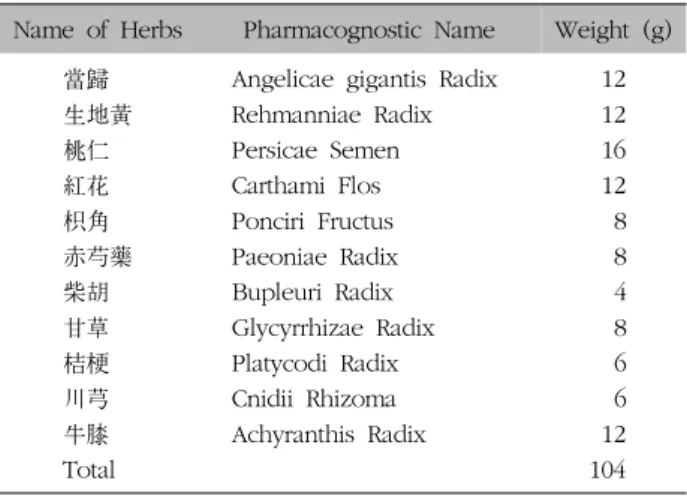
Name of Herbs Pharmacognostic Name Weight (g)
當歸 Angelicae gigantis Radix 12
生地黃 Rehmanniae Radix 12
桃仁 Persicae Semen 16
紅花 Carthami Flos 12
枳角 Ponciri Fructus 8
赤芍藥 Paeoniae Radix 8
柴胡 Bupleuri Radix 4
甘草 Glycyrrhizae Radix 8
桔梗 Platycodi Radix 6
川芎 Cnidii Rhizoma 6
牛膝 Achyranthis Radix 12
Total 104
Table I. Prescription of Hyeolbuchukeo-tang Per Pack 동반질환으로 뇌졸중 관리가 어려워 사회 경제적인 문제
가 되고 있으며2), 연령별 뇌졸중 유병률에서는 50대 1%, 60대 5.9%, 70대 이상 6.5%로 연령 증가에 따라 동반 증 가한다3).
뇌졸중의 위험요인으로 성별, 연령, 유전, 고혈압, 당 뇨, 고지혈증 및 심장질환과 음주, 흡연 등이 있으며, 고 혈압, 당뇨와 고지혈증은 동반질환으로 특히 고혈압이 뇌 졸중 위험요인으로 가장 관련이 높다4).
최근 노인 인구의 증가로 혈관의 노화에 따른 심뇌혈 관질환이 증가하고 있으며, 한의학에서는 관상동맥 질환, 고혈압, 고지혈증, 뇌혈관 질환 등의 병리학적 원인을 瘀 血로 보고 있어 어혈에 대한 병리학적, 임상적인 연구에 대한 의의가 크다고 할 수 있다5).
血府逐瘀湯은 王淸任의 「醫林改錯」6)에서 처음 소개되어 瘀血證을 치료하는 대표적인 처방으로 흉통이나 두통과 같 은 어혈과 관련된 제반 증상에 광범위하게 활용되고 있다7). 혈부축어탕에 대한 실험 연구로는 심근경색 재관류 효 과8), 고지질혈증 억제 효과9,10), 죽상경화증 억제 효과11), 전뇌허혈로 인한 신경세포 손상 보호 효과12), 모세혈관에서 내피 줄기세포의 혈관 형성 촉진 효과13), 혈전색전증으로 뇌경색이 유발된 흰쥐에서 신경세포손상 보호 효과14,15), 죽 상동맥경화증이나 관상동맥 환자를 대상으로 진행된 임
상실험16,17) 등이 있다. 하지만 출혈성 뇌졸중 실험 모델
에서 어혈을 치료하는 약제의 효과에 대한 실험 연구는 적은 실정이다.
이에 본 실험에서는 어혈의 병증에 광범위하게 사용되 는 혈부축어탕이 출혈성 뇌졸중에 미치는 영향을 알아보 고자 교원효소로 유발된 흰쥐의 출혈성 뇌졸중 실험모델 에 혈종, 부종, 뇌연화, 뇌연화 주위세포의 apoptotic cell 및 신생모세혈관증식 등의 변화를 관찰한 결과 다음과 같 은 결과를 얻었기에 보고하는 바이다.
대상 및 방법»»»
1. 재료 및 방법
1) 실험동물
체중 280∼310 g의 수컷 Sprague-Dawley계 흰쥐(샘타 코, 한국)를 사용하였으며 전 실험기간동안 온도 23±1oC,
습도 55±5%로 고정된 항온항습사육장치(대종기기상사, 한국)내에서 사육하였다. 실험기간동안 사료(신촌사료, 한국)와 음수는 자유 급식케 하였다.
2) 약재 및 검액의 제조
실험에 사용한 혈부축어탕의 처방내용은 「醫林改錯」6) 에 의거하였으며, 약재는 ○○대학교 한방병원에서 구입 한 후 엄선하여 사용하였다. 처방의 내용과 분량은 Table I과 같다.
혈부축어탕 4첩 분량을 2,000 ml의 증류수에 1시간 동 안 침지 시킨 후 2시간 30분간 전탕하고, 전탕액을 여과 한 후 감압농축기(EYELA, NE-1001, Japan)로 100 ml로 농축시켰다. 체중 비례로 환산하여 60 kg 성인의 1일 복 용량에 5배에 해당되는 용량인 농축액 1 ml를 흰쥐 마리 당 1일 투여량으로 하였다.
3) 출혈성 뇌졸중 모델 동물의 작성
흰쥐 체중 kg당 50 mg의 pentobarbital sodium (Sigma, USA)을 복강주사하여 마취시킨 후 랫드용 ster- eotaxic appratus (대종기기상사, 한국)에 두부를 고정하 고 두피를 절개하여 두개골을 노출시켰다. Stereotaxic coordinates를 참조하여 결정된 caudate nucleus의 중심 부위에 다음과 같은 방법으로 collagenase (type VII, Sigma Chemical Co., USA)를 주입하였다. 두개골 표면에 서 정수리점(bregma)을 기준으로 우측 3.0 mm, 후측 0.5 mm되는 지점의 두개골을 직경 0.5 mm인 소형 드릴로 천공한 후 미량주입기(10 ul, 26 G, Hamilton, USA)를 깊
이 5.2 mm 자침하여 2 ul의 collagenase (0.5 uint/2 ul) 용액을 주입하여 뇌내 출혈을 유발시켰다. 천공부위 및 두피절개부위를 1% iodine 액으로 소독한 후 봉합하여 항온항습장치내에서 회복하게 하였다.
4) 실험군의 분류
출혈성 뇌졸중 모델 동물의 작성시 정상군(n=12)에는 saline을 주입하였고 대조군(n=12) 및 처치군(n=12)에는 collagenase를 주입하였다. Collagenase 또는 saline 주입 3시간 전에 정상군에는 증류수 1 ml를, 대조군 및 처치군 에는 혈부축어탕 농축액 1 ml를 경구투여 하였으며, 이후 10일간 1일 1회 증류수 또는 혈부축어탕 농축액 1 ml를 경구투여 하였다. 실험 3일 및 실험 10일에 각 군당 6마 리를 희생하여 검사에 이용하였다.
5) 체중의 측정
Collagenase 주입일, 주입 3일 후, 주입 10일 후에 모 든 개체의 체중을 측정하였다.
6) 뇌 무게의 측정
주입 3일 및 주입 10일 후에 각 군당 6마리를 체중 kg 당 50 mg의 pentobarbital sodium을 복강주사하여 마취 시킨 후 0.01 M PBS (pH 7.4)로 10분간 심장관류하고 이어서 4% paraformaldehyde 용액으로 10분간 심장관류 고정 하였다. 고정 후 환추와 후두골 사이의 척수를 절단 하고 뇌를 적출하여 무게를 측정하였다.
7) 혈종의 단면적 측정
주입 3일 후에 적출된 뇌를 4% paraformaldehyde 용 액에 24시간 후고정한 후 자침부위를 기준으로 관상단면 을 만들어 사진촬영하고, 사진상에서의 혈종의 단면적을 칼라영상분석기(Media Cybernetics, Image-Pro Plus, USA)를 이용하여 측정하였다. 주입 10일 후에 적출된 뇌 는 육안적인 병변의 경계가 불명확하여 병변의 단면적 측 정에서는 제외하였다.
8) 뇌연화 면적의 측정
주입 3일 및 주입 10일 후에 적출된 뇌를 일반적인 조 직처리과정을 거쳐 파라핀에 포매하고 4 um의 박절편을 만든 후 hematoxylin-eosin 염색하고, 12.5배의 시야에서
관찰된 뇌 연화(malacia)부위의 면적을 칼라영상분석기를 이용하여 측정하였다.
9) TUNEL 염색 및 연화주위 뇌 실질에서의 apoptotic cell 측정
주입 3일 및 주입 10일 후에 적출된 뇌를 일반적인 조 직처리과정을 거쳐 파라핀에 포매하고 10 um의 박절편 을 만든 후 In Situ Cell Detection Kit (Roche, Germany) 를 이용하여 TUNEL 염색을 실시하였다. 절편을 일반적 인 처리과정을 거친 후 실온에서 proteinase K 용액(20 ug/ml, Roche, Germany)으로 15분간, 3% H2O2 용액으 로 10분간 처리하였다. TUNEL 반응 혼합액으로 1시간동 안 반응시키고 converter-POD 용액으로 30분간 반응시킨 후 DAB (Roche, Germany)로 발색시켰다. 2% methyl green으로 대조염색한 후 polymount로 봉입하여 관찰하 였다. Apoptotic cell의 수는 혈종주변의 연화되지 않은 뇌실질에서 관찰된 세포만을 측정하였다. 연화 병소와 비 교적 뚜렷한 경계를 보이는 뇌실질부위를 1,000×500 um 의 넓이로 두 부위를 선택하여 선택된 영역내에서 TUNEL 염색 양성반응을 보인 세포만을 계수하였다.
10) 병리조직학적 관찰
주입 3일 및 주입 10일 후에 적출된 뇌를 일반적인 조 직처리과정을 거쳐 파라핀에 포매하고 4 um의 박절편을 만든 후 hematoxylin-eosin 염색하여 일반적인 병리조직 학적 변화를 관찰하였다.
11) 통계처리
모든 항목의 결과에 대한 통계는 SPSS (SPSS 9.0 for Windows)를 이용하여 student’s t-test를 실시하여 처리 하였다.
결과»»»
1. 체중의 변화
Collagenase 주입 3일 후의 체중은 대조군 및 처치군 모두 정상군에 비해서 유의하게 감소되었다. 주입 3일 후 의 처치군의 체중은 대조군에 비해서 다소 증가되었으나
Group No. of Animals Body Weight (g)
0 days 3 and 10 days 0 3 10 (days)
Normal 12 6 296.75±7.72* 312.00±7.40 338.83±12.14
Control 12 6 291.83±12.07 285.33±14.50† 311.50±4.76‡
Treated 12 6 294.50±6.20 297.17±9.91§ 328.17±21.22
Normal: Saline injected and distilled water treated. Control: Collagenase injected and distilled water treated. Treated: Collagenase injected and Hyeolbuchukeo-tang treated.
*Mean±SD, †Statistically significant compared with normal group (p<0.01), ‡Statistically significant compared with normal group (p<0.001), §Statistically significant compared with normal group (p<0.05).
Table II. Effect of Hyeolbuchukeo-tang on the Body Weight of Collagenase-Induced Intracerebral Hemorrhagic Rats
Fig. 1. Gross findings of the coronal sectioned rat brains from control group, 3 days after collagenase injection. Note severe hemorrhagic lesions at the right caudate nuclei and adjacent area.
Fig. 2. Gross findings of the coronal sectioned rat brains from treated group, 3 days after collagenase injection. Note severe hemorrhagic lesions at the right caudate nuclei and adjacent area.
Group No. of Animals
Brain Weight (g)
3 10 (Days)
Normal 6 1.93±0.03* 1.98±0.02
Control 6 2.12±0.04† 2.04±0.04‡
Treated 6 1.99±0.04§,∥ 1.97±0.04¶
Normal: Saline injected and distilled water treated. Control:
Collagenase injected and distilled water treated. Treated:
Collagenase injected and Hyeolbuchukeo-tang treated.
*Mean±SD, †Statistically significant compared with normal group (p<0.001), ‡Statistically significant compared with nor- mal group (p<0.01), §Statistically significant compared with normal group (p<0.051), ∥Statistically significant compared with control group (p<0.001), ¶Statistically significant com- pared with control group (p<0.01).
Table III. Effect of Hyeolbuchukeo-tang on the Brain Weight of Collagenase-Induced Intracerebral Hemorrhagic Rats
유의성은 없었다. 주입 10일 후의 체중은 대조군이 정상 군에 비해서 여전히 유의하게 감소되었다. 주입 10일 후 의 처치군의 체중은 대조군에 비해서 증가되었으나 유의 성은 없었다(Table II).
2. 뇌 무게의 변화
Collagenase 주입 3일 후의 뇌 무게는 대조군 및 처치 군 모두 정상군에 비해서 유의하게 증가되었으며, 처치군 은 대조군에 비해서 유의하게 감소되었다. 주입 10일 후 의 뇌 무게는 대조군만이 정상군에 비해서 유의하게 증가 되었으며, 처치군은 대조군에 비해서 유의하게 감소되었 다(Table III).
3. 육안소견 및 뇌 혈종 단면적의 변화
육안소견: collagenase 주입 3일 후의 대조군의 6예 모
두에서 좌침부위의 대뇌피질 및 미상핵과 그 주변에 걸쳐 현저한 출혈이 진행되어 있었으며 출혈의 중심부에는 연 한 황색의 유연한 조직으로 채워져 있었다(Fig. 1). 처치 군의 6예 모두에서도 대조군과 유사한 정도로 출혈이 진
Fig. 3. Gross findings of the coronal sectioned rat brains from control group, 10 days after collagenase injection. Note red hemorrhagic lesions are replaced with brownish colored le- sions at the right caudate nuclei and adjacent area.
Fig. 5. Microscopic findings of the rat brain from control group, 3 days after collagenase injection. Large area of mala- cia(m) with peripheral intact extravasted erythrocytes(e) are shown. H-E stain, Original magnification ×12.5.
Fig. 4. Gross findings of the coronal sectioned rat brains from treated group, 10 days after collagenase injection. Note red hemorrhagic lesions are replaced by brownish colored lesions at the right caudate nuclei and adjacent area. The size and density of brownish colored lesions are decreased than those of control group.
Group No. of
Animals Cross Sectional Area of Brain Hematoma at 3 Days (mm2)
Normal 6 0.00±0.00*
Control 6 16.49±2.22†
Treated 6 14.78±4.71†
Normal: Saline injected and distilled water treated. Control:
Collagenase injected and distilled water treated. Treated:
Collagenase injected and Hyeolbuchukeo-tang treated.
*Mean±SD, †Statistically significant compared with normal group (p<0.001).
Table IV. Effect of Hyeolbuchukeo-tang on the Cross Sectional Area of Brain Hematoma of Collagenase-Induced Intracerebral Hemorrhagic Rats at 3 Days after Collagenase Injection 행되어 있었다(Fig. 2). 주입 10일 후의 대조군의 6예 모
두에서 붉은색의 출혈부위는 모두 사라지고 한계가 불분 명한 갈색의 유연한 조직으로 혈종부위가 대치되었으며 (Fig. 3), 처치군에서는 유연한 조직의 범위 및 색조가 보 다 연하게 관찰되었다(Fig. 4).
뇌 혈종 단면적의 변화: collagenase 주입 3일 후의 육 안 촬영한 뇌 단면의 사진을 칼라영상분석기를 통하여 적 색으로 경계된 혈종의 단면적을 측정한 결과, 처치군이 대조군에 비해서 다소 감소되어 있었으나 통계학적 유의 성은 관찰되지 않았다(Table IV).
4. 뇌 연화 단면적의 변화
Saline을 주입한 정상군의 모든 예에서는 정상적인 조 직학적 소견만이 관찰되었을 뿐, 출혈 또는 연화 등의 소 견은 관찰되지 않았다.
대조군 및 처치군 모두에서 collagenase 주입 3일 후의 뇌 연화 부위는 육안적으로 출혈이 관찰되었던 범위와 유 사하게 관찰되었다(Fig. 5). 대조군 및 처치군 모두에서 주입 10일 후의 뇌 연화 부위는 주입 3일 후의 예에 비해 서 현저히 감소되었다(Fig. 6, 7).
병리조직학적으로 관찰된 뇌 연화 단면적을 칼라영상 분석기를 통해서 측정한 결과, 주입 3일 후의 처치군은 대조군에 비해서 감소되어 있었으나 유의성은 없었다. 주 입 10일 후의 처치군은 대조군에 비해서 유의하게 감소되
Fig. 7. Microscopic findings of the rat brain from treated group, 10 days after collagenase injection. The area of malacia (m) is decreased. Reactive proliferating cells and various sized cavities (*) are shown at the periphery of the malacia. Lateral ventricle (v) is dilated. Compare the area of malacia with Fig.
6. H-E stain, Original magnification ×40.
Fig. 6. Microscopic findings of the rat brain from control group, 10 days after collagenase injection. The area of mala- cia(m) is decreased. Reactive proliferating cells and various sized cavities (*) are shown at the periphery of the malacia.
Lateral ventricle (v) is dilated. H-E stain, Original magnification
×40.
Group No. of Animals
Cross Sectional Area of Brain Malacia (mm2)
3 10 (days)
Normal 6 0.00±0.00* 0.00±0.00
Control 6 10.46±1.14† 1.34±1.19‡
Treated 6 8.81±2.02† 0.27±0.16§,∥
Normal: Saline injected and distilled water treated. Control:
Collagenase injected and distilled water treated. Treated:
Collagenase injected and Hyeolbuchukeo-tang treated.
*Mean±SD, †Statistically significant compared with normal group (p<0.001), ‡Statistically significant compared with nor- mal group (p<0.05), §Statistically significant compared with normal group (p<0.01), ∥Statistically significant compared with control group (p<0.05).
Table V. Effect of Hyeolbuchukeo-tang on the Cross Sectional Area of Brain Malacia of Collagenase-Induced Intracerebral Hemorrhagic Rats
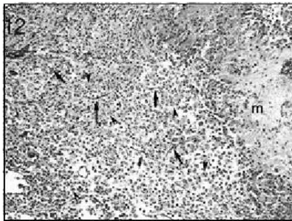
Fig. 9. Photomicrograph of the rat brain from control group, 3 days after collagenase injection. TUNEL positive cells (arrows) are scattered in the central (cm) and peripheral (pm) area of malacia. TUNEL stain, Original magnification ×100.
Fig. 8. Photomicrograph showing positively stained with TUNEL stain. The nuclei of apoptotic cells appear in dark brownish color (arrows). TUNEL stain, Original magnification ×400.
었다(Table V).
5. Apoptotic cell의 수
TUNEL 염색 결과 apoptotic cell은 핵이 진한 갈색으 로 염색되었다(Fig. 8).
주입 3일 후에 대조군 및 처치군의 모든 예에서 뇌연 화가 진행된 중심부 및 변연, 그리고 주위 정상 뇌실질에 걸쳐 TUNEL 염색 양성세포가 산재되어 있었다(Fig. 9).
뇌연화의 중심부 및 변연에서 양성세포가 많이 관찰되었
Group No. of Animals
No. of Apoptotic Cells
3 10 (days)
Control 6 24.33±7.42* 12.33±4.72
Treated 6 20.50±7.01 12.67±4.68
Control: Collagenase injected and distilled water treated. Treated:
Collagenase injected and Hyeolbuchukeo-tang treated.
*Mean±SD.
Table VI. Effect of Hyeolbuchukeo-tang on the Apoptosis in Striatum Adjacent to Malasia of Collagenase-Induced Intracere- bral Hemorrhagic Rats
Fig. 12. Microscopic findings of the rat brain from treated group, 10 days after collagenase injection. Most of proliferative cells are lipid laden macrophages (arrow heads), but the pop- ulation of the cells is more dense than that of the control group. Newly formed capillaries (arrows) are more frequently seen than that of the control group. Compare with the Fig.
11. H-E stain, Original magnification ×100.
Fig. 10. Photomicrograph of the rat brain from control group, 3 days after collagenase injection. Fewer TUNEL positive cells (arrows) are present in the intact brain tissue. The interrupted line indicates the junction of malacia (m) and intact brain tis- sue (b). TUNEL stain, Original magnification ×200.
Fig. 11. Microscopic findings of the rat brain from control group, 10 days after collagenase injection. Most of reactive proliferating cells appeared at the periphery of the malacia are lipid laden macrophages (arrow heads). H-E stain, Original magnification ×100.
으나, 뇌연화 주위의 뇌실질에서는 뇌연화 병소에 인접된 부위에서만 드물게 관찰되었다(Fig. 10).
주입 10일 후에서도 대조군 및 처치군에서의 TUNEL 염색 양성세포의 분포부위는 주입 3일에서와 유사하였으 나 그 수가 감소되어 있었다.
주입 3일 및 10일 모두에서 대조군과 처치군에서의 유 의성 있는 변화는 없었다(Table VI).
6. 병리조직학적 소견
Collagenase 주입 3일 후: 대조군의 모든 예에서 colla- genase 주입부위 뇌실질의 현저한 연화가 진행되어 있었 다. 연화된 중심부위의 대부분의 적혈구들은 흡수되어 있 었으나 연화의 변연부와 정상 뇌실질의 경계부위에는 여 전히 적혈구가 현저히 유출되어 띠를 이루고 있었다(Fig.
5). 처치군의 모든 예에서의 소견도 대조군과 유사한 정 도로 관찰되었다.
Collagenase 주입 7일 후: 대조군의 모든 예에서 혈종 의 중심부는 여전히 연화되어 있었으나 유출된 적혈구의 대부분은 흡수되어 있었다(Fig. 6). 연화소의 주변에서는 지방을 탐식한 대식세포(lipid laden macrophage, gitter cell)가 침윤 증식되어 있었으며, 정상 뇌실질과 연화부의 경계에는 한계가 불분명한 다양한 크기의 동공(cavity)이 형성되어 있었다(Fig. 11). 처치군에서 관찰된 소견도 대
조군에서와 유사한 정도로 관찰되었으나, 대조군에 비해 서 연화소 주변에 증식된 대식세포가 보다 치밀하게 증식 되어 있었고, 성상세포 및 신생모세혈관의 증식이 보다 빈번하게 관찰되었다(Fig. 12).
고찰»»»
뇌졸중은 일반적으로 뇌혈관의 허혈성 또는 출혈성 장 애로 인해 뇌신경이 손상되어 반신마비, 반신의 감각장 애, 구안와사, 실어증, 구음장애, 연하장애, 대소변실금, 시야장애, 의식상태 및 정신상태의 변화, 인지장애 등 여 러 가지 다양한 뇌기능 장애의 임상표현이 나타나는 것을 말한다18).
출혈성 뇌졸중은 지속적인 만성 고혈압과 관련되어 자 발적으로 발생되거나 뇌혈관질환, 뇌종양, 두부외상 등과 관련되어 속발성으로 발생되며, 발생 후 급격한 두개내압 의 상승과 뇌기능부전으로 환자의 40%가 30일 이내에 사 망하거나 회복되더라도 마비, 뇌신경장애 등의 심각한 장 애를 남기게 된다19).
출혈성 뇌졸중의 서양의학적 치료로는 항혈압제로 혈 압을 조절하면서 20% mannitol, furosemide, 고장식염수 등으로 두개내압을 하강시키며 필요시에는 혈종제거술을 시행한다20).
어혈은 전신의 혈액순행이 원활치 못하거나 혈액이 혈 관에서 이탈하고 정체되어 병리적 산물이 된 것으로21), 심뇌혈관질환은 한의학적으로 혈맥의 흐름을 방해하는 어혈과 관련이 깊게 인식하고 있어 어혈증을 치료하는 것 이 그 치료 목표가 된다22). 뇌질환에서는 뇌경색 뿐만 아 니라 뇌출혈도 병변 부위에 혈액이 정체된다는 관점에서 어혈의 일종으로 볼 수 있다. 반면에 서양의학에서는 항 혈전제를 사용하여 혈액의 응집을 예방하고, 뇌졸중 및 심장질환의 위험성을 줄이고자 하지만, 다양한 항혈전제 대부분은 위장 내의 증상과 출혈 등 부작용을 가지고 있 어 안전하고 효과적인 약물의 개발이 필요한 실정이다23). 혈부축어탕은 「醫林改錯」6)에서 처음 기록되어 도인, 당귀, 생지황, 홍화, 우슬, 지각, 적작약, 길경, 천궁, 시 호, 감초로 구성되어 있으며, 一切의 氣滯血瘀證을 通治24) 하여, 임상에서 죽상경화증이나 고지질혈증 등으로 인한 심혈관 질환에 빈용되는 처방 중 하나이다25). 혈부축어탕
이 뇌의 수복에 어떠한 효과가 있는가를 알아보기 위하여 Rosenberg26)의 실험방법에 준한 서27) 등의 방법으로 출 혈성 뇌졸중으로 야기되는 혈종, 부종, 뇌연화, 뇌연화 주 위세포의 apoptotic cell, 신생모세혈관 증식 등의 변화를 관찰하였다.
본 연구에서 collagenase 주입 3일 후 뇌 무게는 대조 군과 처치군 모두 정상군에 비해서 유의하게 증가되었으 며, 처치군은 대조군에 비해서 유의하게 감소되었다.
Collagenase 주입 10일 후의 뇌 무게는 대조군만이 정상 군에 비해서 유의하게 증가되었으며, 처치군은 대조군에 비해서 유의하게 감소되었다. 이는 최소한 3일째부터 혈 부축어탕이 뇌의 수복을 활성화시키는 효과가 인정되며, 혈전 생성을 억제시키고 혈액순환을 촉진시킬 수 있으며 신생모세혈관의 증식을 통한 손상된 뇌조직의 수복에 기 여할 수 있을 것으로 사료된다.
뇌부종은 세포외 공간의 수분이 단순히 세포내로 이동 한 것이나 뇌실질의 수분 함량의 증가없이 뇌혈류량의 증 가나 뇌척수액 공간의 증가로 인한 용적 증가가 아닌, 뇌 실질내 수분 함량의 비정상적인 증가로 인해 뇌조직의 용 적이 증가한 상태라고 정의하는데28), 본 실험에서는 col- lagenase 주입 3일 후의 대조군, 처치군 모두 유사하게 현저한 출혈이 진행되어 있었으며 출혈의 중심부에는 연 한 황색의 유연한 조직으로 채워져 있었다. 주입 10일 후 의 대조군은 붉은색의 출혈부위는 모두 사라지고 한계가 불분명한 갈색의 유연한 조직으로 혈종부위가 대치되었 으며, 처치군에서는 유연한 조직의 범위 및 색조가 보다 연하게 관찰되었다. 육안 촬영한 뇌 단면의 사진을 칼라 영상분석기를 통하여 적색으로 경계된 혈종의 단면적을 측정한 결과, 처치군이 대조군에 비해서 다소간 감소되어 있었으나, 통계학적 유의성은 관찰되지 않았다.
뇌손상에 대한 회복도는 손상 후 혈관 신생, 대사장애 개선, 뇌세포막 펌프작용의 활성화 등에 의해 신경세포의 손상을 가역적으로 소생시켜주는 뇌혈관계 자가조절능을 어느 정도 유지하느냐에 달려있는데29,30), 뇌 연화의 변화 를 관찰하면 대조군과 처치군 모두에서 collagenase 주입 3일 후 뇌 연화 부위는 육안적으로 출혈이 관찰되었던 범 위와 유사하게 관찰되었다. 대조군 및 처치군 모두에서 주입 10일 후의 뇌 연화 부위는 주입 3일 후의 예에 비해 서 현저히 감소되었다.
병리조직학적으로 관찰된 뇌 연화 단면적을 칼라영상
분석기를 통해서 측정한 결과, 주입 3일 후의 처치군은 대조군에 비해서 감소되어 있었으나 유의성은 없었고, 대 조군과 처치군이 유사한 정도로 collagenase 주입부위 뇌 실의 현저한 연화가 진행되어 있었다. 연화된 중심부위의 대부분의 적혈구들은 흡수되어 있었으나 연화의 변연부 와 정상 뇌실질의 경계부위에는 여전히 적혈구가 현저히 유출되어 띠를 이루고 있었다. 주입 10일 후의 처치군은 대조군에 비해서 유의하게 감소되었고, 대조군과 처치군 이 유사한 정도로 혈종의 중심부는 여전히 연화되어 있었 으나 유출된 적혈구의 대부분은 흡수되어 있었고, 연화된 곳의 주변에서는 지방을 탐식한 대식세포가 침윤 증식되 어 있었으며, 정상 뇌실질과 연화부의 경계에는 한계가 불분명한 다양한 크기의 공동이 형성되어 있었으나 처치 군은 대조군에 비해서 연화소 주변에 증식된 대식세포가 보다 치밀하게 증식되어 있었고, 성상세포 및 신생모세혈 관의 증식이 보다 빈번하게 관찰되었다.
뇌연화는 10일 후에 단면적에서 처치군이 대조군에 비 해 유의성있게 감소되었는데 뇌 무게가 3일째부터 처치 군이 대조군에 비해 유의성있게 감소된 변화와 비교하면 꼭 일치하는 변화는 아니라고 생각된다.
정상적인 세포에서는 세포의 성장, 사멸이 세포의 수를 조절하고 노쇠하거나 손상된 세포를 없애는 일상적인 과 정을 가지는데31), 일반적으로 염색질에서는 거의 변화 없 이 세포 크기만 증가하는 괴사세포와 달리 apoptotic cell 의 크기는 오그라들고 초기부터 염색질구조가 대체되는 것이 보이고 기포현상(blebbing)이 보이는 프로그램된 세 포 죽음을 진행한다32).
Apoptotic cell의 관찰에서는 주입 3일 후에 대조군 및 처치군에서 뇌연화의 중심부 및 변연에서 TUNEL 염색 양성세포가 많이 관찰되었으나, 뇌연화 주위의 뇌실질에 서는 뇌연화 병소에 인접된 부위에서만 드물게 관찰되었 다. 주입 10일 후에서도 대조군 및 처치군에서의 TUNEL 염색 양성세포의 분포부위는 주입 3일에서와 유사하였으 나 그 수가 감소되어 있었다. 주입 3일 및 10일 모두에서 대조군과 처치군에서의 유의성 있는 변화는 없었다.
본 실험에서는 출혈성 뇌졸중이 발생한 각 군의 체중 과 단면적, apoptotic cell의 관찰로 임상증상과 예방을 표현하려고 했으나 향후에는 출혈성 뇌졸중 후 임상증상 의 회복의 계획과 함께 혈부축어탕 전탕액 용량의 변화 및 다른 복합합방의 투여를 통해 보다 구체적인 실험이
가능하리라 생각된다. 또한 본 실험은 혈부축어탕 투여 후 apoptosis된 신경세포의 변화 상황을 신경재생과 관련 된 지표와 신경세포의 현미경적 관찰에 그쳤지만 향후 뇌 출혈이 발생된 실험동물의 운동 형태학적 관찰을 통한 평 가 연구가 필요할 것으로 보여진다.
이에 혈부축어탕은 출혈성 뇌졸중 병태모델에서 출혈 로 야기되는 부종을 억제하며, 신생모세혈관의 증식을 통 한 혈종의 흡수와 대식세포 및 성상세포의 증식을 통한 손상된 뇌조직의 수복효과가 인정되므로 임상에서 치료 에 유효하게 적용될 수 있을 것으로 기대된다.
결론»»»
혈부축어탕이 출혈성 뇌졸중에 미치는 영향을 확인하 기 위해서 흰쥐의 오른쪽 미상핵에 collagenase를 주입하 여 뇌내 출혈을 유발시킨 후 혈부축어탕 감압농축액을 투 여하였다. Collagenase 주입 3일 및 10일 후에 체중, 뇌 무게, 혈종의 크기, 연화소의 크기, apoptotic cell의 수 및 병리조직학적 변화를 관찰하여 다음과 같은 결과를 얻 었다.
1. 실험 3일 후 뇌무게(g)는 처치군(1.99±0.04)이 대 조군(2.12±0.04)에 비해서 유의성(p<0.001) 있게 감소 되었으며, 실험 10일 후의 뇌무게도 처치군(1.97±0.04) 이 대조군(2.04±0.04)에 비해서 유의성(p<0.01) 있게 감소되었다.
2. 실험 10일 후의 뇌연화 단면적(mm2)은 처치군 (0.27±0.16)이 대조군(1.34±1.19)에 비해서 유의성(p
<0.05) 있게 감소되었다.
3. 실험 3일 및 10일 후의 연화소 주위 정상 뇌실질에 서의 apoptotic cell의 수는 처치군과 대조군간에 유의성 있는 변화는 없었다.
4. 실험 10일 후의 연화소 주위에서의 대식세포(gitter cell), 성상세포(astrocyte) 및 신생모세혈관 증식이 처치 군이 대조군에 비해서 뚜렷하였다.
이상의 성적으로 미루어 출혈성 뇌졸중 병태모델에서 혈부축어탕은 출혈로 야기되는 부종을 억제하며, 신생모 세혈관의 증식을 통한 혈종의 흡수와 대식세포 및 성상세 포의 증식을 통한 손상된 뇌조직의 수복에 기여하리라 사 료된다.
References»»»
1. Statistics Korea. 2013 Cause of Death Statistics.
2. Reeves MJ, Bushnell CD, Howard G, Gargano JW, Duncan PW, Lynch G, Khatiwoda A, Lisabeth L. Sex dif- ferences in stroke:epidemiology clinical presentation, medical care and outcomes. The Lancet Neurology.
2008;7(10):915-26.
3. Korea Institute for Health and Social Affairs. 2011 Korean National Health and Nutrition Examination Survey. Seoul. 2011.
4. Park TH, Kim MK, Lee KB, Park JM, Lee SJ, Jung KH, Cho YJ, Lee JS, Lee JY, Bae H J. Prevalence of Risk Factors for Ischemic Stroke in Korean:A Systematic Review. Journal of Korean Neurology Assoc. 2009;27(1):
19-27.
5. Park MS, Kim YM. Study on Clinical Diseases of Blood Stasis Pattern. HFS. 2013;21(1):1-15.
6. Wang QR. YiLinGaiCuo. Beijing:China chinese medicine pub. 1995:24-8
7. National Commission of Chinese Pharmacopoeia.
Pharmacopoeia of the People's Republic of China vol. 1.
Beijing:China Medical Science and Technology Press.
2010:693.
8. Tang D, Liu Z, Zhang H, Sun M, Sui Y. Protective ef- fects of xuefu zhuyu decoction on myocardium ischemia reperfusion injury in rats. China J Chin Mater Med.
2010;35:3077-9.
9. Song X, Wang , Wang P, Tian N, Yang M, Kong L. 1H NMR-based metabolomics approach to evaluate the ef- fect of Xue-Fu-Zhu-Yu decoction on hyperlipidemia rats induced by high fat diet. J of Pharmaceutical and Biomedical Analysis. 2013;78-79(5):202-10.
10. Pi CM, Chong MS, Kim HJ, Cho HE, Choi YH, Lee KN.
Effect of Hyolbuchukeo-tang Extracts on Blood and Liver of Hyperlipidemia Rats Induced by High Fat Diet. J of Society of Preventive Korean Medicine. 2009;13(1):41-58.
11. Lee BJ, Yun SY, Park HW, Park JH, Jo IY, Lee JS, Lew JH.
Effects of Concurrent Administration of Hyeolbuchukeo- tang and Aspirin on Atherosclerosis in the ApoE(-/-) Mouse. J Korean Oriental Med. 2011;32(1):164-174.
12. Cho EH, Kim YG, Kwon JN. Neuroprotective Effects of Hyulbuchookau-tang on Global Cerebral Ischemia of the Rats. J of Korean Oriental Medicine. 2007;28(2-70):44-53.
13. Gao D, Wu L, Jiao Y, Chen W, Chen Y, Kaptchuk TJ, Lu B, Song J, Chen K. The effect of Xuefu Zhuyu de- coction on in vitro endothelial progenitor cell tube formation. Chin J Integr Med. 2010;16:50-53.
14. Lee JJ, Hsu WH, Yen TL, Chang NC, Luo YJ, Hsiao G, Sheu JR. Traditional Chinese medicine Xue-Fu-Zhu-Yu de- coction, potentiates tissue plasminogen activator against
thromboembolic stroke in rats, J Ethnopharmacol. 2011;
134:824-30.
15. Cho EH, Kim YG, Kwon JN. Neuroprotective Effects of Hyulbuchookau-tang on Global Cerebral Ischemia of the Rats. J of Korean Oriental Medicine. 2007;28(2-70):44-53.
16. Chu FY, Wang J, Yao KW, Li ZZ. Effect of Xuefu Zhuyu capsule on the symptoms and signs and health-related quality of life in the unstable angina patients with blood-stasis syndrome after percutaneous coronary inter- vention: a randomized controlled trial. Chin J Integr Med. 2010;16:399-405.
17. Hoa B, Cao WD, Yang T. Intervention of Xuefu Zhuyu oral liquid on expression of adhesion molecule CDllb/
CD18 in neutrophils in patients with arteriosclerosis obliterans. Chin J Integr Med. 2006;26:125-7.
18. DeLisa JA. Rehabilitation Medicine Principles and prac- tice (2nd edition). Philadelphia: B. Lippincott company.
1993:183-4, 801, 802.
19. Qureshi AI, Mendelow AD, Hanley DF. Intracerebral haemorrhage. The Lancet. 2009;373(9675):1632-644.
20. Korean Stroke Society. Stroke. Seoul : E-public. 2013 ; 1 : 165-168.
21. Heo J. Donguibogam. Seoul:Namsandang. 1983:372, 373.
22. Choi SH, Ahn KS, Moon JJ, Park JA. A Study on the Effect of Samchilgeun on Thrombosis and elevated Blood Viscosity. J of Oriental Medical Pathology. 1992;
7(1):163-170.
23. Cannon CP, Rhee KE, Califf RM, Boden WE, Hirsch AT, Alberts MJ, Cable G, Shao M, Ohman EM, Steg PG, Eagle KA, Bhatt DL. Current use of aspirin and antith- rombotic agents in the United States among outpatients with atherothrombotic disease(from the Reduction of Atherothrombosis for Continued Health [REACH]
Registry). Am J Cardiol. 2010;105:445-52.
24. Shanghai University of Traditional Chinese Medicine.
Fangjixue. Xianggang:Shangwuyinshuguan. 1977:166, 177.
25. Liu1 L, Cheng Y, Zhang H. Phytochemical Analysis of Anti-atherogenic Constituents of Xue-Fu-Zhu-Yu-Tang Using HPLC-DAD-ESI-MS. Chemical and Pharmaceutical Bulletin. 2004;52(11):1295-301.
26. Rosenberg GA, Mun-Bryce S, Wesley M, Kornfeld M.
Collagenase-induced intracerebral hemorrhage in rat.
Stroke. 1990;21(5):801-7.
27. Seo IB, So GS, Kim GH. Pathologic observation induced by collagenase cerebrum hemorrhage of the rats. Semyung Oriental Medicine Institute. 1998;1:25-36.
28. The Korean Neurosurgical Society. Neurosurgery.
Seoul:Joongang Culture Co. 2001;2:137-8. 263.
29. Hong SS, Son BC, Choi BG, Kim EN, Kim BS, Park CK, Choe BY, Kim MC, Kang JK. Metabolic Changes in Pericontusional Edematous Areas in Mild Head Injury Evaluated by Proton MRS. J Korean Neurosurg Soc.
2000;29(9):1233-7.
30. Seong GG. Bibliographic Study on the Function of the Brain on the Basis of Zangxang Theory. The Journal of Korean Medicine. 1995;16(1):468-74.
31. Kim GS. Structural chartacterization of protein in apopto-
sis and cell cycle. Seoul:Korea Institute of Science and Technology. 2000:75-9.
32. Michael DJ, Nicola JM. Apoptosis. New York:Oxford University Press. 2002:1-16.