호 르 몬 무 첨 가 배 양 액 에 서 생 쥐 Pre-antral Follicles 의 체 외 성 장 과 난 포 강 형 성
대구대학교 축산학과1, 경북대학교병원 산부인과학교실2
박 기 상1,2 김 주 환1 이 택 후2 송 해 범1 전 상 식2
Antrum Formation a n d Outgrowth In Vitro o f M o u s e Pre-antral Follicles Cultured in Media w i t h o u t Hormones
K e e Sang Park1,2, Ju H w a n Kim1, T a e k H o o L e e2, H a i Bum Song1 a n d Sang Sik C h u n2
1Department of Animal Science, Taegu University, Kyungbuk
2Department of Ob/Gyn, Kyungpook National University Hospital, Taegu
이 논 문 은 2 0 0 0 년 도 대 구 대 학 교 학 술 연 구 비 의 지 원 에 의 해 수 행 되 었 음 .
Objective: Mouse pre-antral follicles require the addition of gonadotropins (Gns) to complete maturation and ovulation of oocyte and antrum formation in vitro. However, we tried examination of in vitro growth of mouse pre-antral follicles in medium without Gns and/or phygiological factors. And also, pre-antral follicles were isolated from ovaries by mechanical method. Our present studies were conducted to evaluated on the growth of follicles and intra-follicular oocytes and antrum formation in vitro of mouse pre-antral follicles.
M e t h o d s : Pre-antral follicles (91-120 ㎛) were isolated mechanically by fine 30G needles not using enzymes from ovaries of 3-6 week-old female ICR mice. Isolated pre-antral follicles were cultured in 20 ㎕ droplets of TCM (n=17; follicles: 107.8±1.58 ㎛; oocytes:
57.9±1.2 ㎛) or MEM (n=12; follicles: 109.3±2.53 ㎛; oocytes: 55.4±1.6 ㎛) under mineral oil on the 60 ㎜ culture dish. All experimental media was supplemented with 10%
FBS without Gns and/or physiological factors. Pre-antral follicles were individually cultured for 8 days. Antram formation and outgrowth of pre-antral follicles and intra-follicular oocytes were evaluated using precalibrated ocular micrometer at ×200 magnifications during in vitro culture. Results were analyzed using combination of Student's t-test and Chi-square, and considered statistically significant when P<0.05.
Results: Antrum formation had started in two culture media on day 2. On day 8, antrum formation had occurred in 58.3% (7/12) of pre-antral follicles cultured in DMEM, but only in 23.5% (4/17) of those cultured in TCM (P=0.0364). Outgrowth of pre-antral follicles and intra-follicular oocytes were observed on day 4 and 8. On day 4, follicular diameter was similar (P=0.1338) in TCM (119.4±2.58㎛) and MEM (125.4±4.52㎛). However, on
day 8, diameters of pre-antral follicle cultured in MEM (168.9±17.29㎛) were significantly bigger (P=0.0248) than that in TCM (126.7±4.28㎛). On day 4 and 8, diameters of intra-follicular oocytes were similar in TCM (67.1±1.3 and 72.4±0.9 ㎛) and MEM (65.2±1.7 and 73.3±1.5 ㎛), respectively.
Conclusions: We can conform that medium without Gns and/or physiological factors can be used for in vitro antrum formation and outgrowth of pre-antral follicles and intra-follicular oocytes in mouse. In conclusion, MEM supplemented with FBS can be used for growth in vitro of mouse pre-antral follicles isolated mechanically.
K e y words: Mouse pre-antral follicles, Intra-follicular oocytes, Media, In vitro growth, Antrum formation
생쥐 난소에서 분리한 pre-antral follicles을 체외에서 성장시킬 수 있는 배양기법이 많이 소 개되고 있으며, 신생 생쥐의 난소에 효소 처리를 하여 회수한 pre-antral follicles로부터 산자를 획득하는 배양체계도 보고되고 있다.1 생쥐의 난포를 체외에서 성장시키는 방법은 두 가지로 나눌 수 있는데, 한 가지는 효소를 이용하여 분리된 oocyte-granulosa cell 복합체를 배양하는 것이고, 또 한 가지는 기계적인 방법으로 분리된 pre-antral follicle을 배양하는 방법이다. 첫 번째 밥법 은 Eppig와 Schroeder2가 발정 전 생쥐 난소에 효소를 처리하여 분리한 granulosa-oocyte 복 합체를 collagen membranes에서 10일 동안 배양하여 산자를 생산할 수 있었다. 또한, Torrance 등3과 Carroll 등4은 이것들을 collagen gels에 끼워 넣어 배양하는 복합적인 방법이지만, Eppig 와 Schroeder2의 방법과 비슷하다고 할 수 있다. 두 번째 방법으로는 많은 연구자들5-12에 의해 기계적인 방법으로 분리된 pre-antral follicle의 체외배양이다. 대부분의 연구자들은 단기간 배양 (4-6일)을 하였고, membrane 위에서 개별적으로 또는 그룹으로 배양하거나,5 난포의 형태를 보 존하기 위해서 난포를 transpose 시키면서 배양하였다.7-12
생쥐의 pre-antral follicles을 배란 단계 이상으로 체외성장시킨 다음 배란된 성숙난자를 체 외에서 수정시키는 일련의 일들을 성공적으로 수행하기 위해서는6,10,13 난포 내에 있는 난자의 핵 및 세포질 성숙을 유도해야 되는데, 이를 위해서는 배양액 내에 serum 및/또는 FSH의 첨가가 필 수적이고 이들을 배란시키기 위해서는 LH의 작용이 잇따라야 된다.6
난포의 체외성장에는 혈청, 단백질, growth factors(ITS, EGF 등), gonadotropins(FSH, LH 등) 등의 다양한 물질과 hypoxanthine, dibutyryl-cyclic AMP (dbcAMP) 또는 3-isobutyl-1- methyxanthine (IBMX)와 같은 감수분열 억제인자를 배양액에 첨가하여 체외성장을 시키고 있 다.2,4,8,9,14,15,16
그러나 pre-antral follicle을 체외에서 회수하는 방법과 배양기법이 매우 까다롭고 복잡하기 때문에 재현하기가 쉽지 않을 뿐만 아니라 체외성장에 미치는 효과도 연구자마다 상이하게 나타
나고 있다. 또한 심층적인 연구를 하기 위한 모델로써의 기본 배양액 및 배양환경에 대한 자료가 제한적인 것이 사실이고, 난소에서 분리한 난포를 체외에서 배양하기 위해 연구가 지속적으로 이 루어지고 있으나 체외성장을 위한 최적의 배양 환경이 확립되어 있지 않은 실정이다.
따라서 본 연구에서는 난포의 체외성장에 미칠 수 있는 가장 기초적인 조건인 배양액에 따른 체외성장의 효과를 보기 위하여 실시하였으며, 호르몬이 첨가되지 않은 배양액에서 일어날 수 있 는 난포의 양상을 볼 수 있는 기초 자료가 될 것이다.
연 구 대 상 및 방 법
1. 공시동물
본 연구에서 pre-antral follicle의 회수를 위하여 3-6 주령 사이 ICR 계통의 암 생쥐를 사용 하였다. 이들은 온도(20-22℃) 와 명암(12 hrs : 12 hrs)이 조절되는 곳에서 사료와 물은 무제한 으로 급여하면서 사육하다가 실험에 사용하였다.
2. 배양액의 준비
난소에서 분리한 난포는 Ham's F-10에 10% fetal bovine serum (FBS; 26140-079, Gibco, USA)를 첨가한 배양액에서 세척하였다. 난포의 체외배양은 Tissue Culture Medium 199 (TCM; 11150-059, Gibco, USA) 또는 Dulbecco's Modified Eagle Medium (MEM;
11966-025, Gibco, USA)에 10% FBS 을 첨가한 배양액에서 실시하였다.
모든 배양액은 0.5% antibiotics (Streptomycine sulfate, S-9137; Penicinine-G, P-3032, Sigma, USA)를 첨가한 다음, 삼투압 측정기 (Osmomat 030, Gonatec, Gemany)를 이용하여 삼 투압을 280 mOs㏖/㎏로 보정하고, 0.2㎛의 여과기 (SLGV R25 LS, Millipore, France)로 제균하 면서 14 ㎖ tube (2001, Falcon, USA)에 분주한 다음 4℃로 조절된 냉장고 (3682, Forma, Japan)에서 보관하다가 이용하였다. 제조 후 4주가 경과한 배양액은 실험에서 제외하였다. 배양액 은 최소한 6시간 이상 37℃, 5% CO2 배양기 (3158, Forma, USA)에서 전 배양한 다음 실험에 이용하였다.
3. 난포의 분리
생쥐를 경추탈골법으로 도살한 후 복부를 개복하여 나팔관과 지방이 따라오지 않도록 주의하
면서 난소를 적출하였다. 적출된 난소는 소독된 거즈 위에서 난소 표면에 묻어 있는 이물질과 혈 액을 깨끗이 제거하였다. 1㎖의 washing medium이 들어있는 watch glass로 난소를 옮겨놓은 다 음 현미경 하에서 forceps과 30G syringe needle(320310, BD, USA)을 이용하여 여분의 지방 조직을 제거하고 나서, Fig. 1A와 같이 기계적인 방법으로 난소에서 난포를 분리하였다. 분리된 pre-antral 단계의 난포는 pasteur pipette으로 회수하여 washing medium이 들어 있는 2-well dish에서 3회 세척하고 나서, 체외배양에 사용하였다.17
4. 난포의 배양
(1) 난포의 배양과 관찰
분리된 난포 중에서 91-120 ㎛ 크기의 것만을 선별하여 각각의 배양액으로 옮겨 체외 성장 을 유도하였다. 배양액은 60 ㎜ culture dish (3002, Falcon, USA)의 바닥에 20 ㎕ 소적을 만든 다음mineral oil (M-8410, Sigma, USA)을 덮어 제조하였다. 한 개의 소적에 한 개의 난포 만을 개별적으로 옮겨 배양하였다. 난포강의 형성은 배양 8일까지 매일 관찰하였다.
(2) 난포 및 난포내 난자의 크기측정
난포의 크기는 난포와 theca cell의 경계부분에서부터 반대편 경계까지와, 난포내 난자의 장 축과 단축을 측정한 후 그 길이를 더하고 나서 2로 나눈 값으로 하였다. 난포의 직경은 배양 4일 과 배양 8일째에 현미경 (×200)의 접안렌즈에 부착된 micrometer로 측정하였다.
(3) 배양액의 교환
난포를 체외배양하는 동안 48시간 간격으로 각 소적에서 전체 양의 1/2인 10㎕를 각각의 배 양액과 동일한 신선 배양액(전 배양을 실시한)으로 교체해 주었다. 이때에는 난포 또는 세포에 손
상을 주지 않도록 현미경을 보면서 실시하였다.
5. 통계처리
난포의 체외배양에서 배양액에 따른 난포와 난포내난자의 체외성장 비교는 Student's t-test 와 Chi-square (χ2-test)를 병용하였고, 난포강 형성의 비교는 Chi-square (χ2-test)를 실시하여 5% 유의 수준에서 검정하였다. 표준 오차율은 ±SEM으로 나타내었다.
결 과
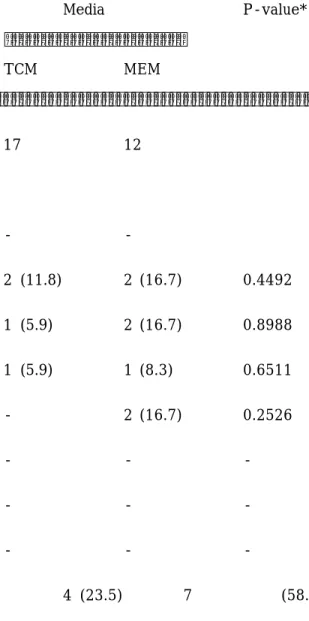
Pre-antral follicle을 각기 다른 배양액에서 체외성장을 유도하면서 난포강 형성에 미치는 영 향을 조사한 결과는 Table 1에 제시하였다.
난포강의 출현 시기는, TCM에서는 배양 2일 (n=2), 3일 (n=1) 및 4일 (n=1), MEM에서는 배양 2일 (n=2), 3일 (n=2), 4일 (n=1) 및 5일 (n=2)로써, 호르몬을 첨가하지 않은 배양액에서 pre-antral follicle의 난포강의 형성을 유도할 수 있었고, 배양 2-5일에 관찰하였다 (Table 1).
배양액에 따른 난포강 형성율을 보면, TCM에서는 17개 중에서 4개 (23.5%)가, MEM에서는 12 개 중에서 7개 (58.3%)가 형성되어, MEM이 TCM 보다 유의하게 (P=0.0364) 높았다. 그러나, 개 별 pre-antral follicle 내에 형성된 난포강의 크기나 난포강의 수에 대한 조사는 본 실험의 결과 에 포함시키지 않았다.
각각의 배양액에서 체외성장을 유도한 pre-antral follicle은 배양 4일과 8일에 난포와 난포 내 난자의 직경을 관찰하여 Figure 2에 각각 나타내었다.
실험에 이용한 pre-antral follicle의 평균 직경은 TCM에서는 107.8 ± 1.58㎛, MEM에서는 109.3 ± 2.53㎛였다. 배양 4일 째에 pre-antral follicle의 직경을 살펴보면, TCM에서는 119.4
± 2.58㎛, MEM에서는 125.4 ± 4.52㎛로써 두 군에서 비슷한 결과를 나타내었다 (P=0.1338).
그러나 배양 8일 째를 보면, TCM에서는 126.7 ± 4.28㎛, MEM에서는 168.9 ± 17.29㎛로 성장 하여 배양기간이 길어지면서 MEM에서 배양하는 것이 TCM에서 보다 성장에 효과적임을 관찰하 였다 (P=0.0248). 배양 당일 난포내 난자의 평균 직경은 TCM에서는 58 ± 1.2㎛, MEM에서는 55 ± 1.6㎛였다. 배양 4일과 8일에 난자의 평균 직경을 보면 TCM에서는 67.1 ± 1.3㎛와 72 ± 0.9㎛였고 MEM에서는 65.2 ± 1.7㎛와 73 ± 1.5㎛로써, 실험군 간에 차이가 나타나지 않았다.
고 찰
Roy와 Greenwald14,15는 hamster에서 small primary와 secondary 단계의 난포를 체외에서 성장시키기 위해서는 FSH와 LH가 필요하고, Katska와 Ry ka16는 FSH, ITS(insulin / transferrin / selenium), L-glutamine 그리고 sodium pyruvate가 난포의 발달에 관여한다고 하 였다. 생쥐 난포를 체외 성장, 배란 및 체외수정을 거쳐 산자 생산까지 이르는 일련의 배양방법이 소개되고 있으나, 7,10,17 체외에서 난포의 성장과 난포내 난자를 성숙시키기 위해서는 serum과 FSH를 첨가해야되고, 체외 배란을 유도하기 위해서는 최종적으로 LH를 첨가해야된다.7 Wright 등18은 난소조직을 체외배양 할 때 10%의 human serum이 함유된 α-minimum essential medium(α-MEM), Waymouth's, Earle's balanced salt solution(EBSS)을 비교한 연구에서 최초 10일 동안의 배양에서 난포는 α-MEM에서 성장의 initiation과 성장율이 증가하였고, α-MEM에 첨가된 300 mIU/㎖의 FSH가 난포의 성장율은 증가시키고, atresia는 현저하게 감소시킨다고 하 였다. 또한 난소 조직 내의 난포는 배양액에 serum만이 첨가된 것보다는 human serum albumin(HSA)과 ITS가 첨가되면 성장이 현저히 증가한다고 하여, 배양액에 FBS 외에 어떠한 물 질도 첨가하지 않고 생쥐 pre-antral follicle에서 난포강 형성(Fig. 1C)과 난포의 성장(Fig. 2A) 을 일으킨 본 실험과는 상반된 결과를 나타내었다. 그러나 배양액에 FSH 또는 ITS를 첨가하지 않아 더 좋은 효과를 낼 수 있을 지에 대해서는 연구가 계속되어야 한다.
사람에서는 처음으로 Roy 와 Treacy19가 난소에 효소를 처리하여 분리한 pre-antral follicle 을 체외 성장시켰고. Abir 등20은 효소처리방법으로 사람의 난소에서 분리한 초기 단계의 난포를 collagen gels에서 배양하였으나 성장하지는 않았다고 하였다. 이 외에도 rat,21,22 hamster,15,23 생쥐24에서도 난소에 효소를 처리하여 pre-antral follicle을 분리하고 있지만, 난소 조직을 효소에 노출시켜 난포를 분리할 때 난포 조직이 효소에 노출되는 시간이 길어지게 되므로 정상적인 체외
성장이 저해되고 E2 생산에 필수적인 theca layer가 손상되어 결과적으로 난포의 발생능력이 떨 어지게 된다.25,26 포유동물 난소에 효소를 처리하여 분리된 난포들은 소적으로 만든 배양액에서 장시간 동안 배양하면 난포조직이 이완되므로 gel을 이용한 체외배양이 많이 이용되고 있는데, theca-free 난포를 체외에서 antral 단계까지 성장시킬 때 agar gel27과 matrigel28이 일반적으로 이용되고 있다. Collagen gels에서는 pre-antral 단계까지 성장이 유도되었지만,29 oocyte를 성숙 시키지는 못하였다.30 또한 2개 또는 여러 개의 granulosa cells 층을 가진 human follicles은 collagen gels에서 자라지 못했다.20 본 실험에서는 저자들이 이전에 연구한 방법17에 따라 생쥐의 난소에서 pre-antral follicle을 회수하였다. 여기에는 크게 효소 처리방법과 기계적인 처리방법 등 두 가지로 나눌 수 있으나, 그 중에서도 처리 시간이 짧고 간단하게 회수할 수 있으며 난포에 주는 자극을 최소화할 수 있는 방법으로 기계적인 방법 중에서 난소를 mincing하는 방법으로 난 포를 회수하였다 (Fig. 1A). 이렇게 회수한 난포(Fig. 1B)는 체외성장이 이루어져서 난포를 회수 하는 방법으로 사용할 수 있는 적절한 방법이라는 것도 확인하였다. 또한 소적으로 제작된 배양액 에 난포를 개별적으로 배양하여 성공적으로 성장시켰고 배양 11일째에는 난포내 난자가 metaphase II까지 성숙하여 granulosa cell 밖으로 도출되어 나오는 것이 관찰되기도 하였는데 (Fig. 1D), 생쥐,31,32 돼지,33 양34 그리고 사람35에서 난포의 크기가 증가하면 난포내 난자의 성숙 단계도 증가한다는 보고와 유사성을 보이고 있다. 본 실험의 결과에는 나타내지 않았으나, 난포가 성장하거나 퇴화되는 것과는 별도로 난포 내 난자는 지속적으로 성숙할 수도 있다는 것이 관찰되 어 이에 대한 연구도 이루어져야 할 것이다.
Cortvrindt와 Smitz36는 F1 hybrid 생쥐 (C57blxCBAca) 난소에서 기계적인 방법으로 회수 한 pre-antral follicle (100˜130㎛)을 5% FCS, 10㎍/㎖ transferrin, 5㎍/㎖ insulin 및 100 mIU/㎖ rFSH가 첨가된 α-MEM에서 성장시킬 때 최초 크기가 56.5±4.4㎛인 난포내 난자는 난포 강이 형성되기 직전까지 67±4.1㎛까지 성장하고 배양 16일째 (배양 마지막 날)에는 72.5±3.2㎛
까지 성장한다고 하여, 본 실험에 사용한 난포내 난자의 최초 직경 (55.4±1.6˜57.9±1.2㎛)과 배 양 4일째 (65.2±1.7˜67.1±1.3㎛)와 배양 8일째 (72.4±0.9˜73.3±1.5㎛)에 관찰된 결과와 거의 유사하게 나타났으나, 본 실험에서는 MEM에 FBS만을 첨가하여 난포내 난자를 성장시켰다는 차 이점이 있다 (Fig. 2B).
생쥐에서 pre-antral follicle의 growth와는 별개로 난포강이 형성 발달하기 위해서는 고농 도의 FSH가 필수적이지만,37 난포강이 발달하는 데에는 초기 난포강 형성 시기보다는 FSH의 농 도가 낮아야된다5고 하여 호르몬의 첨가 농도와 첨가 시기는 연구자에 따라 견해차가 있었지만 난포강이 형성되고 발달하기 위해서는 호르몬의 첨가가 필수적인 것으로 알려져 있다. 본 실험에 서는 호르몬을 전혀 첨가하지 않았으나 난포강의 형성을 관찰할 수 있었다 (Fig 1C). 그러나 granulosa cell이나 cumulus cell의 확장은 관찰되지 않았다. Pre-antral follicle의 granulosa cell layer 에 형성된 난포강의 질(quality)에 대한 지수로써 개별 난포강의 수와 크기를 어떻게 계산에 이용하고, 난포강의 질과 난포(또는 난포내 난자) 성장과는 어떤 상관관계에 있는지에 대 한 조사도 이루어져한다.
결과를 요약하면, 생쥐 난소에서 기계적인 방법으로 분리한 pre-antral follicle을 호르몬 또 는 어떠한 성장 조절인자도 첨가하지 않은 배양액에서 성공적으로 체외성장을 유도할 수 있는데, 난포강은 배양 2-5일 사이에 형성되었고 배양 8일에 난포의 직경이 급격히 증가하였고 난포내 난자는 배양기간이 진행되면서 지속적으로 성장하였다. 결론적으로, 생쥐 난포를 단순 체외배양할 때 FBS를 첨가한 MEM을 효과적으로 이용할 수 있을 것이다.
참 고 문 헌
1. Eppig JJ, O'Brien MJ. Development in vitro of mouse oocytes from primordial follicles.
Biol Reprod 1996; 54: 197-207.
2. Eppig DG, Schroeder AC. Capacity of mouse oocytes from preantral follicles to undergo embryogenesis and development to live young after growth, maturation, and fertilization in vitro. Biol Reprod 1989; 41: 268-76.
4. Caroll J, Whittingham DG, Wood MJ, Telfer E, Gosden RG. Extra-ovarian production of mature viable mouse oocytes from frozen primary follicles. J Reprod Fert 1991; 92:
197-207.
5. Nayudu PL, Osborn SM. Factors influencing the rate of preantral and antral growth of mouse ovarian follicles in vitro. J Reprod Fert 1992; 95: 349-62.
6. Boland NI, Humpherson PG, Leese HJ, Gosden RG. Pattern of lactate production and steroidogenesis during growth and maturation of mouse ovarian follicles in vitro. Biol.
Reprod 1993; 48: 798-806.
7. Boland NI, Gosden RG. Effects of epidermal growth factor on the growth and differentiation of cultured mouse ovarian follicles. J Reprod Fert 1994; 101: 369-74.
8. Hartshorne GM, Sargent IL, Barlow DH. Growth rates and antrum formation of mouse ovarian follicles in vitro in response to FSH, relaxin, cyclic AMP and hypoxanthin.
Hum Reprod 1994; 9: 1003-12.
9. Hartshorne GM, Sargent IL, Barlow DH. Meiotic progression of mouse oocytes throughout follicle growth and ovulation in vitro. Hum Reprod 1994; 9: 352-9.
10. Spears N, Boland NI, Murray AA, Gosden RG. Mouse oocytes derived from in vitro grown primary ovarian follicles are fertile. Hum Reprod 1994; 9(suppl 3): 527-32.
11. Almahbobi G, Nagodavithane A, Trounson AO. Effects of epidermal growth factor, transforming growth factor α and androstenedione on follcular growth and aromatization in culture. Hum Reprod 1995; 10: 2767-72.
12. Johnson LD, Albertini DF, McGinnis LK, Biggers JD. Chromatin organization, meiotic status and meiotic competence acquisition in mouse oocytes from cultured ovarian follicles. J Reprod Fert 1995; 104: 277-84.
13. Gosden RG, Boland NI, Spears N, Murray AA, Chapman M, Wade JC, et al. The biology of follicular oocyte development in vitro. Reproductive Medicine Review 1993;
2: 129-52.
14. Roy SK, Greenwald GS. In vitro steroidogenesis by primary to antral follicles in the hamster during the periovulatory period: effects of follicle-stimulating hormone, luteinizing hormone and prolactin. Biol Reprod 1987; 37: 39-46.
15. Roy SK, Greenwald GS. In vitro effects of follicle-stimulating hormone, luteinizing hormone and prolactin on follicular deoxyribonucleic acid synthesis in the hamster.
Endocrinology 1988; 122: 952-8.
16. Katska L, Ry ka B. The isolation and in vitro culture of bovine preantral and early follicles of different size classes. Theriogenology 1998; 50: 213-22.
17. Kim JH, Park KS, Song HB, Chun SS. A simple isolating method of preantral follicles from mouse ovaries. Fertil Steril 2000; 3 (Suppl 1): 145.
18. Wright CS, Hovatta O, Margara R, Trew G, Winston RML, Franks S, et al. Effects of
follicle-stimulating hormone and serum substitution on the in vitro growth of human ovarian follicles. Hum Reprod 1999; 14: 1555-62.
19. Roy SK, Treacy BJ. Isolation and long-term culture of human preantral follicles. Fertil Steril 1993; 59 (Suppl 4): 783-90.
20. Abir R, Roizman P, Fisch B, Nitke S, Okon E, Orvieto R, et al. Pilot study of isolated early human follicles cultured in collagen gels for 24 hours. Hum Reprod 1999; 14:
1299-301.
21. Gore-Langton RE, Daniel SAJ. Follicle-stimulating hormone and estrogen regulate antrum-like reorganization of grenulosa cells in rat preantral follicle cultures. Biol Reprod 1990; 43: 65-72.
22. Cain L, Chatterjee S, Collins TJ. In vitro folliculogenesis of rat preantral follicles.
Endocrinology 1995; 136: 3369-77.
23. Roy SK, Greenwald GS. Methods of separation and in-vitro culture of pre-antral follicles from mammalian ovaries. Hum Reprod Update 1996; 2: 236-45.
24. Torrance C, Telfer E, Gosden RG. Quantitative study of the development of isolated mouse preantral follicles in collagen gel culture. J Reprod Fert 1989; 87: 367-74.
25. Gougeon A. Regulation of ovarian follicular development in primates: facts and hypotheses. Endocr Rev 1996; 17: 121-54.
26. Gilling-Smith C, Willis DS, Beard RW, Franks S. Hypersecretion of androstenedione by isolated thecal cells from polycystic ovaries. J Clin Endocrinol Metab 1994; 79:
1158-65.
27. Qvist R, Blackwell LF, Bourne H, Brown JB. Development of mouse ovarian follicles
from primary to preovulatory stages in vitro. J Reprod Fert 1990; 89: 169-180.
28. Hartshorne GM, Clark C. Growth and endocrine responses of mouse ovarian follicles in vitro. 48th Annual Meeting of the American Fertility Society, New Orleans, Louisiana, USA, 31 October-5 November 1992; P-0286: p 77.
30. Merriman JA, Carroll J, Whittingham DG. Maturation and fertilization of oocytes grown in vitro and in vivo. J Reprod Fert Abstr Ser 1993; 11: 1.
31. Erickson GF, Sorensen RA. In vitro maturation of mouse oocytes isolated from late, middle, and preantral graafian follicles. J Exp Zoo 1974; 190: 123-7.
32. Sorensen RA, Wassarman PM. Relationship between growth and meiotic maturation of the mouse oocyte. Dev Biol 1976; 50: 531-6.
33. Tsafriri A, Channing C. 1975. Influence of follicular maturation and culture conditions on the meiosis of pig oocytes in vitro. J Reprod Fert 1975; 43: 149-52.
34. Moor RM, Trounson AO. Hormonal and follicular factors affecting maturation of sheep oocytes in vitro and their subsequent developmental capacity. J Reprod Fert 1977;
49: 101-9.
35. Kiyoshi T, Masanori S, Ryosuke N. Relationship between human oocyte maturation and different follicular sizes. Bio Reprod 1985; 32: 413-7.
36. Cortvrindt R, Smitz J. Early preantral mouse follicle in vitro maturation: oocyte growth, meiotic maturation and granulosa-cell proliferation. Theriogenology 1998; 49:
845-59.
37. Zeleznik AJ, Hiller SG. The role of gonadotropins in the selection of the preovulatory follicle. Clin Obstet Gynecol 1984; 27: 927-40.
Table 1. Formation of antrum-like cavity of mouse pre-antral follicles following in vitro growth day.
Viable Media P-value*
TCM MEM
No. of examined pre-antral follicles 17 12 Formation of antrum-like cavity
On day 1 - -
2 2 (11.8) 2 (16.7) 0.4492
3 1 (5.9) 2 (16.7) 0.8988
4 1 (5.9) 1 (8.3) 0.6511
5 - 2 (16.7) 0.2526
6 - - -
7 - - -
8 - - -
Total 4 (23.5) 7 (58.3)
0.0364
*χ2-test