미토콘드리아 질환에서 웨스트 증후군 환자의 경련 발생 연령에 따른 임상 양상 비교
연세대학교 의과대학 세브란스 어린이 병원 소아청소년과
최영하ᆞ백민성ᆞ나지훈ᆞ강훈철ᆞ이준수ᆞ김흥동ᆞ이영목
We presented this paper as an oral presentation at the 67th Fall Conference of the Korean Pediatric Society, 2017.
Submitted: 1 September, 2018 Revised: 4 October, 2018 Accepted: 5 October, 2018
Correspondence to Young-Mock Lee, MD, PhD Department of Pediatrics, Gangnam Severance Hospital, Yonsei University College of Medicine, 211, Eonju-ro, Gangnam-gu, Seoul 06273, Korea Tel: +82-2-2019-3354, Fax: +82-2-2019-4881 E-mail: [email protected]
Age-Based Characteristics of West Syndrome in Patients with Mitochondrial Disease
Purpose: West syndrome is a severe form of age-specific epilepsy that typically af-
fects infants younger than 2 years of age with mitochondrial disease. We aimed to examine age-specific characteristics of the syndrome in these patients.
Methods: We retrospectively analyzed 54 patients with West syndrome diagnosed
with mitochondrial disease between March 2006 and March 2016. We compared treatment strategies and diagnostic and clinical variables between patients with early-onset (<6 months of age) and late-onset (≥6 months of age) seizures.
Results: Seizure was the first symptom in 30 (90.9%) and 13 (65%) patients of the
early-onset and late-onset groups, respectively (P=0.046). Delayed development was observed in 3 (9.1%) and 7 (35%) patients of the early-onset and late-onset groups, respectively ( P=0.023). Lactate levels were normal in 17 patients (55%) of the early-onset group and 5 (25%) of the late-onset group (P=0.036), while initial brain magnetic resonance imaging (MRI) findings were normal in 23 (67.6%) and 8 (40%) patients of the early-onset and late-onset groups, respectively. Final MRI fin- dings were abnormal in 32 patients (94.1%) of the early-onset group and 18 (90%) of the late-onset group ( P=0.036). Although ketogenic diets reduced seizure frequ- ency in both groups, the difference was not significant.
Conclusion: There is no significant difference in epilepsy-related variables when
patients are divided based on a cut-off age of 6 months. However, differences in the first symptom at onset and MRI findings were observed. Although lactate levels were not of significant diagnostic value in the early-onset group, they may be in the late- onset group.
Key Words: Mitochondrial disease, West syndrome, Epilepsy, Lactic acidosis, Spasm
Young Ha Choi, MD, Min-Seong Baek, MD, Ji- Hoon Na, MD, Hoon-Chul Kang, MD, PhD, Joon Soo Lee, MD, PhD, Heung Dong Kim, MD, PhD, Young-Mock Lee, MD, PhD
Department of Pediatrics, Severance Children’s Hospital, Yonsei University College of Medicine, Seoul, Korea
Copyright © 2018 by The Korean Child Neurology Society
http://www.cns.or.kr
Introduction
Mitochondrial disease is characterized by defects in mitochondrial energy me
tabolism (e.g., insufficient production of adenosine triphosphate (ATP) via the re
spiratory chain) as well as abnormal oxidative phosphorylation1,2). Mitochondrial disease is a clinically heterogeneous, multisystem disorder that represents a major cause of neurometabolic disorders during childhood3). Many patients with mito
chondrial disease exhibit central nervous system (CNS) dysfunction, particularly
in the form of epilepsy4).
West syndrome is a severe form of encephalopathy/agespecific epilepsy that typically affects infants under 2 years of age. It is characterized by spasms, hypsarrhythmia on electroencephalo
graphy (EEG), and delayed development. The incidence of West syndrome is 2 to 5 cases per 10,000 live births, with a prevalence rate of 1.5–2 per 10,000 children57). Firstline treatment options include adrenocorticotropic hormone, highdose prednisolone and vigabatrin; secondline options include the adoption of a ketogenic diet or the use of other antiepileptic drugs (AEDs)810). Patients with West syndrome diagnosed with mitochondrial dis
ease show poor prognosis; 75–90% of these patients present with neurologic and developmental regression, and 50–60% present with recurrent seizures.
While several studies have investigated mitochondrial disease and West syndrome individually, few studies have examined these two together in children1,2,11). Previous studies have revealed that the proportion of patients with West syndrome is high among patients with mitochondrial disease who experience seizures, and that prognosis is poor in this population11). In the present study, we aimed to examine agespecific characteristics of West synd
rome in patients with mitochondrial disease. We also reviewed clinical/diagnostic features and treatment options based on age.
Materials and Methods
1. Patients and inclusion criteria
We conducted a retrospective analysis of 54 patients with West syndrome diagnosed with mitochondrial disease at the Depart
ment of Pediatrics of Gangnam Severance Hospital between March 2006 and March 2016. Among 372 patients who met the modified criteria for mitochondrial disease proposed by Bernier et al.12), 248 patients with diagnoses of epilepsy were selected. A total of 54 selected patients were diagnosed with West syndrome based on the following three features, in accordance with criteria out
lined by the International League Against Epilepsy (ILAE): epi
leptic spasms, developmental delay, and characteristic EEG pat
terns (i.e., hypsarrhythmia)13). We then compared diagnostic and clinical variables between patients with earlyonset (<6 months of age) and lateonset (≥6 months of age) seizures. Leigh synd
rome (LS) that fulfilled the following criteria were included: 1) characteristic features of LS on neuroimaging, i.e., symmetrical hyperintense lesions in the basal ganglia and/or brainstem on T2weighted magnetic resonance imaging; 2) abnormal energy metabolism indicated by a severe defect in oxidative phosphory
lation or pyruvate dehydrogenase complex activity, and 3) genetic
analysis of whole mtDNA performed at a diagnostic workup14). The study was approved by the Institutional Review Board of the Yonsei University Gangnam Severance Hospital (420110463).
2. Data collection for mitochondrial disease
Diagnostic evaluations for mitochondrial disease were per
formed based on detailed clinical features, laboratory studies, imaging studies, histology studies, and enzymatic analyses. La
boratory studies included serum lactic acid level. The degree of serum lactic acidosis was defined as mild, moderate, or severe if the increase relative to the normal reference values that is 0 to 2.0 mmol/L was at least two, three, or more than threefold, respectively. Imaging studies included brain magnetic resonance imaging (MRI) and magnetic resonance spectroscopy. Histo
logical and enzymatic analyses were performed using muscle biopsy specimens obtained from the quadriceps femoris muscle.
His tological findings associated with mitochondrial disease were categorized as either specific (e.g., the presence of ragged red fibers or succinate dehydrogenase (SDH) staining) or nonspecific (e.g., percentages of muscle fiber types and sizes on light micro
scopy). Biochemical enzyme analysis was performed to evaluate mitochondrial respiratory chain enzyme activity, which was re
garded as defective when residual enzyme activity was less than 10% of the reference value. In the present study, the clinical status of mitochondrial disease was classified according to severity, as follows: 0, mild (i.e., the patient is independently ambulatory, but may or may not be dependent on others during daily activities);
1, moderate (i.e., the patient is confined to a wheelchair fulltime or partially dependent on others during daily activities, with li
mited communication abilities); 2, severe (i.e., the patient is bed
ridden and totally dependent on others during daily activities; 3, expired.
3. Data collection regarding epileptic features of West syndrome We investigated features of West syndrome, including seizure type, EEG pattern, and treatment strategies. Seizure type was classified based on the first symptom (head drop, spasms, gene
ralized seizures), in accordance with ILAE criteria13). EEG studies were graded based on the presence of generalized slowing or focal slowing of background rhythms, focal sharp waves, multi
focal sharp waves, generalized epileptiform discharge, and classic/
modified hypsarrhythmic background activity. Treatment stra
tegies included antiepileptic drugs, ketogenic diets, and surgery.
Patients were evaluated for resistance to antiepileptic drugs based on the number of drugs utilized15). Ketogenic diets were assessed based on the frequency of seizures 6 months after initiation of the diet.
2. Statistical analysis
To assess the impact of age at the onset of the first seizure, pa
tients were divided into earlyonset (<6 months of age) and late
onset ( ≥ 6 months of age) groups. We then compared variables associated with mitochondrial disease and West syndrome bet
ween the two groups. All data were coded and analyzed using SPSS Statistics 22.0 (SPSS Inc, Chicago, IL, USA). Data were ana
lyzed using descriptive statistics, including the mean, standard deviation, median, and range. Bivariate analyses were performed using ttests and chisquare tests to evaluate differences between the groups. The level of statistical significance was set at P<0.05.
Results
1. Patient characteristics
A total of 54 pediatric patients with West syndrome were diag
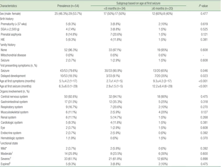
nosed with mitochondrial disease between January 2006 and January 2016, among whom 25 were male (46.3%) and 29 were female (53.7%) (Table 1). Prenatal asphyxia and hypoxicischemic encephalopathy (HIE) injuries at birth were noted in 14.8% and 9.3% of patients, respectively. Family history of mitochondrial disease was absent in 96.0% of patients. Initial symptoms included seizures (81.1%) and delayed development (18.9%). Mean age at first seizure onset was 6.3±6.0 months (range: 1 to 17 months).
Most patients (92.6%) exhibited CNS involvement, although va
rious organ involvement was also noted. Functional state at the
Table 1. Clinical Characteristics of the 54 Included Patients
Characteristics Prevalence (n=54) Subgroup based on age at first seizure
P-value
<6 months (n=34) ≥6 months (n=20)
Sex (male: female) 25 (46.3%):29 (53.7%) 17 (50%):17 (50%) 12 (60%):8 (40%) 0.477
Birth history
Prematurity (<37 wks) 5 (9.3%) 3 (8.8%) 2 (10%) 0.619
SGA (<2,500 g) 4 (7.4%) 3 (8.8%) 1 (5%) 0.525
Prenatal asphyxia 8 (14.8%) 7 (20.6%) 1 (5%) 0.121
HIE 5 (9.3%) 4 (11.8%) 1 (5%) 0.381
Family history
None 52 (96.3%) 33 (97.1%) 19 (95%) 0.608
Mitochondrial disease 0 (0%) 0 (0%) 0 (0%) -
Seizure 2 (3.7%) 1 (2.9%) 1 (5%) 0.608
First presenting symptoms (n, %)
Seizure 43/53 (79.6%) 30/33 (90.9%) 13/20 (65%) 0.046
Delayed development 10/53 (18.5%) 3/33 (9.1%) 7/20 (35%) 0.023
Age at first symptoms (months) 5.1±4.2 (1–17) 2.7±1.4 (1–5) 9.3±4.2 (3–17) <0.001
Age at first seizure (months) 6.3±6.0 (1–29) 2.9±1.5 (1–5) 12.2±6.4 (6–29) <0.001
Organs involvement (n, %)
Central nervous system 50 (92.6%) 32 (94.1%) 18 (90%) 0.475
Gastrointestinal system 17 (31.5%) 12 (35.3%) 5 (25%) 0.318
Respiratory system 9 (16.7%) 7 (20.6%) 2 (10%) 0.270
Musculoskeletal system 6 (11.1%) 2 (5.9%) 4 (20%) 0.127
Renal system 6 (11.1%) 5 (14.7%) 1 (5%) 0.268
Cardiologic system 5 (9.3%) 4 (11.8%) 1 (5%) 0.381
Eye 2 (3.7%) 1 (2.9%) 1 (5%) 0.608
Endocrine system 2 (3.7%) 2 (5.9%) 0 (0%) 0.392
Hematologic system 1 (1.9%) 0 (0%) 1 (5%) 0.370
Functional state
Mild* 2 (3.7%) 2 (5.9%) 0 (0%) 0.392
Moderate† 14 (25.9%) 8 (23.5%) 6 (30%) 0.600
Severec‡ 33 (61.1%) 21 (61.8%) 12 (60%) 0.898
Expired§ 5 (9.3%) 3 (8.8%) 2 (10%) 0.475
SGA, Small for gestational age; HIE, Hypoxic-ischemic encephalopathy.
*The patient is independently ambulatory, but may or may not be dependent on others during daily activities.
†The patient is confined to a wheelchair full-time or partially dependent on others during daily activities, with limited communication abilities.
‡The patient is bedridden and totally dependent on others during daily activitie.
§Expired.
final followup visit was classified as mild, moderate, and severe in 3.7%, 25.9%, and 61.1% of patients, respectively.
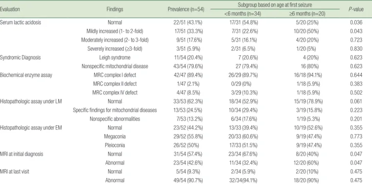
2. Diagnostic evaluations for mitochondrial disease
The results of diagnostic evaluations for mitochondrial disease are presented in Table 2. Twentynine patients exhibited increases in serum lactic acid levels. Serum levels of lactic acid were normal, mildly increased, moderately increased, and severely increased in 22 (43.1%), 17 (33.3%), 9 (17.6%), and 3 (5.9%) patients, respec
tively. Muscle biopsy findings and mitochondrial respiratory chain enzyme activity are also shown in Table 2. Mitochondrial respi
ratory chain (MRC) complex I, II, and IV defects were noted in 42 (89.4%), 1 (2.1%), and 4 (8.5%) patients, respectively. Specific findings for mitochondrial disease were noted 13 patients (24.5
%) based on light microscopy analyses. Electron microscopy analyses revealed megaconia and pleioconia in 29 (55.8%) and 26 (50.0%) patients, respectively. Initial MRI findings were normal in 31 patients (57.4%) and abnormal in 23 patients (42.6%). MRI findings at the last visit were normal in 5 patients (9.3%) and ab
normal in 49 (90.7%). Abnormalities included abnormal signals in different areas of the brain or atrophy. Specifically, abnormal signals were observed in the basal ganglia in 6 patients (11.1%), in the thalamus in 8 patients (14.8%), and in the brainstem in 9 patients (16.7%). Atrophy of the cerebellum and cortex was ob
served in 28 patients (51.8%), while diffuse cerebral atrophy was observed in 47 patients (87.1%).
3. Diagnosis and treatment of West syndrome
In patients with West syndrome diagnosed with mitochondrial disease, seizure was the first symptom in 34 (63%) patients be
fore the age of 6 months and in 20 (37.1%) patients after age of 6 months. The latest age for seizure onset was 29 months (Table 3). The first seizure type was spasm in 43 patients (79.6%), head drop in 4 patients (7.4%), and generalized seizure in 7 patients (13%). EEG revealed classic hypsarrhythmia in 37 patients (68.5
%), generalized slowing of background rhythms in all patients, and focal slowing in 10 patients (18.9%). The epileptiform dis
charge was multifocal in 49 patients (92.5%). Within the first 3 months after diagnosis, the mean number of AEDs used was 1.7±
0.9, although this value increased to more than 2 AEDs after the age of 6 months, which indicates drug resistance. Patients whose seizures remained uncontrolled despite the use of more than 2 AEDs were started on a ketogenic diet, which was effective in reducing seizures by 50% in 22 patients (71%). In addition, 20 pa
tients (64.5%) experienced sustained reductions in the incidence of spasms for more than 6 months. Of the two patients who re
ceived surgery, one underwent total callostomy, while the other underwent right frontal lobectomy with temporal disconnection.
4. Analysis of clinical characteristics by subgroup
Clinical characteristics according to subgroup are presented in Table 1. The age at first symptom onset was 2.7±1.4 months in the earlyonset group and 9.3±4.2 months in the lateonset group.
Table 2. Results of Diagnostic Evaluations for Mitochondrial Disease
Evaluation Findings Prevalence (n=54) Subgroup based on age at first seizure
P-value
<6 months (n=34) ≥6 months (n=20)
Serum lactic acidosis Normal 22/51 (43.1%) 17/31 (54.8%) 5/20 (25%) 0.036
Mildly increased (1- to 2-fold) 17/51 (33.3%) 7/31 (22.6%) 10/20 (50%) 0.043
Moderately increased (2- to 3-fold) 9/51 (17.6%) 5/31 (16.1%) 4/20 (20%) 0.723
Severely increased (≥3-fold) 3/51 (5.9%) 2/31 (6.5%) 1/20 (5%) 0.830
Syndromic Diagnosis Leigh syndrome 11/54 (20.4%) 7 (20.6%) 4 (20%) 0.623
Nonspecific mitochondrial disease 43/54 (79.6%) 27 (79.4%) 16 (80%) 0.623
Biochemical enzyme assay MRC complex I defect 42/47 (89.4%) 26/29 (89.7%) 16/18 (94.1%) 0.644
MRC complex II defect 1/47 (2.1%) 0/29 (0%) 1/18 (5.9%) 0.383
MRC complex IV defect 4/47 (8.5%) 3/29 (10.3%) 1/18 (5.9%) 0.502
Histopathologic assay under LM Normal 33/53 (62.3%) 18/34 (52.9%) 15/19 (78.9%) 0.061
Specific findings for mitochondrial diseases 13/53 (24.5%) 10/34 (29.4%) 3/19 (15.8%) 0.223
Nonspecific abnormalities 7/53 (13.2%) 6/34 (17.6%) 1/19 (5.3%) 0.201
Histopathologic assay under EM Normal 23/52 (44.2%) 13/33 (39.4%) 10/19 (52.6%) 0.355
Megaconia 29/52 (55.8%) 20/33 (60.6%) 9/19 (47.4%) 0.773
Pleioconia 26/52 (50%) 17/33 (51.5%) 9/19 (47.4%) 0.355
MRI at initial diagnosis Normal 31/54 (57.4%) 23/34 (67.6%) 8/20 (40%) 0.047
Abnormal 23/54 (42.6%) 11/34 (32.4%) 12/20 (60%) 0.047
MRI at last visit Normal 5/54 (9.3%) 2/34 (5.9%) 2/20 (10%) 0.475
Abnormal 49/54 (90.7%) 32/34(94.1%) 18/20 (90%) 0.475
MRC, mitochondrial respiratory chain; LM, light microscopy; EM, electron microscopy; MRI, magnetic resonance imaging.
The age at first seizure was 2.9±1.5 months in the earlyonset group and 12.2±6.4 months in the lateonset group. As classifi
ca tion criteria were based on age, significant differences in these criteria were observed between the groups. There were 30 pa
tients (90.9%) with earlier seizure onset who experienced seizure as the first symptom, relative to 13 patients (65%) with lateonset seizures (P=0.046). Delayed development was observed in 3 pa
tients (9.1%) with earlyonset seizures and 7 patients with late
onset seizures (35%) (P=0.023). No significant differences in organ involvement, functional state, respiration, or feeding status were observed. The duration between the first diagnosis of West synd
rome and the final followup was 8.0±5.1 years in the earlyonset group and 9.8±5.1 years in the lateonset group.
5. Diagnostic analysis of mitochondrial disease by subgroup We evaluated serum levels of lactic acid based on the age at the onset of the first seizure (Table 2). Serum levels of lactic acid were normal in 17 patients (54.8%) of the earlyonset group and
5 patients (25%) of the lateonset group (P=0.036). Significant dif
ferences in these abnormalities were observed only among pa
tients with mild increases in serum lactic acid levels (earlyonset group: 22.6% (n=7) vs. lateonset group: 50% (n=10) (P=0.043)).
Initial MRI findings were normal in 23 patients (67.6%) of the earlyonset group and 8 patients (40%) of the lateonset group (P=0.047). There was a statistically significant difference in the initial MRI findings between the two groups according to the age at onset. MRI findings at the last visit were abnormal in 32 patients (94.1%) of the earlyonset group and 18 patients (90%) of the lateonset group, with no significant differences between the two groups (P=0.475).
6. Diagnosis and treatment of West syndrome
Because we classified first seizure onset based on age, signifi
cant differences in this variable were observed between the two groups (P<0.001). No significant differences in seizure type or EEG findings (i.e., hypsarrhythmia, background rhythm, epileptiform
Table 3. Diagnosis and Treatment of Infantile Spasms
Clinical Feature Specification Prevalence (n=54) Subgroup based on age at first seizure
P-value
<6 months (n=34) ≥6 months (n=20)
First Seizure Type Head drop 4/54 (7.4%) 1/34 (2.9%) 3/20 (15%) 0.138
Spasm
Flexor dominant 18/54 (33.3%) 12/34 (35.3%) 6/20 (30%) 0.690
Extensor dominant 9/54 (16.7%) 5/34 (14.7%) 4/20 (20%) 0.441
Indeterminant 16/54 (29.6%) 9/34 (26.5%) 7/20 (35%) 0.507
Generalized seizure
GTC 4/54 (7.4%) 4/34 (11.8%) 0/20 (0%) 0.147
GT 3/54 (5.6%) 3/34 (8.8%) 0/20 (0%) 0.241
EEG
Background rhythm Generalized slowing 53/53 (100%) 34/34 (100%) 19/19 (100%) -
Focal slowing 10/53 (18.9%) 8/34 (23.5%) 2/19 (10.5%) 0.217
Epileptiform Focal sharp/spike waves 4/53 (7.5%) 3/34 (8.8%) 1/19 (5.3%) 0.547
Multifocal sharp waves 49/53 (92.5%) 31/34 (91.2%) 18/19 (94.7%) 0.547
Generalized epileptiform discharge 36/53 (67.9%) 22/34 (64.7%) 14/19 (73.7%) 0.502
Hypsarrhythmia Classic 37/53 (68.5%) 22/34 (64.7%) 15/20 (75%) 0.432
Atypical 17/53 (31.5%) 12/34 (35.3%) 5/20 (25%) 0.432
Treatment options
Antiepileptic drugs 3 months after diagnosis 1.7±0.9 (1–4) 1.8±0.8 (1–3) 1.7±1.1 (1–4) 0.916
6 months after diagnosis 2.6±0.9 (1–4) 2.7±0.9 (1–4) 2.4±0.9 (1–4) 0.282
1 year after diagnosis 2.6±1.0 (1–5) 2.6±1.0 (1–5) 2.5±1.1 (1–4) 0.614
2 years after diagnosis 2.6±1.3 (0–6) 2.7±1.4 (0–6) 2.6±1.1 (1–5) 0.942
Ketogenic diet Seizure reduction rate
Reduction >50% 22/31 (71.0%) 13/18 (72.2%) 9/12 (69.2%) 0.583
Reduction <50% 2/31 (6.5%) 0/18 (0%) 2/13 (15.4%) 0.168
No effect 7/31 (22.6%) 5/18 (27.8%) 2/12 (16.7%) 0.358
Retention rate
Retention >6 months 20/31 (64.5%) 13/18 (72.2%) 7/12 (53.8%) 0.334
Retention <6 months 11/31 (35.5%) 5/18 (27.8%) 6/12 (46.2%) 0.263
Epilepsy surgery Yes 2/54 (3.7%) 1/34 (2.9%) 1/20 (5%) 0.400
GTC, generalized tonic clonic; GT, generalized tonic; EEG, electroencephalography.
hibit a sensitivity between 34–62%, and a specificity between 83–100%28,29). Previous studies have also indicated that increases in lactate levels are more significant in children than in adults30). In the present study, lactate levels were not of significant diagno
stic value in the earlyonset group. However, significant changes were observed in patients of the lateonset group, indicative of greater diagnostic value in this group. These findings suggest that, even though lactate levels are more sensitive in children than adults, these levels are less useful prior to the age of 6 months.
MRI abnormalities are reported in approximately 70–80% of patients with West syndrome31,32). The duration of spasms tends to be shorter in patients with normal MRI findings than in those with abnormal MRI findings. Developmental outcomes are also better for patients with normal MRI findings and worse for pati
ents with perinatal injuries33,34). In the present study, MRI findings were normal in 67.6% of patients with earlyonset seizures but only 40% of patients with lateonset seizures. However, at the last visit, findings were normal in 5.9% and 10% of patients with early and lateonset seizures, respectively. These findings sug
gest that the timing of the MRI evaluation is important, and that continued MRI evaluations are necessary as brain development progresses.
The ketogenic diet is accepted as a potent antiepileptic treat
ment for intractable childhood epilepsy35,36). Eun et al. reported that ketogenic diets exhibit dramatic efficacy even among pati
ents with intractable infantile spasms37). However, some studies have suggested that ketogenic diets can be lethal in patients with underlying metabolic diseases38). The ketogenic diet is also known to promote metabolic stress in patients with MRC complex de
fects, and its use is typically avoided in such cases39,40). However, Kang et al. reported that patients with such defects can tolerate the ketogenic diet when supplementary mitochondrial cocktail therapy is administered41). In the present study, we observed that ketogenic diets were effective in treating patients with West synd
rome and mitochondrial disease, although there was no statisti
cally significant difference between the two age groups. Nonethe
less, clinicians should remain aware of the potential compli cations associated with their use in this patient population.
The present study is limited in that prospective randomization was not used to select participants or set other research para
meters, as mitochondrial disease is incredibly rare. Despite these limitations, our analyses provide valuable data regarding a homo
genous group of patients with West syndrome and mitochondrial disease. Our study is also advantageous in that patients were compared and analyzed according to the age at symptom onset.
Previous studies have revealed that the proportion of patients with West syndrome is high among patients with mitochondrial discharge) were observed between the groups. In addition, we
observed no significant differences in surgery rates or the use of AEDs or ketogenic diets between the two groups (Table 3).
Discussion
In the present study, we examined the agespecific characteri
stics of West syndrome in 54 pediatric patients who were diag
nosed with mitochondrial disease. West syndrome is usually clas sified as either cryptogenic or symptomatic, although ap
proximately 60–80% of cases are symptomatic16,17) . While some studies have indicated that spasm is typically the first major symptom, followed by developmental delay, others have indi
cated the reverse18,19). These differences are considered to be due to differences in the criteria for categorizing symptoms, as well as differences among patient groups and observers. In the present study, most patients in the earlyonset group experienced spasm as the first symptom, while most patients in the lateonset group experienced developmental delay as the first symptom.
These findings suggest that West syndrome should be considered when patients with mitochondrial disease present with develop
mental delay after the age of 6 months, and that patients prior to this age should be evaluated for spasms, since developmental assessments may be ambiguous.
West syndrome is an epilepsy syndrome characterized by early onset, usually presenting between the ages of 4 and 7 months20). Bednarek et al. studied West syndrome by dividing patients into groups of those younger or older than 1 year . While there were similar characteristics, such as developmental delay and hypsarr
hythmia, the etiology and EEG findings were different in each group21,22). The lateonset group was characterized by lesspro
minent slow waves, morenumerous multifocal spikes, and more
marked interhemispheric synchronization on interictal EEG. The treatment responses to vigabatrin and steroids are lower in pati
ents with lateonset23,24). Gul Mert et al. reported that neurode
velopmental outcomes are worse in patients with earlyonset in
fantile spasms25). In the present study, patients were categorized based on the cutoff age of 6 months, which was the highest in
cidence. Although we observed no significant differences in seizure type, EEG findings, or treatment based on age, further studies are required to determine whether our findings were due to variables associated with mitochondrial disease or to West syndrome.
Lactic acidosis is the most recognized laboratory abnormality in patients with mitochondrial disease26,27). In patients with pri
mary mitochondrial disease, true elevations in lactate levels ex
disease who experience seizures.
In conclusion, our findings indicated that there was no signifi
cant difference in epilepsyrelated variables when patients with West syndrome and mitochondrial disease were divided based on a cutoff age of 6 months. However, differences in the first symptom at onset and MRI findings were observed according to age at onset. In addition, MRI findings were more specific in the lateonset group. Although our findings indicate that lactate levels were not of significant diagnostic value in the earlyonset group, they were of diagnostic value in the lateonset group. Our findings also indicated that ketogenic diets are effective in redu
cing symptoms in patients with West syndrome and mitochond
rial disease. Taken together, our results can be applied to the diagnosis and treatment of West syndrome based on the age at disease onset. Future largescale studies are required to develop a clinical protocol for agerelated diagnosis and treatment.
Acknowledgments
The authors are grateful to all staff members, doctors, and statistical consultants who were involved in this study.
요약
목적: 미토콘드리아 질환은 산화적 인산화의 결함으로 인한 세포 에너지의 부족으로 발생하는 이질적인 질환이다. 이 질환은 소아에서 대사질환의 중요한 원인이며 임상증상으로는 경련이 대표적이다. 웨 스트 증후군은 영아기에 특징적으로 보이는 뇌전증 증후군이다. 우리 는 미토콘드리아 질환에서 웨스트 증후군 환자의 임상 양상을 비교 분석하였다.
방법: 본 연구는 2006년부터 2016년까지의 의무기록을 통해 후향 적 연구를 진행하였다. 미토콘드리아 질환으로 진단 된 환자 중 웨스 트 증후군으로 확인 된 54명의 소아를 대상으로 하였다. 환자군을 경 련 발생 연령 6개월을 기준으로 조기 발병 그룹과 후기 발병 그룹으 로 나누었고 이들의 임상 양상을 비교분석하였다.
결과: 첫 임상증상으로 경련을 보이는 경우가 조기 발병 그룹에서 90.9%, 후기 발병 그룹에서는 65% 였으며(P=0.046) , 발달 지연은 조 기 발병 그룹에서는 9.1%, 후기 발병 그룹에서는 35% 였다(P=0.023).
또한 조기 발병 그룹에서 젖산 혈증은 45%, 초기 MRI 이상 소견은 67.6%, 마지막 MRI 이상 소견은 94.1%에서 나타났고 후기 발병 그룹 에서 젖산 혈증은 75%, 초기 MRI 이상 소견은 40%, 마지막 MRI 이 상 소견은 90%에서 나타났다. 케톤생성 식이요법은 미토콘드리아 질 환을 가진 웨스트 증후군 환자 31명에서 시행하였고 22명의 환자에 서 경련 횟수가 50% 이상 감소하는 효과가 있었다.
결론: 미토콘드리아 질환에서 웨스트 증후군 환자들을 경련 발생
연령을 기준으로 비교분석 했을 때 경련 관련 요인에 대해서는 큰 차 이를 보이지 않았다. 하지만 미토콘드리아 질환 관련 요인과 MRI에 대 해서는 일부 의미 있는 차이가 있었다. 또한 케톤생성 식이요법은 미 토콘드리아 질환을 가진 웨스트 증후군 환자에서도 효과가 있었다.
References
1) Lee YM. Epilepsy in various metabolic disorders. Korean J Pediatr 2008;51:1290-4.
2) Eom S, Lee HN, Lee S, Kang HC, Lee JS, Kim HD, et al. Cause of death in children with mitochondrial diseases. Pediatr Neurol 2017;66:82-8.
3) Kisler JE, Whittaker RG, McFarland R. Mitochondrial diseases in childhood: a clinical approach to investigation and management.
Dev Med Child Neurol 2010;52:422-33.
4) Eom S, Lee YM. Preliminary study of neurodevelopmental out- comes and parenting stress in pediatric mitochondrial disease.
Pediatr Neurol 2017;71:43-9.
5) Riikonen R. Long-term outcome of patients with West syndrome.
Brain Dev 2001;23:683-7.
6) Lúthvígsson P, Olafsson E, Sigurthardóttir S, Hauser WA.
Epidemiologic features of infantile spasms in Iceland. Epilepsia 1994;35:802-5.
7) Sidenvall R, Eeg-Olofsson O. Epidemiology of infantile spasms in Sweden. Epilepsia 1995;36:572-4.
8) Baram TZ, Mitchell WG, Tournay A, Snead OC, Hanson RA, Hor- ton EJ. High-dose corticotropin (ACTH) versus prednisone for infantile spasms: a prospective, randomized, blinded study. Pe- diatrics 1996;97:375-9.
9) Hussain SA, Shinnar S, Kwong G, Lerner JT, Matsumoto JH, Wu JY, et al. Treatment of infantile spasms with very high dose pre- dnisolone before high dose adrenocorticotropic hormone. Epi- lepsia 2014;55:103-7.
10) Go CY, Mackay MT, Weiss SK, Stephens D, Adams-Webber T, Ashwal S, et al. Evidence-based guideline update: medical treat- ment of infantile spasms. Report of the guideline development subcommittee of the American academy of neurology and the practice committee of the child neurology society. Neurology 2012;78:1974-80.
11) Lee YM, Kang HC, Lee JS, Kim SH, Kim EY, Lee SK, et al. Mito- chondrial respiratory chain defects: underlying etiology in vari- ous epileptic conditions. Epilepsia 2008;49:685-90.
12) Bernier FP, Boneh A, Dennett X, Chow CW, Cleary MA, Thorburn DR. Diagnostic criteria for respiratory chain disorders in adults and children. Neurology 2002;59:1406-11.
13) Fisher RS, Cross JH, D’Souza C, French JA, Haut SR, Higurashi N, et al. Instruction manual for the ILAE 2017 operational classifi- cation of seizure types. Epilepsia 2017;58:531-42.
14) Lake NJ, Compton AG, Rahman S, Thorburn DR. Leigh syndrome:
one disorder, more than 75 monogenic causes. Ann Neurol 2016;
79:190-203.
15) Kwan P, Arzimanoglou A, Berg AT, Brodie MJ, Allen Hauser W, Mathern G, et al. Definition of drug resistant epilepsy: consensus proposal by the ad hoc task force of the ILAE commission on therapeutic strategies. Epilepsy 2010;51:1069-77.
16) Hamano S, Yoshinari S, Higurashi N, Tanaka M, Minamitani M, Eto Y. Developmental outcomes of cryptogenic West syndrome.
J Pediatr 2007;150:295-9.
17) Hamano S, Tanaka M, Mochizuki M, Sugiyama N, Eto Y. Long- term follow-up study of West syndrome: differences of outcome among symptomatic etiologies. J Pediatr 2003;143:231-5.
18) Lagae L, Verhelst H, Ceulemans B, De Meirleir L, Nassogne MC, De Borchgrave V, et al. Treatment and long term outcome in West syndrome: the clinical reality. A multicentre follow up study.
Seizure 2010;19:159-64.
19) Kaushik JS, Patra B, Sharma S, Yadav D, Aneja S. Clinical spectrum and treatment outcome of West syndrome in children from Nor- thern India. Seizure 2013;22:617-21.
20) Pellock JM, Hrachovy R, Shinnar S, Baram TZ, Bettis D, Dlugos DJ, et al. Infantile spasms: a U.S. consensus report. Epilepsia 2010;51:2175-89.
21) Gastaut H, Roger J, Ouahchi S, Timsit M, Broughton R. An electro- clinical study of generalized epileptic seizures of tonic expression.
Epilepsia 1963;4:15-44.
22) Hrachovy RA, Frost JD Jr, Kellaway P. Hypsarrhythmia: variations on the theme. Epilepsia 1984;25:317-25.
23) Chiron C, Dulac O, Beaumont D, Palacios L, Pajot N, Mumford J.
Therapeutic trial of vigabatrin in refractory infantile spasms. J Child Neurol 1991;Suppl 2:S52-9.
24) Aicardi J, Mumford JP, Dumas C, Wood S. Vigabatrin as initial therapy for infantile spasms: a European retrospective survey.
Epilepsia 1996;37:638-42.
25) Gul Mert G, Herguner MO, Incecik F, Altunbasak S, Sahan D, Unal I. Risk factors affecting prognosis in infantile spasm. Int J Neurosci 2017;127:1012-8.
26) Koenig MK. Presentation and diagnosis of mitochondrial dis- orders in children. Pediatr Neurol 2008;38:305-13.
27) Parikh S, Goldstein A, Koenig MK, Scaglia F, Enns GM, Saneto R, et al. Diagnosis and management of mitochondrial disease: a consensus statement from the mitochondrial medicine society.
Genet Med 2015;17:689-701.
28) Tarnopolsky M, Stevens L, MacDonald JR, Rodriguez C, Mahoney D, Rush J, et al. Diagnostic utility of a modified forearm ischemic exercise test and technical issues relevant to exercise testing.
Muscle Nerve 2003;27:359-66.
29) Suomalainen A, Elo JM, Pietiläinen KH, Hakonen AH, Sevastia- nova K, Korpela M, et al. FGF-21 as a biomarker for muscle-mani- festing mitochondrial respiratory chain deficiencies: a diagnostic study. Lancet Neurol 2011;10:806-18.
30) Davis RL, Liang C, Edema-Hildebrand F, Riley C, Needham M, Sue CM. Fibroblast growth factor 21 is a sensitive biomarker of mitochondrial disease. Neurology 2013;81:1819-26.
31) Khatami A, Sell E, Aggag M, Miller E. Brain MRI findings in in- fantile spasm: outcome correlations in a patient cohort. Open J Med Imaging 2016;6:80-92.
32) Saltik S, Kocer N, Dervent A. Magnetic resonance imaging fin- dings in infantile spasms: etiologic and pathophysiologic aspects.
J Child Neurol 2003;18:241-6.
33) Saltik S, Kocer N, Dervent A. Informative value of magnetic res- onance imaging and EEG in the prognosis of infantile spasms.
Epilepsia 2002;43:246-52.
34) Nordli DR Jr, De Vivo DC. The ketogenic diet revisited: back to the future. Epilepsia 1997;38:743-9.
35) Freeman JM, Vining EP, Pillas DJ, Pyzik PL, Casey JC, Kelly LM.
The efficacy of the ketogenic diet-1998: a prospective evaluation of intervention in 150 children. Pediatrics 1998;102:1358-63.
36) Kang HC, Chung DE, Kim DW, Kim HD. Early- and late-onset complications of the ketogenic diet for intractable epilepsy. Epi- lepsia 2004;45:1116-23.
37) Eun SH, Kang HC, Kim DW, Kim HD. Ketogenic diet for treat- ment of infantile spasms. Brain Dev 2006;28:566-71.
38) Kang HC, Kim HD. Diet therapy in refractory pediatric epilepsy:
increased efficacy and tolerability. Epileptic Disord 2006;8:309- 16.
39) Freeman J, Veggiotti P, Lanzi G, Tagliabue A, Perucca E. The ke- togenic diet: from molecular mechanisms to clinical effects. Epi- lepsy Res 2006;68:145-80.
40) Nordli DR Jr, Kuroda MM, Carroll J, Koenigsberger DY, Hirsch LJ, Bruner HJ, et al. Experience with the ketogenic diet in infants.
Pediatrics 2001;108:129-33.
41) Kang HC, Kim YJ, Kim DW, Kim HD. Efficacy and safety of the ke- togenic diet for intractable childhood epilepsy:Korean multi- centric experience. Epilepsia 2005;46:272-9.