아급성기 뇌졸중 환자의 인지기능 회복
서울대학교 의과대학 분당서울대학교병원 재활의학교실, 1경상대학교병원 재활의학과 오민균1ㆍ임종엽ㆍ백남종
Recovery of Post-stroke Cognitive Function during Subacute Stage Min-Kyun Oh, M.D.1, Jong Youb Lim, M.D. and Nam-Jong Paik, M.D., Ph.D.
Department of Rehabilitation Medicine, Seoul National University College of Medicine, Seoul National University Bundang Hospital,
1Department of Rehabilitation Medicine, Gyeongsang National University Hospital
Objective: The purpose of this study is to find the change in post-stroke cognitive function during
subacute phase, and to investigate factors associated with this change.Method: The cognitive function was evaluated using Mini-Mental Status Examination (MMSE) at the
beginning of rehabilitation (mean 23.1 ± 12.6 days after onset) and discharge (mean 88.1 ± 29.8 days after onset) in 56 stroke patients. At the same time functional status was also evaluated using modified Rankin Scale (mRS), National Institutes of Health Stroke Scale (NIHSS) and modified Barthel Index (MBI). Then we compared the improvements of MMSE scores between two time period according to age, sex, stroke profile, and initial functional status. We also determined the correlation between cognitive and functional gain.Results: MMSE scores improved from rehabilitation start to discharge (3.14 ± 5.4, p = 0.000). MMSE
scores improved more when patients were male, older than 65 years, and had hemorrhagic and cortical lesions (p<0.05). And, MMSE scores improved more when patients had a higher functional level initially (mRS≤2, NIHSS≤1, MBI≥75), (p<0.05) and improvement of MMSE score was significantly correlated with initial MBI score (r = 0.351, p<0.05 ).Conclusion: Cognitive function recovered during subacute phase of stroke, and improvement of cognitive
function at this phase were associated with sex, age, stroke profiles, initial functional status. These results would provide us an information to plan cognitive rehabilitation in these patients. (Brain &NeuroRehabilitation 2009; 2: 71-77)
Key Words: cognition, function, recovery, rehabilitation, stroke
접수일: 2009년 1월 23일, 1차 심사일: 2009년 2월 25일 게재승인일: 2009년 3월 9일
교신저자: 백남종, 경기도 성남시 분당구 구미로 166
463-707, 분당서울대학교병원 재활의학과 Tel: 031-787-7731, Fax: 031-787-4056 E-mail: [email protected]
서 론
뇌졸중은 신체적 기능장애 뿐만 아니라 인지기능의 저 하도 동반되어1,2 일상 생활 동작과 사회 생활에 제한을 주 고 삶의 질을 감소시키는 결과를 초래할 수 있다.3,4 그러 므로 뇌졸중 환자의 치료 초기부터 정확한 인지 기능 평가 와 치료가 필요한데5 뇌졸중 후 인지기능 장애의 유병률 은 인지기능 장애를 정의하는 기준, 뇌졸중 발병으로부터 의 시간, 선택된 환자 군에 따라 11.6∼56.3%로 다양하게 보고되고 있다.1,6-9 뇌졸중 후 인지 장애와 연관된 위험 인
자로는 여성, 고령, 낮은 교육 수준, 우울증, 뇌졸중의 중 증도(severity), 좌측 뇌병변(left hemispheric lesion), 피질 하 병변(subcortical lesion), 편측 무시(unilateral neglect), 구음장애(dysarthria), 요실금(urinary incontinence) 등이 보고되고 있으나,1,6,10-13 아직 통일된 의견이 없는 실정이 다.14
뇌졸중 후 인지기능 저하의 자연적 경과와 회복에 대한 몇몇 연구에 의하면 시간경과에 따라 인지기능이 향상될 수 있다고 하였으나,15-17 뇌졸중 후 아급성기에 인지기능 회복이 어느 정도 일어나는지 또한 그러한 회복에 미치는 요인은 어떠한 것들이 있는지에 대하여는 아직 연구가 미 미한 실정이다. 이는 향후 지속적인 인지 재활치료가 필요 한 환자나 이차적으로 혈관성 치매가 발생할 수 있는 환자 군을 조기에 발견하고 치료 방침을 정하는데 있어 중요한 문제이기도 하다.
이에 본 연구에서는 뇌졸중 후 아급성기에서 인지기능
Table 1. Characteristics of Patients
Number of patients (n = 56) Age (years)
Sex
Stroke type
Lesion location
Lesion site
<65
≥65 Male Female Infarct Hemorrhage Right hemisphere Left hemisphere Bilateral hemisphere Cortical
Subcortical Cortical-subcortical Infratentorial
16 40 31 25 40 16 25 25 6 6 25 14 11
의 변화 양상을 살펴보고, 이에 영향을 미치는 요인들을 알아보고자 하였다.
연구대상 및 방법
1) 연구대상
뇌졸중 초회 발병으로 입원 치료를 받았던 환자를 대상 으로 하였으며 이 중 시력 및 청력 장애, 실어증, 전신 상 태 불량과 그밖에 여러 가지 이유로 협조가 되지 않아 MMSE의 수행이 불가능하였던 환자를 제외한 56명을 대 상으로 하였으며 대상자의 연령분포는 30세에서 107세로 연령은 68.8 ± 13.2세였고, 성별분포는 남자 31명, 여자 25명이었다. 뇌졸중의 유형은 뇌출혈 16명, 뇌경색 40명 이었으며, 뇌병변의 위치는 우측 대뇌반구에 병변이 있는 경우가 25명, 좌측 대뇌반구에 병변이 있는 경우가 25명, 양측 병변인 경우가 6명이었다. 뇌병변의 병소는 피질 병 변이 6명, 피질하 병변이 25명, 피질-피질하 병변이 14명, 천막하 병변이 11명이었다(Table 1). 본 연구의 프로토콜 은 임상심의 윤리위원회의 승인을 받았다.
2) 연구방법
연구 대상 환자 모두를 대상으로 인지기능과 기능적 수 준을 평가 하였다. 인지기능은 MMSE (Mini-Mental Status Examination)을 이용하여 숙련된 재활의학과 의사 가 뇌졸중 후 재활 치료 시작 시 (Time 1, 발병후 23.1 ± 12.6일, 범위 6∼55일)와 재활 치료 후 퇴원 시 혹은 발병 후 3개월째 (Time 2, 발병후 88.1 ± 29.8일, 범위 44∼181 일) 2회 평가하였고, 같은 시기에 mRS (modified Rankin Scale), NIHSS (National Institutes of Health Stroke Scale), MBI (modified Barthel Index)를 이용하여 환자의 기능적
수준과 신경학적 회복정도를 평가하였다.
3) 통계처리
통계분석은 SPSS 12.0을 사용하였다. 뇌졸중 후 Time 1에 평가된 MMSE의 총점에 대하여 성별, 연령별, 뇌졸중 의 유형별, 뇌병변의 위치별, 뇌병변의 병소별로 독립표본 t 검정과 분산분석(ANOVA)을 이용하여 비교하였고 뇌졸 중 후 Time 1과 Time 2, 2회 평가된 MMSE의 5가지(지남 력, 기억등록, 주의집중과 계산, 기억회상, 언어능력) 각 항목별 점수와 총점에 대하여 대응표본 t 검정을 시행하여 비교하였고, 성별, 연령, 뇌졸중의 유형 및 위치, 초기의 기능적 수준과 MMSE 점수의 변화와의 연관성은 공분산 분석(ANCOVA)을 사용하여 분석하였다. 또한 초기의 mRS, NIHSS 및 MBI, 그리고 NIHSS 및 MBI의 향상정도 와 MMSE 점수의 향상 정도와의 상관성은 Pearson 상관 분석을 통하여 알아보았다.
결 과
1) 재활치료 시작 시 성별, 연령별, 뇌졸중의 유형별, 뇌병변의 위치별, 뇌병변의 병소별 MMSE 점수 비교 재활치료 시작 시 Time 1시기의 각 군간의 MMSE 점수 는 성별로는 남자는 18.6 ± 9.8점, 여자는 17.0 ± 9.5점 이었고 성별간 차이는 통계적으로 유의하지는 않았다(p
= 0.548). 연령별로는 65세 이상인 경우 16.0 ± 9.1점, 65세 미만인 경우 22.5 ± 9.4점 이었고 통계적으로 유의 한 차이가 있었다(p = 0.022). 뇌졸중의 유형별로는 뇌출 혈은 15.7 ± 11.2점, 뇌경색은 18.7 ± 8.8점 이었고 뇌 졸중 유형별 차이는 통계적으로 유의하지는 않았다(p = 0.129). 뇌병변의 위치별로는 우측 대뇌반구에 병변이 있 는 경우 16.9 ± 10.7점, 좌측 대뇌반구에 병변이 있는 경우 19.0 ± 8.2점, 양측 병변인 경우 17.3 ± 11.2점 이었고 뇌병변의 위치별 차이는 통계적으로 유의하지는 않았다(p = 1.000). 뇌병변의 병소별로는 피질 병변이 7.6 ± 10.5점, 피질하 병변이 20.3 ± 8.7점, 피질-피질하 병변이 14.5 ± 9.4점 4명, 천막하 병변이 22.2 ± 6.0점 이었고 피질 병변과 피질하 병변, 피질 병변과 천막하 병 변 간에 통계적으로 유의한 차이가 있었다(p = 0.014, 0.010) (Table 2).
2) 재활치료 시작 시와 퇴원 시 MMSE의 향상 MMSE의 총점은 Time 1 시기에 17.9 ± 9.6점에서 Time 2 시기에 21.0 ± 7.4점으로 3.1 ± 5.4점 증가하였 다(p = 0.000). 각 항목별로는 지남력은 0.9 ± 2.3 (p =
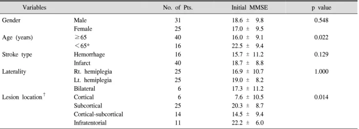
Table 2. Initial MMSE Score according to Demographic Variables and Stroke Features
Variables No. of Pts. Initial MMSE p value
Gender
Age (years)
Stroke type
Laterality
Lesion location†
Male Female
≥65
<65*
Hemorrhage Infarct Rt. hemiplegia Lt. hemiplegia Bilateral Cortical Subcortical Cortical-subcortical Infratentorial
31 25 40 16 16 40 25 25 6 6 25 14 11
18.6 ± 9.8 17.0 ± 9.5 16.0 ± 9.1 22.5 ± 9.4 15.7 ± 11.2 18.7 ± 8.8 16.9 ± 10.7 19.0 ± 8.2 17.3 ± 11.2 7.6 ± 10.5 20.3 ± 8.7 14.5 ± 9.4 22.2 ± 6.0
0.548
0.022
0.129
1.000
0.014
Values are mean ± standard deviations. *p<0.05 by independent t-test, †Group difference: subcortical = infratentorial>cortical = cortical-subcortical, MMSE: Mini-Mental Status Examination.
Table 3. Improvement of Mini-Mental Status Examination Score From Rehabilitation Admission to Discharge
Rehabilitation
admission Discharge p value Orientation
Registration
Attention and alculation Language
Recall Total
6.6 ± 3.7 2.5 ± 1.1 2.1 ± 2.0 6.0 ± 2.9 0.7 ± 1.1 17.9 ± 9.6
7.4 ± 3.2*
2.9 ± 0.4*
2.4 ± 1.9 7.1 ± 1.9*
1.2 ± 1.3*
21.0 ± 7.4*
0.006 0.002 0.135 0.001 0.001 0.000 Values are mean ± standard deviation. *p<0.05 by paired t-test.
0.006), 기억등록은 0.4 ± 0.9 (p = 0.002), 기억회상은 0.5 ± 1.2 (p = 0.001), 언어능력은 1.0 ± 2.2 (p = 0.001)점으로 통계적으로 유의하게 증가하였다. 그러나 주의집중과 계산은 평균 0.3 ± 1.4 (p = 0.135)으로 증가 하였으나 통계적으로 유의하지는 않았다(Table 3).
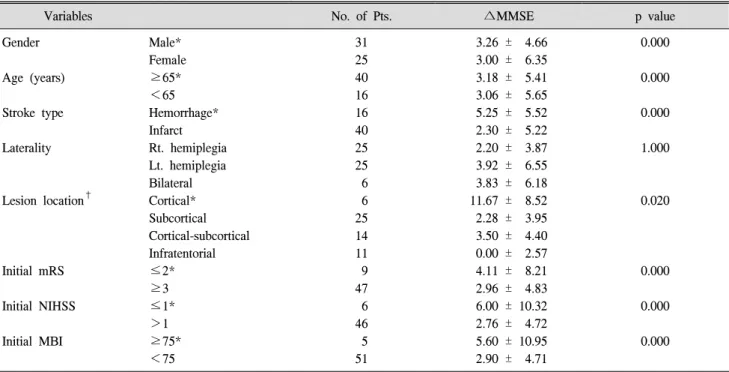
3) 성별, 연령, 뇌졸중의 유형 및 위치, 초기 기능적 수준에 따른 MMSE 점수의 변화
성별 및 연령에 따른 MMSE 총점의 변화량은 여성보다 남성에서, 65세 미만보다 65세 이상의 연령에서 통계적으로 유의하게 증가하였다(p<0.05). 뇌졸중의 유형별로는 뇌 경색보다 뇌출혈에서 MMSE가 더 증가 되었으며(p<0.05), 초기의 기능적 수준에 따라서는 초기mRS (mRS≤2)와 NIHSS가 좋았을 경우(NIHSS≤1) 더 유의하게 증가되었 다(p<0.05). MBI는 초기 점수가 90점 이상인 경우가 1명 밖에 없어서 75점을 기준으로 나누어 분석하였는데, 초기 기능상태가 좋았을 경우(MBI≥75)가 그렇지 않았을 경우
에 비해 MMSE가 통계적으로 유의하게 더 많이 증가되었 다(p<0.05). 그러나, 뇌졸중의 좌우 병소 위치에 따른 MMSE 점수의 증가 정도는 차이가 없었다. 뇌졸중의 병 소별로는, 피질 병변이 피질하 병변, 피질-피질하 병변 및 천막하 병변에 비해 통계적으로 유의하게 MMSE가 증가 되었다(p<0.05). 그 외 나머지 병소 간에는 MMSE 변화 의 차이는 관찰되지 않았다(Table 4).
4) 초기 기능적 수준 및 기능적 향상 정도와 MMSE 점수의 향상과의 상관관계
초기 mRS, NIHSS, MBI 점수 중에서는 초기 MBI 점수 만이 MMSE 향상 정도와 통계적으로 유의한 상관관계가 있었다(r = 0.351, p<0.05). mRS 변화량, NIHSS 변화량 및 MBI 변화량 중에서는 NIHSS 변화량만이 MMSE 향상 정도와 통계적으로 유의한 상관관계가 있었다 (r = −0.548, p<0.05) (Table 5).
고 찰
뇌졸중 후 인지기능의 저하는 흔한 합병증 중의 하나로, 재활치료에 있어 불량한 예후 인자로 알려져 있다.1,18 인 지기능 저하와 뇌졸중은 밀접한 관련이 있어 인지기능 저 하 환자에서 뇌졸중 발생이 더 흔하고19 더 심한 증상을 나타내며20 뇌졸중의 재발빈도 역시 더 흔한 것으로 알려 져 있고,21 뇌졸중환자에서 치매의 위험도가 3배 정도 높 은 것으로 알려져 있다.22 여러 연구에서 기저 인지 상태 (baseline cognitive status)가 뇌졸중 환자의 치료 후 기능 적 회복에 주요한 영향을 미친다고 알려져 있고,23-25 또한
Table 5. Pearson’s Correlation between MMSE gain and Initial Functional Level and Functional Gain Orientation Registration Attention and
calculation Recall Language Total
Initial mRS Initial NIHSS Initial MBI
∆mRS
∆NIHSS
∆MBI
−0.057
−0.004
−0.172 0.045
−0.475*
0.427*
0.374*
0.506*
−0.543*
0.069
−0.524*
0.125
0.075 0.088
−0.152
−0.035
−0.253 0.097
−0.243
−0.199 0.154
−0.043 0.200
−0.063
0.097 0.238
−0.400*
−0.015
−0.590*
0.171
0.049 0.174 0.351*
0.001
−0.548*
0.292 Values are γ by Pearson correlation. *p<0.05 by Pearson correlation. ∆: Difference between time 1 and time 2 (Functional gain).
MMSE: Mini-Mental Status Examination, mRS: modified Rankin Scale, NIHSS: National Institutes of Health Stroke Scale, MBI: modified Barthel Index.
Table 4. Improvement of MMSE Score according to Demographic Variables, Stroke Features and Initial Functional Levels
Variables No. of Pts. ∆MMSE p value
Gender
Age (years)
Stroke type
Laterality
Lesion location†
Initial mRS
Initial NIHSS
Initial MBI
Male*
Female
≥65*
<65 Hemorrhage*
Infarct Rt. hemiplegia Lt. hemiplegia Bilateral Cortical*
Subcortical Cortical-subcortical Infratentorial
≤2*
≥3
≤1*
>1
≥75*
<75
31 25 40 16 16 40 25 25 6 6 25 14 11 9 47 6 46 5 51
3.26 ± 4.66 3.00 ± 6.35 3.18 ± 5.41 3.06 ± 5.65 5.25 ± 5.52 2.30 ± 5.22 2.20 ± 3.87 3.92 ± 6.55 3.83 ± 6.18 11.67 ± 8.52 2.28 ± 3.95 3.50 ± 4.40 0.00 ± 2.57 4.11 ± 8.21 2.96 ± 4.83 6.00 ± 10.32 2.76 ± 4.72 5.60 ± 10.95 2.90 ± 4.71
0.000
0.000
0.000
1.000
0.020
0.000
0.000
0.000
Values are mean ± standard deviations. *p<0.05 by ANCOVA (Analysis of Covariance). Covariate is an initial MMSE score. †Group difference: cortical>subcortical = cortical-subcortical = infratentorial, MMSE: Mini-Mental Status Examination, mRS: modified Rankin Scale, NIHSS: National Institutes of Health Stroke Scale, MBI: modified Barthel Index.
이때의 인지기능이 재활 치료 후 퇴원 시 기능적 상태를 예측할 수 있는 결정 가치(determination value)가 있어 뇌 졸중 후 조기에 그리고 기본적인 인지기능 평가가 이루어 져야 한다고 권고되고 있다.18
본 연구에서는 뇌졸중 후 인지기능 평가를 위해 간이 정신상태 검사(mini-mental status examination; MMSE)를 사용하였다. MMSE는 침상에서의 인지기능 평가에 있어 표준화된 도구로, 인지기능을 간편하고 객관적으로 측정 할 수 있어 널리 사용되어지며, 타당도 역시 잘 정립되어 있다.26 또한 연속적인 평가를 할 수 있어 시간경과에 따른 인지기능의 악화 및 호전여부를 평가할 수 있다.27 지남력
(orientation), 기억등록(registration), 주의집중 및 계산 (attention and calculation), 기억회상(recall), 언어능력 (language)으로 구성되어 있으며, 최고 점수는 30점으로 점수가 많을수록 인지기능이 잘 보존되고 있음을 의미하 며 24점 미만이면 인지기능 장애가 있음을 의미한다.26 뇌졸중 후 인지기능의 변화에 대해 Appelros 등28에 따 르면 뇌졸중 후 1주부터 1년 사이에는 MMSE 점수가 1점 증가한다고 한 반면, Sachdev 등29에 따르면 뇌졸중 후 3∼6 개월부터 1년 사이에는 MMSE 점수가 0.83점 감소한다고 하였고, Ballard 등17은 뇌졸중 후 3개월에서 15개월까지 1년간의 추적 관찰 시 50%의 환자에서 인지기능의 향상
이 있었고 16%에서 MMSE 점수가 2점 이상 향상되었다 고 보고하였다. 이렇듯 연구간 인지기능 변화의 차이는 뇌 졸중 발병으로부터의 시간, 뇌졸중의 중증도, 선택된 환자 군 등에 따른 차이로 생각되는데, 본 연구에서는 뇌졸중 발병 후 약 1개월 (23.1 ± 12.6일)에서 약 3개월 (88.1 ± 29.8일) 사이 아급성기에 MMSE의 총점은 평균 3.14점 증 가되었다. 따라서 뇌졸중 후 아급성기에는 어느 정도의 인 지기능의 호전을 기대할 수 있을 것이라 사료되나, 그 이 후의 변화에 대하여는 본 연구에서는 다루지 못하였고 MMSE의 5가지(지남력, 기억등록, 주의집중과 계산, 기억 회상, 언어능력) 각 항목별 변화에 대한 추가적인 연구가 필요할 것으로 생각된다. 또한 본 연구에서 성별, 연령에 따른 MMSE 점수의 향상은 남성과 65세 이상의 연령에서 더 유의하게 관찰되었다. 65세 이상의 연령에서 MMSE 점수의 향상이 유의한 것은 예상 밖의 결과였지만, 재활치 료 시작 시 MMSE 점수가 65세 미만인 경우 보다 낮아 점수의 향상 폭이 더 컷을 것으로 여겨지나 기존의 여러
연구들10,30-32에 의하면 성별 및 연령과 인지기능의 연관성
에 대해서는 아직도 논란이 있는 상태이다. 뇌졸중의 유형 별로는 Nys 등33은 뇌출혈에서 뇌경색 보다 인지기능 장 애의 발생빈도가 6배정도 높다고 보고 하였으며 이는 광 범한 두개내압의 영향일 것으로 고찰 하였고 Kelly 등34의 연구에 의하면 재활치료 입원 당시 뇌출혈에서 뇌경색 보 다 인지기능 장애와 기능적 장애가 더 심하였으나 회복의 정도는 뇌출혈에서 더 많았다고 보고 하였다. 본 연구 에 서는 뇌경색 보다 뇌출혈에서 MMSE가 더 유의하게 향상 되어 Kelly 등34의 연구와 비슷한 결과를 보였다.
Pohjasvaara 등30의 연구에서는 뇌졸중의 아형(subtype) 과 인지기능 사이에 상관관계가 없었고, Kase 등12의 연구 에서는 뇌졸중 후 6개월 추적관찰에서 깊은 병변(deep lesion)과 얕은 병변(superficial lesion)의 비교 시 얕은 병 변이 깊은 병변에 비해 뇌졸중 후 MMSE 점수 차이가 컸 지만 통계적으로 유의하지는 않았다고 보고하였다. 반면 Szatmari등35은 다발성 피질하 경색(multiple subcortical infarct)에서 뇌졸중 후 인지기능 저하의 위험도가 높다고 보고하였는데 이는 피질하 경색 환자에서 인지기능의 장 애는 피질하 구조 자체의 직접적인 영향, 즉 피질하 구조 의 저관류(hypoperfusion) 또는 기능적 분리(functional disconnection)로 설명되고 있다.33 본 연구에서는 피질 병 변이 다른 병변에 비해 통계적으로 유의하게 MMSE 변화 가 커서 Kase 등12의 연구와 비슷한 결과를 보였다.
또한, 인지기능 장애가 기능적 회복 수준에 크게 영향을 미치는 것으로 알려져 있는데,36 Appelros37의 연구에서는 뇌졸중 후 1년간 추적관찰에서 인지기능 장애가 있는 환
자에서 mRS 점수가 1.8점 더 높게 나타나 기능적 회복이 불량하였다고 하였고, 김 등38의 연구에서는 뇌졸중 환자 에서 재활치료 시작시와 퇴원시 MMSE의 증가값은 FIM (functional independence measure)과 MBI의 증가값과 통 계학적으로 유의한 연관성이 있어서 인지기능의 향상이 기능적 회복과 관련이 있다고 보고하였다. 본 연구에서는 초기의 기능적 수준에 따라서 인지기능의 향상이 어떻게 다른가를 알아보고자 하였는데 초기 기능적 수준 이 좋을 경우(mRS≤2, MBI≥75, NIHSS≤1) 그렇지 않을 경우보 다 통계적으로 유의하게 보다 많은 MMSE 점수의 향상이 관찰되었다(p<0.05). 또한, 초기의 MBI와 MMSE의 향상 정도가 상관관계가 있었다. 이는 뇌졸중 초기의 기능적 수 준이 아급성기의 뇌졸중 환자의 인지기능 회복과 연관이 있다는 것을 의미하며, 초기의 기능적 수준이 좋을수록 인 지기능의 회복 정도가 많을 것으로 기대 된다고 볼 수 있 다.
그러나 본 연구는 인지장애에 중요한 영향을 미치는 것 으로 알려진 교육 수준,30,39 우울증6 여부를 고려하지 못했 다는 점과 표본수가 작았다는 제한점이 있다. 그러나 본 연구 결과를 통하여 뇌졸중 후 아급성기에 인지기능이 향 상되는 것을 확인하였고 그 향상에 미치는 요인들을 알아 낼 수 있었다.
결 론
결론적으로 본 연구에 의하면 아급성기의 뇌졸중 환자 에서 인지기능은 향상되며, 남자, 65세 이상, 뇌출혈, 피질 병변에서 인지기능이 보다 많이 향상되며, 초기의 기능적 상태가 좋을수록 인지기능의 향상이 더욱 많다. 이러한 정 보는 향후 뇌졸중 환자에서 인지 기능의 회복을 예측하고 이와 관련된 재활 치료 계획을 수립하는데 많은 도움을 줄 수 있을 것이라 생각된다.
참 고 문 헌
1) Tatemichi TK, Desmond DW, Stern Y, Paik M, Sano M, Bagiella E. Cognitive impairment after stroke: frequency, patterns, and relationship to functional abilities. J Neurol Neurosurg Psychiatry. 1994;57:202-207
2) Barba R, Martinez-Espinosa S, Rodriguez-Garcia E, Pondal M, Vivancos J, Del Ser T. Poststroke dementia: clinical features and risk factors. Stroke. 2000;31:1494-1501 3) Shimoda K, Robinson RG. The relationship between social
impairment and recovery from stroke. Psychiatry. 1998; 61:
101-111
4) Anderson CS, Linto J, Stewart-Wynne EG. A population-
based assessment of the impact and burden of caregiving for long-term stroke survivors. Stroke. 1995;26:843-849 5) Diamond PT, Felsenthal G, Macciocchi SN, Butler DH,
Lally-Cassady D. Effect of cognitive impairment on rehabili- tation outcome. Am J Phys Med Rehabil. 1996;75:40-43 6) Ebrahim S, Nouri F, Barer D. Cognitive impairment after
stroke. Age Ageing. 1985;14:345-348
7) Tatemichi TK, Desmond DW, Mayeux R, Paik M, Stern Y, Sano M, Remien RH, Williams JB, Mohr JP, Hauser WA, Figueroa M. Dementia after stroke: baseline frequency, risks, and clinical features in a hospitalized cohort. Neurology.
1992;42:1185-1193
8) Pohjasvaara T, Erkinjuntti T, Vataja R, Kaste M. Dementia three months after stroke. Baseline frequency and effect of different definitions of dementia in the Helsinki Stroke Aging Memory Study (SAM) cohort. Stroke. 1997;28:785-792 9) Rockwood K, Wentzel C, Hachinski V, Hogan DB,
MacKnight C, McDowell I. Prevalence and outcomes of vascular cognitive impairment. Vascular Cognitive Impair- ment Investigators of the Canadian Study of Health and Aging. Neurology. 2000;54:447-451
10) Patel MD, Coshall C, Rudd AG, Wolfe CD. Cognitive impairment after stroke: clinical determinants and its associations with long-term stroke outcomes. J Am Geriatr Soc. 2002;50:700-706
11) Patel M, Coshall C, Rudd AG, Wolfe CD. Natural history of cognitive impairment after stroke and factors associated with its recovery. Clin Rehabil. 2003;17:158-166
12) Kase CS, Wolf PA, Kelly-Hayes M, Kannel WB, Beiser A, D'Agostino RB. Intellectual decline after stroke: the Framing- ham Study. Stroke. 1998;29:805-812
13) Toole JF, Bhadelia R, Williamson JD, Veltkamp R. Progre- ssive cognitive impairment after stroke. J Stroke Cerebrovasc Dis. 2004;13:99-103
14) Gorelick PB. Status of risk factors for dementia associated with stroke. Stroke. 1997;28:459-463
15) Desmond DW, Moroney JT, Sano M, Stern Y. Recovery of cognitive function after stroke. Stroke. 1996;27:1798-1803 16) Tham W, Auchus AP, Thong M, Goh ML, Chang HM, Wong
MC, Chen CP. Progression of cognitive impairment after stroke: one year results from a longitudinal study of Singaporean stroke patients. J Neurol Sci. 2002;203-204:
49-52
17) Ballard C, Rowan E, Stephens S, Kalaria R, Kenny RA.
Prospective follow-up study between 3 and 15 months after stroke: improvements and decline in cognitive function among dementia-free stroke survivors >75 years of age.
Stroke. 2003;34:2440-2444
18) Paolucci S, Antonucci G, Gialloreti LE, Traballesi M, Lubich S, Pratesi L, Palombi L. Predicting stroke inpatient rehabilitation outcome: the prominent role of neuropsy- chological disorders. Eur Neurol. 1996;36:385-390
19) Zhu L, Fratiglioni L, Guo Z, Winblad B, Viitanen M.
Incidence of stroke in relation to cognitive function and dementia in the Kungsholmen Project. Neurology.
2000;54:2103-2107
20) Appelros P, Nydevik I, Seiger A, Terent A. Predictors of severe stroke: influence of preexisting dementia and cardiac disorders. Stroke. 2002;33:2357-2362
21) Appelros P, Nydevik I, Viitanen M. Poor outcome after first-ever stroke: predictors for death, dependency, and recurrent stroke within the first year. Stroke. 2003;34:
122-126
22) Zhu L, Fratiglioni L, Guo Z, Aguero-Torres H, Winblad B, Viitanen M. Association of stroke with dementia, cognitive impairment, and functional disability in the very old: a population-based study. Stroke. 1998;29:2094-2099
23) Sisson RA. Cognitive status as a predictor of right hemisphere stroke outcomes. J Neurosci Nurs. 1995;27:152-156 24) Mysiw WJ, Beegan JG, Gatens PF. Prospective cognitive
assessment of stroke patients before inpatient rehabilitation.
The relationship of the Neurobehavioral Cognitive Status Examination to functional improvement. Am J Phys Med Rehabil. 1989;68:168-171
25) MacNeill SE, Lichtenberg PA. Home alone: the role of cognition in return to independent living. Arch Phys Med Rehabil. 1997;78:755-758
26) Tombaugh TN, McIntyre NJ. The Mini-Mental State Exami- nation: a comprehensive review. J Am Geriatr Soc. 1992;
40:922-935
27) Oczkowski WJ, Barreca S. The functional independence measure: its use to identify rehabilitation needs in stroke survivors. Arch Phys Med Rehabil. 1993;74:1291-1294 28) Appelros P, Andersson AG. Changes in mini mental state
examination score after stroke: lacunar infarction predicts cognitive decline. Eur J Neurol. 2006;13:491-495
29) Sachdev PS, Brodaty H, Valenzuela MJ, Lorentz LM, Koschera A. Progression of cognitive impairment in stroke patients. Neurology. 2004;63:1618-1623
30) Pohjasvaara T, Erkinjuntti T, Ylikoski R, Hietanen M, Vataja R, Kaste M. Clinical determinants of poststroke dementia.
Stroke. 1998;29:75-81
31) Madureira S, Guerreiro M, Ferro JM. Dementia and cognitive impairment three months after stroke. Eur J Neurol.
2001;8:621-627
32) Fure B, Bruun Wyller T, Engedal K, Thommessen B.
Cognitive impairments in acute lacunar stroke. Acta Neurol Scand. 2006;114:17-22
33) Nys GM, van Zandvoort MJ, de Kort PL, Jansen BP, de Haan EH, Kappelle LJ. Cognitive disorders in acute stroke:
prevalence and clinical determinants. Cerebrovasc Dis. 2007;
23:408-416
34) Kelly PJ, Furie KL, Shafqat S, Rallis N, Chang Y, Stein J.
Functional recovery following rehabilitation after hemor- rhagic and ischemic stroke. Arch Phys Med Rehabil. 2003;84:
968-972
35) Szatmari S, Fekete I, Csiba L, Kollar J, Sikula J, Bereczki D.
Screening of vascular cognitive impairment on a Hungarian cohort. Psychiatry Clin Neurosci. 1999;53:39-43
36) Zwecker M, Levenkrohn S, Fleisig Y, Zeilig G, Ohry A,
Adunsky A. Mini-Mental State Examination, cognitive FIM instrument, and the Loewenstein Occupational Therapy Cognitive Assessment: relation to functional outcome of stroke patients. Arch Phys Med Rehabil. 2002;83:342-345 37) Appelros P. Characteristics of Mini-Mental State Examination
1 year after stroke. Acta Neurol Scand. 2005;112:88-92 38) Kim HW, KO YJ, Kang SY, Chang YA. The relationship of
MMSE to functional improvement in brain injured patients.
J Korean Acad Rehab Med. 1998;22:1179-1184
39) Tatemichi TK, Desmond DW, Paik M, Figueroa M, Gropen TI, Stern Y, Sano M, Remien R, Williams JB, Mohr JP, Mayeux R. Clinical determinants of dementia related to stroke. Ann Neurol. 1993;33:568-575