Hepatic veno-occlusive disease may develop in secondary iron overloaded mice after allogeneic hematopoietic stem cell
transplantation with total body irradiation
Mi Young Yeom
1, Yoo Jin Kim
2, Nack Gyun Chung
3, Jae Wook Lee
3, Pil Sang Jang
3, Bin Cho
3, Chul Seung Kye
4, Dae Chul Jeong
3,51Department of Clinical Research Laboratory, Incheon St. Mary's Hospital, Incheon, Departments of 2Internal Medicine, 3Pediatrics,
4Radiation Oncology, 5Vaccine Bio Research Institute, College of Medicine, The Catholic University of Korea, Seoul, Korea
p-ISSN 2287-979X / e-ISSN 2288-0011 http://dx.doi.org/10.5045/br.2015.50.3.140 Blood Res 2015;50:140-6.
Received on July 3, 2015 Revised on August 11, 2015 Accepted on August 11, 2015
Background
The outcome of hematopoietic stem cell transplantation (HSCT) is poor in patients with secondary iron overload (SIO). We evaluated the relationship between SIO and veno-oc- clusive disease (VOD) in an animal model with radiation for HSCT.
Methods
We used a 6-week-old female BDF1 (H-2b/d) and a male C57/BL6 (H-2b) as recipient and donor, respectively. Recipient mice were injected intraperitoneally with 10 mg of iron dex- tran (cumulative doses of 50 mg, 100 mg, and 200 mg). All mice received total body irradi- ation for HSCT. We obtained peripheral blood for alanine transaminase (ALT) and liver for pathologic findings, lipid hyperoxide (LH) as reactive oxygen species (ROS), and liver iron content (LIC) on post-HSCT day 1 and day 7. The VOD score was assessed by patho- logic findings.
Results
ALT levels increased depending on cumulative iron dose, with significant differences be- tween days 1 and 7 for mice loaded with 200 mg of iron (P<0.01). LH levels significantly increased in mice loaded with 200 mg of iron compared to those in other groups (P<0.01).
For mice loaded with 100 mg of iron, the LH level depended on the radiation dose (P
<0.01). There was a statistically significant relationship among ALT, LH, and LIC parame- ters (P<0.05). Pathologic scores for VOD correlated with LIC (P<0.01).
Conclusion
Livers with SIO showed high ROS levels depending on cumulative iron dose, and correla- tions with elevated liver enzyme and LIC. The pathologic score for VOD was associated with the LIC. Our results suggest that SIO may induce VOD after HSCT with irradiation.
Key Words Iron overload, Hepatic veno-occlusive disease, Radiation, Reactive oxygen species
Correspondence to Dae Chul Jeong, M.D., Ph.D.
Department of Pediatrics, Seoul St. Mary’s Hospital, College of Medicine,
The Catholic University of Korea, 222, Banpodaero, Seocho-gu, Seoul 06591, Korea
Tel: +82-2-2258-6180 Fax: +82-2-537-4544 E-mail: dcjeong@catholic.ac.kr
Ⓒ 2015 Korean Society of Hematology
INTRODUCTION
Patients with hematologic malignancies or hemoglobino- pathies may receive multiple red blood cell transfusions dur- ing treatment. As a result, these patients may have secondary iron overload (SIO) [1]. SIO leads to high infection rates, and increased morbidity from hepatic, cardiac, or endocrino- logic diseases [2]. Patients with transfusion dependency should
be monitored for SIO status using markers including liver biopsy findings, MRI findings, or serum ferritin level [1, 3, 4].
Hematopoietic stem cell transplantation (HSCT) is a cura- tive treatment for hematologic malignancies or hemoglobi- nopathies. In patients with transfusion dependency, the mor- tality and morbidity after HSCT were related to the iron burden [5]. HSCT in patients with SIO resulted in high infection rates, acute graft-versus-host disease (GVHD), and hepatic veno-occlusive disease (VOD) in the early post-trans-
plant period, and high infection, cardiac fibrosis, and abnor- mal liver function in the late post-transplant period [6-9].
High level of serum ferritin as a surrogate marker of SIO was related to risk of acute GVHD and VOD [7, 10]. VOD is one of the life threatening complications after HSCT [10, 11]. VOD develops in about 11–31% in HSCT recipients, with a mortal- ity rate up to 50% [11-13]. Hepatic VOD after HSCT is associated with hepatotoxic conditioning regimen and pre- transplant liver damage [11, 12, 14].
Iron accumulation in the liver results in the generation of reactive oxygen species (ROS) via the Fenton and Haber- Weiss reaction, and oxidant damage of cellular structures and molecules. In the animal model, livers containing ex- cessive iron stimulated hepatic stellate cells to produce colla- gen protein, and subsequently induced hepatic fibrosis in the periportal area [14, 15]. In HSCT, radiation, as part of the conditioning regimen, injured the sinusoidal and central vein endothelium, and initiated activation of the coagulation cascade [16]. This pro-coagulant status induced fibrin deposit and clot formation, and eventually occluded the vessel [8, 16]. Hence, both hepatic iron excess and pre-transplant irra- diation might be associated with the development of post-transplant VOD, possibly in an additive manner. We evaluated the relationship between an iron excessive state and the development of VOD in a SIO animal model treated with irradiation-based HSCT.
MATERIALS AND METHODS
Animals
We used 6-week-old female BDF1 (H-2b/d) mice as recipi- ents, and C57B/6 (H-2b) mice as donor for HSCT. All mice were acclimatized for 1 week before commencing the experi- ments, and were housed in temperature- and humidity-con- trolled rooms with light-dark cycles. All mice were given irradiated food and sterile water ad libitum. The Institutional Animal Care and Use Committee, Incheon St. Mary’s Hospital, College of Medicine, The Catholic University of Korea approved these experiments. All mice were purchased from OrientBio (Gapyung, Gyeonggi do, Korea).
Establishment of secondary iron overload
We used the SIO animal model, as previously described [17].
Recipient mice were injected intraperitoneally with 300 L (10 mg) of iron dextran (Sigma-Aldrich, St. Louis, MO) in PBS (phosphate buffer solution) for five consecutive days per week according to the cumulative iron dose. The ex- perimental design was for cumulative iron dose of 50 mg, 100 mg, and 200 mg, respectively. The placebo control group received the same volume of PBS. The mice were observed for 2 weeks in order to monitor their viability after iron loading until the next experiment. All experimental groups used at least six mice according to study design.
HSCT
All recipient mice received total body irradiation using
the linear accelerator (Linac, Varion, CA) with 75 cGy/min of dose rate according to different doses. Mice were trans- planted with donor bone marrow mononuclear cells and splenocytes, 1×10⁷ and 5×10⁶ respectively, per mouse, by tail vein within 4 hours after irradiation. Under 1,100 cGy with the same dose rate, mice were transplanted with bone marrow and splenocytes, according to the cumulative iron dose (N=6). Mice with 100 mg iron overload received 500 cGy, 700 cGy, 900 cGy, and 1,100 cGy under the same dose rate (N=6). HSCT was done as both syngeneic and allogeneic type (N=6). We assessed the mice daily for survival and clinical manifestations of graft-versus-host disease.
Blood sampling and analysis
Murine peripheral blood was collected from the retro-orbi- tal sinus using blood collection capillary tubes under anesthesia. Blood sampling was done at post-HSCT day 1 and 7. The collected peripheral blood was transferred to 1.5-mL Eppendorf tubes and centrifuged. The obtained plas- ma was stored in a -80oC freezer until the measurement of alanine transaminase (ALT) and creatinine (Cr). An auto- matic biochemical analyzer (Hitachi 7600-110, Hitachi, Tokyo, Japan) measured plasma ALT (IU/L) and Cr (mg/dL).
Liver iron content (LIC) and lipid hyperoxide (LH)
We obtained liver specimens just before death at around post-HSCT day 10. The hepatic iron concentration (mg/dry 1 g of liver) was analyzed by atomic absorbance spectropho- tometer. We assessed lipid hyperoxide (LH) as a surrogate marker of reactive oxygen species (ROS) in fresh liver by the BCA protein assay kit (Thermoscientific Co., Rockford, IL). The analysis of LH was performed as follows. The liver (0.05–0.1 g) was homogenized with 1 mL of sucrose buffer composed of 210 mM mannitol, 70 mM sucrose, 1 mM EDTA, and 10 mM Hepes solution. Lysate from the liver was quanti- fied for protein. Lysate (100 g) was added with 100 L of distilled water. We prepared standard H2O2 according to dilution, from 100 M to 0 M. The sample was mixed with standard H2O2 and subsequently added to a FOX sol- ution, which included 1 mM xylenol orange, 25 mM ferrous ammonium sulfate, 1 mM sorbitol, and 0.25 M H2SO4. The sample (200 L) was incubated at room temperature for 30 minutes and analyzed by spectrophotometer under 560 nm.
Pathology of the liver
The liver was fixed in 10% formalin and stained by the hematoxylin-eosin and trichrome staining method. We eval- uated the pathologic score using eight parameters including endothelial damage of central venule (CV), coagulation ne- crosis of hepatocytes, subendothelial hemorrhage, sinusoidal hemorrhage, subendothelial fibrosis of CV, adventitial fibrosis of CV, inflammation of the CV, and lobular inflammation [18]. Each parameter was graded on a 4-point system.
Statistical analysis
We analyzed the values of the LH, cumulative iron amount, and ALT according to the radiation dose, one-way ANOVA,
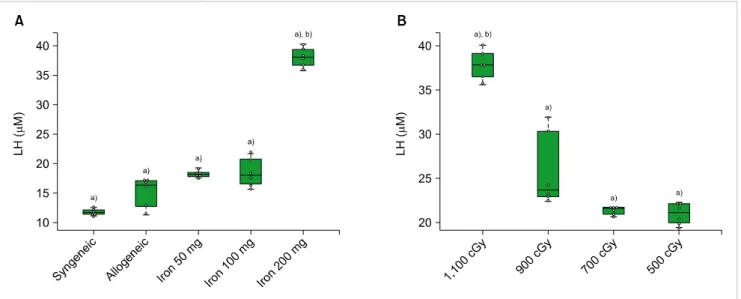
Fig. 2. The lipid hyperoxide (LH) levels according to the cumulative iron dose (A) and radiation dose (B). (A) LH (M) was highest in the mice that received 200 mg of iron at post-HSCT day 7 (P=0.01). All mice (N=6) treated with allogeneic HSCT received 1,100 cGy. (B) LH was highest in the recipients of 1,100 cGy (P<0.01) of radiation. All mice (N=6) treated with allogeneic HSCT had cumulative iron levels of 100 mg. Beeswarm boxplots show the median values and the quartiles, and single measurements are shown as open circles. The middle bar in the box denotes mean value. a)P<0.01 in t-test, b)P<0.01 in ANOVA.
Fig. 1. A comparison of the levels of alanine transaminase (ALT) at post-HSCT days 1 and 7. ALT (IU/L) increased according to the cumula- tive iron dose. The ALT in mice that have been loaded with 200 mg of iron was highest among all the other groups at post-HSCT day 7 (P<
0.01). All mice received 1,100 cGy (N=6). a)P<0.01 in t-test, b)P<0.01 in ANOVA.
and experimental design by repeated measures ANOVA. We identified correlations among the ALT, LH, LIC, and the pathologic score, respectively. The statistical significance was set at a P-value below 0.05.
RESULTS
Elevation of liver enzyme in mice with different cumulative iron doses after allogeneic HSCT with irradiation
The ALT was not significantly different between post- HSCT days 1 and 7 in the allogeneic group without iron and the syngeneic group. The level of ALT at post-HSCT days 1 and 7 in mice with iron were dependent on cumulative iron dose after 1,100 cGy irradiation (P<0.01). The level of ALT in mice that have been loaded with 200 mg of iron was highest among the experimental groups (P<0.01).
Although there was a trend towards higher ALT levels on post-HSCT day 7 than on post-HSCT day 1 in the other iron loaded groups (P>0.05), the ALT on post-HSCT day 7 was higher than that on day 1 (P<0.05) in mice with 200 mg of iron (Fig. 1). The creatinine levels in all ex- perimental groups were within normal limits.
Elevation of lipid hyperoxide levels in mice with different cumulative iron and radiation doses
We obtained liver tissue in order to check the LH levels at post-HSCT day 7. In mice that received 1,100 cGy, the LH levels in iron-loaded mice were higher than in those without iron, including syngeneic transplantation (P<0.05).
LH levels were highest among mice that have been loaded with 200 mg of iron (P<0.01) (Fig. 2A). For analysis of the radiation effects, we evaluated LH levels in the liver
in mice with 100 mg of iron, according to the radiation dose. The LH levels in mice that received 1,100 cGy were highest among the radiation dose groups (P<0.01). There were rapid increments of LH levels in mice that received radiation doses of over 900 cGy compared to those that re- ceived less than 700 cGy (P<0.01). LH levels were not sig- nificantly different between iron-loaded mice, which received doses of 700 and 500 cGy, respectively (P>0.05) (Fig. 2B).
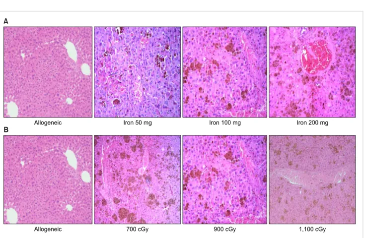
Fig. 3. The pathologic findings in the liver (H-E stain; ×200). (A) The findings show the hepatic pathology according to cumulative iron dose. There is no evidence of veno-occlusion in allogeneic transplantation without iron load. However, there are iron deposits, sinusoidal hemorrhages, hepatic necrosis, and endothelial damage in the iron loaded group. (B) The hepatic findings in mice that received 100 mg of iron are shown according to radiation dose. Damage to hepatocytes and sinusoidal hemorrhages are not significantly different according to radiation dose.
Pathologic findings and scores in the liver
There was sinusoidal hemorrhage and endothelial damage in all mice with iron load (Fig. 3). These pathologic findings in mice were of greater severity and showed a higher score according to a greater cumulative iron dose (Fig. 3A). However, pathologic findings and scores were not significantly different in mice that have been loaded with 100 mg of iron with different irradiation doses (data was not shown) (Fig. 3B).
These results suggest that the severity of pathologic findings may be related to the iron-loading dose and not to the radia- tion dose.
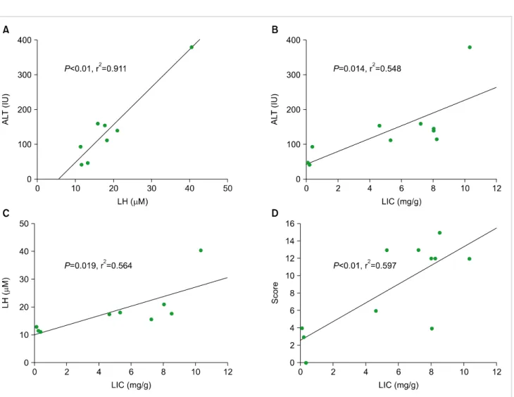
The correlation among lipid hyperoxide, liver enzyme, liver iron content, and pathologic scores
We analyzed correlation between the laboratory and pathologic findings for risk of VOD in iron-loaded mice that received allogeneic HSCT. The levels of ALT were corre- lated with LH (Fig. 4A, P<0.01, r2=0.911), and LIC (Fig.
4B, P=0.014, r2=0.548). The LH levels were significantly correlated with the LIC levels (Fig. 4C, P=0.019, r2=0.564).
We evaluated the relationship among the pathologic findings and other parameters. Significant correlation was only found between the pathologic scores and LIC (Fig. 4D, P<0.01, r2=0.597). The other correlations were not significant.
DISCUSSION
VOD may be an obstacle to successful outcomes in patients who receive HSCT [11]. Many patients who have been treated with HSCT received multiple transfusions before trans- plantation [4]. SIO due to repeated transfusions might lead to several problems including infection, acute GVHD, and hepatotoxicity during and after HSCT [7, 8]. The hepatotoxic effects of HSCT are associated with the use of radiation as part of the conditioning regimen, and may worsen with an abnormal liver state, including iron overload before trans- plant [7, 8, 10, 16]. In our study, the development of pathol- ogy was closely related to LIC and radiation. The excessive hepatic iron induced high levels of LH as a ROS and the elevation of ALT levels. LH increased according to radiation dose. Higher amounts of ROS due to iron overload and radia- tion may induce hepatic inflammation, and the development of VOD.
In our results, the ALT levels in SIO mice were higher than those in non-iron loaded mice (syngeneic and alloge- neic), and increased according to a cumulative iron dose.
The SIO contributed to liver damage through ROS. Mice with chronic dietary iron overload showed increased ALT and hepatocyte apoptosis [19]. Excess intracellular iron leads to oxidative injury to the liver by ROS [1, 15, 20]. In this
Fig. 4. A comparison of the individual correlations between lipid hyperoxide (LH), alanine transaminase (ALT), liver iron content (LIC) levels, and pathologic score, respectively. (A) Correlation between LH and ALT levels (P<0.01, r2=0.911). (B) Correlation between ALT and LIC (mg/gm) (P=0.014, r2=0.548). (C) Correlation between LIC and LH (P=0.019, r2=0.564), and (D) pathologic score (P<0.01, r2=0.0597), respectively.
study, the level of ALT after radiation increased according to cumulative iron dose, although ALT showed no significant differences between recipients of 50 mg iron and non-iron loaded mice. Radiation induces hepatic damage and serious injury to hepatic sinusoidal and central vein endothelium [16, 21]. The ALT levels in mice treated with HSCT and radiation have been reported to be higher than normal, with high levels persisting until 30 days post-transplant [22].
Therefore, the ALT levels in SIO increased according to the cumulative iron dose due to oxidative damage by radia- tion and cumulative iron.
We evaluated the change of LH as a surrogate marker for ROS according to doses of cumulative iron and radiation.
In this study, the level of LH in recipients of 200 mg iron and 1,100 cGy was highest among the groups. The level of LH was similar in the allogeneic HSCT group with different cumulative iron dose under the same radiation exposure, except for recipients of 200 mg iron. There were no significant differences between the syngeneic and allogeneic groups. These results may represent high ROS production due to the accumu- lation of iron and exposure to high doses of radiation [2, 5, 16]. The ROS play a role in hepatic damage by oxidative stress
through iron overload and radiation [2, 15, 16, 19, 20]. Ionizing radiation leads to the generation of ROS and free radicals, and results in oxidative injury to normal tissue [23].
The pathologic scores for VOD, including sinusoidal hem- orrhage and endothelial damage, were higher in SIO mice than in non-iron loaded mice (Fig. 3). Pathologic findings were particularly significantly related to the cumulative iron dose regardless of radiation dose. This result may be very important for hepatic damage in SIO mice that has been treated with HSCT. Our data suggested that radiation played a role in the production of ROS. However, pathologic findings suggested that cumulative iron dose mainly contributed to liver damage in SIO with HSCT. Higher pretransplant serum ferritin levels as well as mortality rates during transplant in HSCT patients have been reported with VOD than without VOD [10]. Jastanish et al suggested that VOD after HSCT was associated with liver inflammation and elevated LIC [5].
Pre-transplant risk factors for developing VOD included ele- vations of the liver enzyme and HLA disparity including performance status, more intensive alkylator use, and prior abdominal radiation [12]. Iron chelation therapy has been previously recommended for successful outcomes in HSCT
[7, 8, 10, 24]. A prospective cohort study reported that iron overload was not associated with HSCT outcome including VOD and mortality. However, this data has limitations in the evaluation of iron content because of the use of R2-MRI for measurement of liver iron content [25].
We investigated for common factors between VOD and SIO. Correlations were found among ALT, LH, LIC, and the pathologic score, respectively. The LIC showed correla- tions with ALT, LH, and the pathologic score, respectively.
The pathologic score may represent the severity of hepatic VOD [18, 26]. These results suggest that the level of LIC may be strongly associated with severity of hepatic VOD in HSCT, including radiation. Hepatic iron overload induced liver damage by ROS [1, 2, 9, 19, 23], and radiation in HSCT contributed to hepatic injury caused by ROS and free radicals [16, 21, 23]. In HSCT, iron accumulation in the liver plays a major role in the development of hepatic VOD, while radiation has also been associated with VOD [6-8]. Our study has some limitations in terms of the evaluation for long-term biological effects between iron overload and hepatic injury in SIO mice after allogeneic HSCT. As previously mentioned, we need to study the relationship between iron overload and graft-versus-host disease, and whether iron overload induces inflammation and aggravates GVHD in allogeneic HSCT [6, 7].
In this study, SIO in the liver induced ROS production, according to the cumulative iron dose. ROS also showed correlations with elevated liver enzyme and LIC. The patho- logic findings of hepatic VOD were strongly associated with LIC. We suggest that SIO may induce hepatic VOD after HSCT with irradiation, and iron chelation therapy should be considered for improvement of HSCT outcomes in patients with SIO due to prior multiple transfusions [8].
ACKNOWLEDGMENTS
The authors are grateful to Hee Chul Lee and Eun Sun Kang, Department of Clinical Research Laboratory, Incheon St. Mary’s Hospital, Incheon, South Korea, for technical their support and assistance.
AuthorsÊ Disclosures of Potential Conflicts of Interest
No potential conflicts of interest relevant to this article were reported.
REFERENCES
1. Shander A, Cappellini MD, Goodnough LT. Iron overload and toxicity: the hidden risk of multiple blood transfusions. Vox Sang 2009;97:185-97.
2. Porter JB, Garbowski M. The pathophysiology of transfusional iron overload. Hematol Oncol Clin North Am 2014;28:683-701.
3. Shander A, Sazama K. Clinical consequences of iron overload from
chronic red blood cell transfusions, its diagnosis, and its manage- ment by chelation therapy. Transfusion 2010;50:1144-55.
4. Jensen PD. Evaluation of iron overload. Br J Haematol 2004;124:
697-711.
5. Jastaniah W, Harmatz P, Pakbaz Z, Fischer R, Vichinsky E, Walters MC. Transfusional iron burden and liver toxicity after bone marrow transplantation for acute myelogenous leukemia and hemoglobinopathies. Pediatr Blood Cancer 2008;50:319-24.
6. Deeg HJ, Spaulding E, Shulman HM. Iron overload, hematopoietic cell transplantation, and graft-versus-host disease. Leuk Lymphoma 2009;50:1566-72.
7. Kanda J, Kawabata H, Chao NJ. Iron overload and allogeneic hematopoietic stem-cell transplantation. Expert Rev Hematol 2011;4:71-80.
8. Majhail NS, Lazarus HM, Burns LJ. Iron overload in hematopoietic cell transplantation. Bone Marrow Transplant 2008;41:997-1003.
9. Fan CQ, Crawford JM. Sinusoidal obstruction syndrome (hepatic veno-occlusive disease). J Clin Exp Hepatol 2014;4:332-46.
10. Lee SH, Yoo KH, Sung KW, et al. Hepatic veno-occlusive disease in children after hematopoietic stem cell transplantation: inci- dence, risk factors, and outcome. Bone Marrow Transplant 2010;
45:1287-93.
11. Coppell JA, Richardson PG, Soiffer R, et al. Hepatic veno- occlusive disease following stem cell transplantation: incidence, clinical course, and outcome. Biol Blood Marrow Transplant 2010;16:157-68.
12. Wadleigh M, Ho V, Momtaz P, Richardson P. Hepatic veno- occlusive disease: pathogenesis, diagnosis and treatment. Curr Opin Hematol 2003;10:451-62.
13. McDonald GB, Hinds MS, Fisher LD, et al. Veno-occlusive disease of the liver and multiorgan failure after bone marrow transplan- tation: a cohort study of 355 patients. Ann Intern Med 1993;118:
255-67.
14. Helmy A. Review article: updates in the pathogenesis and therapy of hepatic sinusoidal obstruction syndrome. Aliment Pharmacol Ther 2006;23:11-25.
15. Ramm GA, Ruddell RG. Hepatotoxicity of iron overload: mecha- nisms of iron-induced hepatic fibrogenesis. Semin Liver Dis 2005;25:433-49.
16. Guha C, Kavanagh BD. Hepatic radiation toxicity: avoidance and amelioration. Semin Radiat Oncol 2011;21:256-63.
17. Moon SN, Han JW, Hwang HS, et al. Establishment of secondary iron overloaded mouse model: evaluation of cardiac function and analysis according to iron concentration. Pediatr Cardiol 2011;32:
947-52.
18. DeLeve LD, McCuskey RS, Wang X, et al. Characterization of a reproducible rat model of hepatic veno-occlusive disease. Hepa- tology 1999;29:1779-91.
19. Liu D, He H, Yin D, et al. Mechanism of chronic dietary iron overload-induced liver damage in mice. Mol Med Rep 2013;7:
1173-9.
20. Fujita N, Miyachi H, Tanaka H, et al. Iron overload is associated with hepatic oxidative damage to DNA in nonalcoholic steato- hepatitis. Cancer Epidemiol Biomarkers Prev 2009;18:424-32.
21. Ingold JA, Reed GB, Kaplan HS, Bagshaw MA. Radiation Hepa- titis. Am J Roentgenol Radium Ther Nucl Med 1965;93:200-8.
22. Qi K, Li H, An L, et al. The correlation between platelet activation
and liver injury by conditioning and bone marrow transplanta- tion. Transplant Proc 2014;46:1523-30.
23. Zhao W, Robbins ME. Inflammation and chronic oxidative stress in radiation-induced late normal tissue injury: therapeutic impli- cations. Curr Med Chem 2009;16:130-43.
24. McDonald GB. Hepatobiliary complications of hematopoietic cell transplantation, 40 years on. Hepatology 2010;51:1450-60.
25. Trottier BJ, Burns LJ, DeFor TE, Cooley S, Majhail NS. Association of iron overload with allogeneic hematopoietic cell transplan- tation outcomes: a prospective cohort study using R2-MRI- measured liver iron content. Blood 2013;122:1678-84.
26. Chen Z, Huo JR, Yang L, Zhu HY. Effect of ligustrazine on mice model of hepatic veno-occlusive disease induced by Gynura sege- tum. J Gastroenterol Hepatol 2011;26:1016-21.