Received on December 9, 2008. Revised on December 19, 2008. Accepted on December 24, 2008.
*Corresponding Author. Tel: 82-43-261-2826; Fax: 82-43-268-2732; E-mail: [email protected] Keywords: macrophage, M-CSF, APC function, TGF-β1
Production of TGF-β1 as a Mechanism for Defective
Antigen-presenting Cell Function of Macrophages Generated in vitro with M-CSF
Jae Kwon Lee1, Young-Ran Lee2, Young-Hee Lee2, Kyungjae Kim3 and Chong-Kil Lee2*
1School of Science Education (Biology), 2College of Pharmacy, Chungbuk National University, Cheongju, 3College of Pharmacy, Sahmyook University, Seoul, Korea
Background: Macrophages generated in vitro using macro- phage-colony stimulating factor (M-CSF) and interleukin (IL)-6 from bone marrow cells (BM-Mp) are defective in anti- gen presenting cell (APC) function as shown by their ability to induce the proliferation of anti-CD3 mAb-primed synge- neic T cells. However, they do express major histocompati- bility (MHC) class I and II molecules, accessory molecules and intracellular adhesion molecules. Here we demonstrate that the defective APC function of macrophages is mainly due to production of TGF-β1 by BM-Mp. Methods:
Microarray analysis showed that TGF-β1 was highly ex- pressed in BM-Mp, compared to a macrophage cell line, B6D, which exerted efficient APC function. Production of TGF-β1 by BM-Mp was confirmed by neutralization experi- ments of TGF-β1 as well as by real time-polymerase chain reaction (PCR). Results: Addition of anti-TGF-β1 mono- clonal antibody to cultures of BM-Mp and anti-CD3 mAb-primed syngeneic T cells efficiently induced the pro- liferation of syngeneic T cells. Conversely, the APC function of B6D cells was almost completely suppressed by addition of TGF-β1. Quantitative real time-PCR analysis also con- firmed the enhanced expression of TGF-β1 in BM-Mp.
Conclusion: The defective APC function of macrophages generated in vitro with M-CSF and IL-6 was mainly due to the production of TGF-β1 by macrophages.
[Immune Network 2009;9(1):27-33]
INTRODUCTION
Macrophages can be generated from bone marrow (BM) pro- genitor cells by culturing in the presence of macro-
phage-colony stimulating factor (M-CSF). Although M-CSF alone can induce differentiation of BM progenitor cells into macrophages, the combination of M-CSF and interleukin-6 (IL-6) significantly enhances macrophage production from BM progenitors (1). The macrophages generated in vitro using M-CSF or M-CSF plus IL-6, however, appeared to be different from normal macrophages isolated from tissues. Although macrophages generated in vitro with M-CSF do express MHC class II molecules, numerous accessory molecules and inter- cellular adhesion molecules, they are defective in APC func- tion (2-4). It has also been shown that some members of the macrophage family suppress antigen presentation by dendritic cells (DCs) (5), or induce T cell anergy (6), suppression (7) or apoptosis (8). Thus it has been suggested that differ- entiation along divergent pathways influences the APC func- tion of various cell types. For instance, thymic macrophages are a specialized subset of macrophages that can phagocytose apoptotic cells very efficiently, but have poor APC functioning (9).
The APC function of mature macrophages may be affected by a variety of factors including the micro environmental con- dition of its development. As shown recently, acquisition of APC functioning of mature macrophages was significantly in- fluenced during development by factors such as the presence of CC chemokines like Lkn-1, MIP-1α and RANTES together with M-CSF (10). The defective APC functioning of macro- phages generated in vitro with M-CSF may also be linked to the intrinsic inability of the macrophages to produce cyto- kines such as IL-12 or to express an invariant (Ii) chain of class II MHC molecules (2,3).
In the present study, we demonstrate that macrophages generated in vitro with M-CSF and IL-6 from BM cells (BM-Mp) produce TGF-β1 in a quantity that is sufficient to suppress the proliferation of anti-CD3 mAb-primed syngeneic T cells. A macrophage cell line B6D, which exerts efficient APC function, produces much less TGF-β1 compared to BM-Mp. The present study, together with other studies which demonstrated that TGF-β1 inhibits the proliferation of T and B-lymphocytes, thymocytes and NK cells (11-15), indicates that the defective APC function of BM-Mp is mainly attribut- able to the production of TGF-β1 by BM-Mp.
MATERIALS AND METHODS Cells and cell culture
The mouse thymic stromal cell line, TFGD, producing M-CSF and IL-6, was obtained from a thymoma mass that sponta- neously developed in a p53-/- mouse system as described previously (4). A macrophage cell line, B6D cells, was ob- tained by subculturing BM-Mp in a culture medium supple- mented with the culture supernatant of TFGD (50%, final con- centration) for a year. The cells were cultured in Dulbecco's modified Eagle's minimum essential medium supplemented with 100 U/ml penicillin, 100μg/ml streptomycin, and 10%
heat-inactivated fetal bovine serum (Hyclone, Logan, USA).
Cytokines and monoclonal antibodies
Recombinant human (rh) G-CSF, rhM-CSF, rhGM-CSF, rhTGF- β1 and mouse IFN-γ were purchased from PeproTech (Rocky Hill, NJ). The monoclonal antibodies (mAbs) recog- nizing murine cell surface markers, anti-CD11c (clone HL3) and anti-CD69 (clone H1.2F3) were purchased from Pharmingen (San Diego, CA). Anti-Dec-205 and anti-CSF1R were provided by Dr. K. Komschlies and Dr. J. Keller (National Cancer Institutes, Frederick, MD), respectively.
Generation of BM-Mp
BM cells were isolated from femurs of C57BL/6 mice, cultured in a medium containing the culture supernatant of TFGD cells (50%, final concentration) in a 100 mm petri dish overnight.
Non-adherent cells were collected after gentle shaking, count- ed, adjusted to 2×10⁵ cells/ml with the same medium, dis- tributed in each well of a 6-well tissue culture plate (5 ml/well), and then incubated for 3 more days. At days 4 and 6 from the initiation of culture, non-adherent cells were re- moved after vigorous shaking, and then 5 ml of the same
medium was added to the culture. After 8 days of culture, the adherent cells were harvested by gentle pipetting with ice-cold phosphate buffered saline (PBS) containing 0.5 mM ethylenediaminetetraacetic acid (EDTA).
Assessment of cell proliferation (XTT assay)
XTT (Sodium 3’-[1-(phenylaminocarbonyl)-3,4-tetrazolium]-bis (4-methoxy-6-nitro) benzene sulfonic acid hydrate) (Sigma- Aldrich) was dissolved in PBS (1 mg/ml), and stored at 4oC.
XTT working solution was prepared just prior to use by mix- ing 1 ml of XTT stock solution with 5μl of PMS (N-methyl dibenzo-pyrazine methyl sulfate, 5 mM in PBS) (Sigma- Aldrich). The XTT working solution was added to wells of cell culture (50μl/well), incubated at 37oC for 4 h, and the absorbance was measured using an ELISA plate reader at 460 nm (DynaTech MR5000).
Phenotypic analysis
Cells were stained with monoclonal antibodies recognizing murine cell surface markers as described previously (4), and flow cytometric analysis was performed using a FACSCalibur (Becton-Dickinson). Dead cells were gated out by their low forward angle light scatter intensity. In most analysis, 10,000 cells were scored.
Measurement of APC function
The APC function of macrophages was determined by testing their ability to stimulate proliferation of anti-CD3 mAb-primed syngeneic T cells, as described previously (4). Briefly, puri- fied T cells (1×10⁶ cells/ml) were mixed with anti-CD3 mAb (50 ng/ml; PharMingen), and then 100 μl of the cell suspen- sion was added to each well of 96-well plates. Macrophages were treated with mitomycin-C (Sigma) for 20 minutes at 37oC, washed, and then indicated amounts of the cells were added to each well. DNA synthesis was measured by [3H]-thymidine (DuPont Pharmaceuticals, Wilmington, DE) in- corporation (0.5 μCi/well) for the final 8 h of the 3-day cul- ture period.
Microarray analysis
RNA was isolated from BM-Mp and B6D cells using an RNA extraction system (RNeasy: Qiagen, Valencia, CA). Prepara- tion of probes and hybridization processes were performed essentially as previously described (16) Briefly, cDNAs were synthesized from total RNAs by random-primed reverse tran- scription in the presence of Cy3-UTP or Cy5-UTP. The
Figure 1. Effects of various cytokines on the growth of B6D cells. B6D cells were cultured in the presence of the indicated amounts of the culture supernatant of TFGD cells (A) or with the indicated cytokines (B) for 3 days. The proli- feration of B6D cells was determined by an XTT assay. The results show the mean±S.D. of three independent experi- ments.
full-length enriched mouse cDNA microarrays were hybri- dized with labeled cDNA probes overnight at 65oC, and then washed in 2×SSC/0.1% SDS, washed in 1×SSC, and finally washed in 0.1×SSC. Then, these slides were scanned on a Scan Array 5000 confocal laser scanner, and the images were analyzed using ImaGene (BioDiscovery).
Real-time PCR
One microgram of RNA isolated from BM-Mp and B6D cells was used to prepare cDNA with a TaqMan Reverse Transcrip- tion kit (Applied Biosystems, Branchburg, NJ). One microliter of each cDNA sample was then used for quantification using the SYBR Green PCR master mix (Applied Biosystems) and reactions were run on the ABI Prism 7700 Sequence Detector (Applied Biosystems). The results were normalized to GAPDH using the Quantum RNA universal 18S (Ambion, Austin, TX) and were also used to determine relative quantities. The probe, TGCACAGCTCACGGCACCGG, was labeled at the 5’
end with 6-carboxyfluorescein (6-FAM) and contained the quencher dye 6-carboxy-N,N,N’,N’-tetramethylrhodamine (TAMRA) at the 3’ end. The forward primer was 5’-TGGAAAGGGCC- CAGCAC-3’, and the reverse primer was 5’-GCAATAGTTG- GTATCCAGGGCT-3’. The relative level of TGF-β1 mRNAs was calculated as described by Liu and Saint (17).
RESULTS
Establishment and characterization of a macro- phage cell line, B6D
BM-Mp, which was generated from BM cells of C57BL/6 mice by culturing in a medium containing the culture supernatant of TFGD cells (50%, final concentration), was continuously
subcultured in a medium supplemented with the culture su- pernatant of TFGD. After a year of subculture, the macro- phages, named B6D, exhibited M-CSF-dependency as well as TFGD-supernatant-dependency in the growth pattern (Fig. 1).
Flow cytometric analysis showed that B6D cells expressed CD11b, CD24, CD44, CD45, CD54, MHC class II (I-Ab), CD80, CD86 and CD40 to a level similar to that of BM-Mp which was freshly generated from BM cells using TFGD-supernatant (data not shown). However, the expression levels of DEC205, CSF-R1, CD69 and CD11c were significantly higher in B6D cells compared to that of freshly generated BM-Mp (Fig. 2).
Comparison of the APC function of B6D cells and BM-Mp
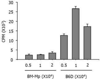
The APC function of B6D cells and BM-Mp was comparatively studied by testing their ability to induce proliferation of syn- geneic anti-CD3 mAb-primed T cells. Consistent with pre- vious observations (4), BM-Mp cells were defective in induc- ing proliferation of syngeneic anti-CD3 mAb-primed T cells.
However, B6D cells efficiently enhanced the proliferation of syngeneic anti-CD3 mAb-primed T cells (Fig. 3). The pro- liferation-inducing activity of B6D cells was most potent when the ratio of B6D cells and syngeneic anti-CD3 mAb-primed T cells was 1:10.
Microarray analysis of the differential gene expres- sion between B6D cells and BM-Mp
To examine any differences in gene expression between B6D cells and BM-Mp, mRNAs were isolated from each cell type, and then cDNAs were synthesized from total mRNAs by ran- dom-primed reverse transcription in the presence of Cy3-UTP (green color) or Cy5-UTP (red color). The microanalysis plate
Figure 2. Phenotypic differences between BM-Mp and B6D cells. The cells were stained with the indicated mAbs, washed and analyzed by flow cytometry. Levels of expression (thin line) were illustrated in comparison to isotype control (dotted line).
Table I. Microarray analysis for the differential expression of genes between B6D cells and BM-Mp
Green-Cy3-B6D
Red-Cy5-BM-Mp Green-Cy3-BM-Mp
Red-Cy5- B6D
Genes R/G ratioa Genes R/G ratio
Mannose receptor, C type1 7.57 Glycoprotein 49 B 0.11 TGF-β1 7.10 Mannose receptor, C type 10.11 Fc receptor. IgG 6.59 Fc receptor, IgG 0.10 Fatty acid binding protein 6.27 Fatty acid binding protein 0.08
Interleukin 7 receptor 5.35 TGF-β1 0.07
aR/G ratio indicates the fluorescence ratio of red (Cy5) vs green (Cy3).
Figure 3. Comparison of the APC functions of BM-Mp and B6D cells.
Syngeneic anti-CD3 mAb-primed T cells (1×10⁵ cells/well) were cultured with the indicated number of BM-Mp or B6D cells.
Proliferation of T cells was measured by [3H]-thymidine incorporation for the final 8 h of the culture period of 3 days. The results show the mean±S.D. of three independent experiments.
contained 1,200 genes. To confirm the differential expression of genes, microarray analysis was repeated with the cDNAs reversely labeled with the dyes. Microarray analysis identified approximately 89 genes that were differentially expressed (data not shown). The genes that were profoundly different in expression are shown in Table I. Among these, TGF-β1, which was highly expressed in BM-Mp compared to B6D cell, was selected for further experiments, because TGF-β1 has been known to inhibit immune responses in a variety of sys- tems (11-15).
Effects of blocking or addition of TGF-β1 on the APC function of macrophages
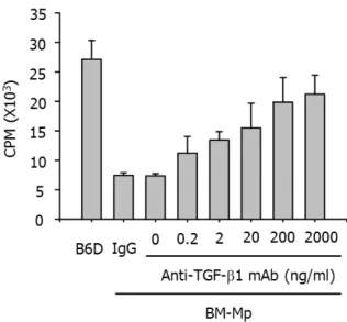
To confirm that TGF-β1 produced from BM-Mp was respon- sible for the defective APC function of BM-Mp, two experi- ments, blocking of TGF-β1 with anti-TGF-β1 mAbs and ad- dition of TGF-β1, were performed. As shown in Fig. 4, addi- tion of anti-TGF-β1 mAbs to mixed cultures of BM-Mp and anti-CD3 mAb-primed syngeneic T cells dose-dependently in- creased the proliferation of T cells. Conversely, addition of TGF-β1 to mixed cultures of B6D cells and anti-CD3 mAb-primed syngeneic T cells dose-dependently inhibited the proliferation of T cells (Fig. 5). These results indicated that TGF-β1 produced from BM-Mp was responsible for the de- fective APC function of BM-Mp.
Quantitative comparison of the expression of TGF-β1 mRNA
In order to compare the expression of TGF-β1 mRNA more accurately, real time PCR analysis was performed with the cDNAs obtained from BM-Mp and B6D cells. The quantity of mRNA was compared by the values of 2−ΔΔCT (relative level). As shown in Table II, BM-Mp expressed a 2.8-fold higher level of TGF-β1 mRNA compared to B6D cells under unstimulated conditions. The ratio of difference in expression of TGF-β1 mRNA was not significantly altered after stim- ulation with LPS (100 ng/ml), or IFN-γ (100 U/ml). The vari- ability between triplicate assays of the same cDNA sample was typically less than 5%.
DISCUSSION
The present study investigated the possible cause of defective
Figure 5. Inhibition of the APC function of B6D cells by addition of TGF-β1. Syngeneic anti-CD3 mAb-primed T cells (1×10⁵ cells/well) were culture with B6D cells (1×10⁴ cells/well) in the presence of the indicated amounts of rhTGF-β1. The results show the mean±S.D. of three independent experiments.
Table II. Expression of TGF-β1 mRNA
ΔCT ΔΔCT 2−ΔΔCT (Relative level)
No stimulation BM-macrophage 1.93 0 1
B6D cell 3.43 1.509 0.351
LPS (100 ng/ml) BM-macrophage 1.89 0 1
B6D cell 3.30 1.408 0.377
IFN-γ (100 U/ml) BM-macrophage 1.90 0 1
B6D cell 3.62 1.715 0.305
RNA samples were isolated from BM-Mp and B6D cells that were stimulated with LPS or IFN-γ for 48 h, and then used to generate the corresponding cDNA samples. Real time PCR reactions were run on an ABI Prism 7700 Sequence Detector (Applied Biosystems) with the probes described in the Methods section. Relative gene expression levels were calculated as described by Liu and Saint (17).
Figure 4. Recovery of APC function of BM-Mp by blocking with anti-TGF-β1 mAb. Syngeneic anti-CD3 mAb-primed T cells (1×10⁵ cells/well) were cultured with BM-Mp (1×10⁴ cells/well) in the presence of the indicated amounts of anti-TGF-β1 mAb. IgG is an isotype control for anti-TGF-β1 mAb. The results show the mean±
S.D. of three independent experiments.
APC functioning of macrophages that were generated in vitro using M-CSF. Macrophages are usually strongly phagocytic for IgG-opsonized sheep red blood cells (SRBCs), produce nitric oxide (NO) in response to interferon IFN-γ plus LPS, and express cell surface molecules that are known to be asso- ciated with mouse macrophages (2-4). However, macro- phages generated from BM cells in vitro using M-CSF alone or in combination with IL-6 are defective in APC functions.
Macrophages generated from CD34+ progenitors by cytokines produced from a renal carcinoma cell line were also shown to be defective in APC function, although they exerted pow- erful phagocytic activity and expressed the same surface phe-
notype markers with peripheral blood macrophages (18). The renal carcinoma cell line was shown to produce M-CSF and IL-6 (18). This feature is unique, because DCs generated from BM cells or peripheral blood monocytes with GM-CSF exert strong APC functions (19,20).
To address the possible reasons for defective APC function- ing of the macrophages generated in vitro using M-CSF, we compared differences in gene expression between a macro- phage cell line B6D, which exerted efficient APC function, and that of macrophages generated from BM cells with M-CSF and IL-6. Microarray analysis showed that TGF-β1 was highly expressed in macrophages generated from BM cells with M-CSF, compared to B6D cells. Blocking experiments with
anti-TGF-β1 mAbs as well as additional experiments with TGF-β1 confirmed that TGF-β1 produced by macrophages that were generated from BM cells in vitro using M-CSF was responsible for the defective APC function. Real time PCR analysis also confirmed that TGF-β1 was highly expressed in the macrophages generated by BM cells in vitro using M-CSF and IL-6.
Macrophages can be generated from CD34+ progenitors in vitro using M-CSF, which is a hematopoietic glycoprotein that stimulates the proliferation and differentiation of BM progeni- tor cells into myeloid cells. M-CSF plays an important role in monocyte/macrophage homeostasis (21,22). M-CSF, how- ever, by itself is not effective in inducing macrophage differ- entiation from BM progenitors (1,18). Earlier studies showed that M-CSF synergies with IL-6 in the generation of macro- phages from BM progenitor cells (33,34,38). We used the cul- ture supernatant of TFGD cells, which was shown to contain large amounts of M-CSF and IL-6 (4), to produce macro- phages from BM progenitor cells in vitro. The macrophages generated by the culture supernatant of TFGD cells were de- fective in APC function, as shown by the present study as well as by an earlier study (4).
One of the most potent activities of TGF-β1 on lympho- cytes is its anti-proliferative effect. TGF-β1 inhibits the pro- liferation of T lymphocytes and B-lymphocytes, thymocytes, large granular lymphocytes, and NK cells (11-15). Studies us- ing peripheral blood mononuclear cells, monocytes and T lymphocytes suggest that TGF-β1 may function as a strong inhibitor of the expression of many cytokines involved in the effector functions of activated cells (11,15). TGF-β1 was shown to inhibit the effects and/or the production of IFN-γ, TNF-α, TNF-β, IL-1, IL-2 and IL-3, as well as the expression of IL-2 receptor (22-25). Thus, the inhibition of cytokine activ- ity is presumably a major factor in TGF-β1-induced immunosuppression. Further evidence of the strong im- munosuppressive effect of TGF-β1 on lymphocytes is the re- ported downregulation of IFN-γ-induced MHC class II anti- gen expression by TGF-β1 in both lymphoid and non-lym- phoid cells (26). Taken together, the present study demon- strated that TGF-β1 produced from the M-CSF-generated macrophages was responsible for the defective APC function- ing of the macrophages.
ACKNOWLEDGEMENTS
This work was supported by the research grant of the
Chungbuk National University in 2007.
CONFLICTS OF INTEREST
Disclosures: The authors have no financial conflict of interest.
REFERENCES
1. Jansen JH, Kluin-Nelemans JC, van Damme J, Weintjens GJ, Willemze R, Fibbe WE: Interleukin 6 is a permissive factor for monocytic colony formation by human hematopoietic cells. J Exp Med 175;1151-1154, 1992
2. Khalili H, Deshpande R, Chang MY: The defective anti- gen-presenting activity of murine fetal macrophage cell lines. Immunol 92;487-493, 1997
3. Smith W, Feldmann M, Londei M: Human macrophages in- duced in vitro by macrophage colony-stimulating factor are deficient in IL-12 production. Eur J Immunol 28;2498-2507, 1998
4. Lee CK, Kim JK, Kim Y, Lee MK, Kim K, Kang JK, Hofmeister R, Durum SK, Han SS: Generation of macro- phages from early T progenitors in vitro. J Immunol 166;
5964-5969, 2001
5. Holt PG, Oliver J, Bilyk N, McMenamin C, McMenamin PG, Kraal G, Thepen T: Downregulation of the antigen present- ing cell function(s) of pulmonary dendritic cells in vivo by resident alveolar macrophages. J Exp Med 177;397-407, 1993
6. Miyazaki T, Suzuki G, Yamamura K: The role of macro- phages in antigen presentation and T cell tolerance. Int Immunol 5;1023-1033, 1993
7. Poulter LW, Janossy G, Power C, Sreenan S, Burke C:
Immunological/physiological relationships in asthma: po- tential regulation by lung macrophages. Immunol Today 15; 258-261, 1994
8. Munn DH, Pressey J, Beall AC, Hudes R, Alderson MR:
Selective activation-induced apoptosis of peripheral T cells imposed by macrophages. A potential mechanism of anti- gen-specific peripheral lymphocyte deletion. J Immunol 156;523-532, 1996
9. Surh CD, Sprent J: T-cell apoptosis detected in situ during positive and negative selection in the thymus. Nature 372;100-103, 1994
10. Lee JK, Kim JK, Lee YR, Kim HS, Im SA, Kim K, Lee CK:
Exposure to chemokines during maturation modulates anti- gen presenting cell function of mature macrophages. Cell Immunol 234;1-8, 2005
11. Kehrl JH, Roberts AB, Wakefield LM, Jakowlew S, Sporn MB, Fauci AS: Transforming growth factor beta is an im- portant immunomodulatory protein for human B lymphocytes. J Immunol 137;3850-3860, 1986
12. Ristow HJ: BSC-1 growth inhibitor/type beta transforming growth factor is a strong inhibitor of thymocyte prolifera- tion. Proc Natl Acad Sci U S A 83;5531-5533, 1986 13. Rook AH, Kehrl JH, Wakefield LM, Roberts AB, Sporn MB,
Burlington DB, Lane HC, Fauci AS: Effects of transforming
growth factor beta on the functions of natural killer cells:
depressed cytolytic activity and blunting of interferon responsiveness. J Immunol 136;3916-3920, 1986
14. Kuppner MC, Hamou MF, Bodmer S, Fontana A, de Tribolet N: The glioblastoma-derived T-cell suppressor fac- tor/transforming growth factor beta 2 inhibits the gen- eration of lymphokine-activated killer (LAK) cells. Int J Cancer 42;562-567, 1988
15. Wahl SM, Hunt DA, Bansal G, McCartney-Francis N, Eliingsworth L, Allen JB: Bacterial cell wall-induced immunosuppression. Role of transforming growth factor beta. J Exp Med 168;1403-1417, 1988
16. Miki R, Kadota K, Bono H, Mizuno Y, Tomaru Y, Carninci P, Itoh M, Shibata K, Kawai J, Konno H, Watanabe S, Sato K, Tokusumi Y, Kikuchi N, Ishii Y, Hamaguchi Y, Nishizuka I, Goto H, Nitanda H, Satomi S, Yoshiki A, Kusakabe M, DeRisi JL, Eisen MB, Iyer VR, Brown PO, Muramatsu M, Shimada H, Okazaki Y, Hayashizaki Y:
Delineating developmental and metabolic pathways in vivo by expression profiling using the RIKEN set of 18,816 full-length enriched mouse cDNA arrays. Proc Natl Acad Sci U S A 98;2199-2204, 2001
17. Liu W, Saint DA: A new quantitative methods of real time reverse transcription polymerase chain reaction assay based on stimulation of polymerase chain reaction kinetics. Anal Biochem 302;52-59, 2002
18. Menetrier-Caux C, Montmain G, Dieu MC, Bain C, Favrot MC, Caux C, Blay JY: Inhibition of the differentiation of dendritic cells from CD34(+) progenitors by tumor cells:
role of intereukin-6 and macrophage colony-stimulating factor. Blood 92;4778-4791, 1998
19. Cella M, Sallusto F, Lanzavecchia A: Origin, maturation and antigen presenting function of dendritic cells. Curr Opin Immunol 9;10-16, 1997
20. Sallusto F, Lanzavecchia A: Efficient presentation of soluble antigen by cultured human dendritic cells is maintained by granulocyte/macrophage colony-stimulating factor plus in- terleukin 4 and downregulated by tumor necrosis factor alpha. J Exp Med 179;1109-1118, 1994
21. Wong GG, Temple PA, Leary AC, Witek-Giannotti JS, Yang CY, Ciarletta AB, Chung M, Murtha P, Kriz R, Kaufman RJ, Ferenz CR, Sibley BS, Tunrner KJ, Hewick RM, Clark SC, Yanai N, Yamada M, Saito M, Motoyoshi K, Takaku F:
Human CSF-1: molecular cloning and expression of 4-kb cDNA encoding the human urinary protein. Science 235;1504-1508, 1987
22. Metcalf D: The molecular biology and functions of the granulocyte-macrophage colony-stimulating factors. Blood 67;257-267, 1986
23. Espevik T, Figari I, Ranges GE, Palladino MA Jr:
Transforming growth factor-beta 1 (TGF-beta 1) and re- combinant human tumor necrosis factor-alpha reciprocally regulate the generation of lymphokine-activated killer cell activity. Comparison between natural porcine plate- let-derived TGF-beta 1 and TGF-beta 2, and recombinant human TGF-beta 1. J Immunol 140;2312-2316, 1988 24. Ohta M, Greenberger JS, Anklesaria P, Bassols A, Massaqué
J: Two forms of transforming growth factor-beta dis- tinguished by multipotential haematopoietic progenitor cells. Nature 329;539-541, 1987
25. Ranges GE, Figari IS, Espevik T, Palladino MA Jr: Inhibition of cytotoxic T cell development by transforming growth factor beta and reversal by recombinant tumor necrosis fac- tor alpha. J Exp Med 166;991-998, 1987
26. Czarniecki CW, Chiu HH, Wong GH, McCabe SM, Palladino MA: Transforming growth factor-beta 1 modulates the ex- pression of class II histocompatibility antigens on human cells. J Immunol 140;4217-4223, 1988