Synthesis and Characterization of Silica/Polystyrene Composite Nanoparticles by in situ Miniemulsion Polymerization
Archana S. Patole, S. P. Patole*, Mi-Hyang Song, Joo-Young Yoon**, Jinhwan Kim, and Tae-Ho Kim†
Department of Polymer Science & Engineering, Sungkyunkwan University, Suwon, 440-746, Korea
*SAINT, Sungkyunkwan University, Suwon, 440-746 Korea
**M&B GreenUs Co. Ltd. Seoul, 152-021 Korea E-mail:[email protected]
(Received February 10, 2009, Revised & Accepted March 2, 2009)
In situ 미니에멀젼중합에 의한 실리카/폴리스타이렌 복합체 나노입자의 합성과 특성
Archana S. Patole ․ S. P. Patole*․ 송 미 향 ․ 윤 주 영**․ 김 진 환 ․ 김 태 호† 성균관대학교 공과대학 고분자시스템공학과, *SAINT, 성균관대학교, **㈜엠엔비그린어스
(2009년 2월 10일 접수, 2009년 3월 2일 수정 및 채택)
ABSTRACT:New coupling agent or surface modified agent (9-decenoic acid) was used to enhance the compatibility between silica and polystyrene in silica/polystyrene hybrid nanocomposite, synthesized by in situ miniemulsion polymerization.
Composites contain well dispersed nanosize silica particles. Related tests and analyses confirmed the success of synthesis.
Functionalization of silica by 9-decenoic acid and silica on the polystyrene was confirmed by FTIR. TGA showed presence and amount of silica in final latex. The glass transition temperature of the hybrid nanocomposite was increased with the silica amount. SEM and TEM analysis showed the spherical morphology of PS and composite with an average diameter of 55 nm. The presence of silica within composite was confirmed by EDS attached to the existing TEM.
요 약:In situ 미니에멀젼 중합으로 실리카/폴리스타이렌 하이브리드 나노복합체를 만들기 위하여 9-데세노 산을
실리카 표면 개질제로 처음 도입하였다. 복합체는 나노크기의 잘 분산된 실리카를 함유하고 있었다. 실리카의 표면 개질 및 폴리스타이렌의 합성 등은 FTIR로 확인하였고 생성 라텍스 중에 존재하는 실리카의 양은 TGA 분석으로 확인하였다. 생성된 하이브리드는 실리카의 양 증가에 따라 유리전이온도가 상승하였다. SEM과 TEM으로 확인한 결과 하이브리드 복합체는 평균 직경이 55 nm 정도로 나타났다. 복합체내의 실리카의 존재는 EDS와 연결된 TEM 등으로 확인되었다.
Keywords:silica, polystyrene, 9-decenoic acid, miniemulsion polymerization, nanocomposite
Ⅰ. Introduction
In recent years, considerable effort has been devoted to the design and controlled preparation of hybrid organic-inorganic composites using colloidal particles as building blocks. The hybrid composite often exhibits properties that are substan- tially different from those of host particles thus making them attractive from both scientific and technological viewpoints.
Among colloidal materials, silica has been studied extensively because of its well known uses in a variety of applications including high performance coatings, optics, electronics, pho- tonic devices, magnetic fluids, and paper coating.1-5 Further- more, the incorporation of colloidal silica at the nanoscale level can increase the fire retardancy, abrasion resistance, mechan-
ical strength of organic polymers.6-9 Many methods have been studied to encapsulate silica particles to give organic-inorganic hybrid dispersions, such as intercalative polymerization, emul- sion polymerization, hybrid latex polymerization, pickering emulsion polymerization etc.10-14 Recently, miniemulsion poly- merization has been found to be attractive way to obtained silica nanocomposite particles.15 In the miniemulsion polymer- ization, the particle nucleation occur preliminary within the sub micrometer monomer droplet making it more advanta- geous to make nanocomposite particles with various morpho- logies.16,17
Depending on the synthesis strategy and the nature of the inorganic particle, a wide range of polymer morphologies have been obtained.17 Silica/polymer composite colloids with vari-
Although the surface of inorganic particles are usually modi- fied by using so-called coupling agents or oleic acid to improve the compatibility between polymeric phase and inorganic phase, these agents only have good effect in dispersing and encapsulating of sol-gel silica particles, and it may not be easy to obtain stable organic-inorganic hybrid dispersion by encap- sulating of commercial nanosilica powder.19
In order to obtained high performance hybrid material, the inorganic component must be dispersed minutely and homoge- nously in the organic polymer matrix, and there must be also good adhesion at the interface between inorganic and organic component.20 The reduced chemical affinity between the in- organic particles (of hydrophilic nature) and the monomer (predominantly hydrophobic) is an important aspect to be con- sidered in the preparation of hybrid composite. The compati- bility of the particles with the polymeric matrix can be im- proved by the surface chemical modification of the particles.
To do so, normally use is made of an agent which promotes chemical compatibilization via hydrogen bonding, electrostatic interactions or covalent bonds within the inorganic/organic interface.21
In this study, 9-decenoic acid was implemented as the sur- face modifier to get the better compatibility between silica and polystyrene matrix. The reactive silica nanoparticles were prepared by reaction between the Si-OH group of silica par- ticles and carboxylic group of 9-decenoic acid. With this meth- od, polymerizable group could be introduced on to the silica particles. The reactive silica particles were co-polymerized with polystyrene using 2, 2'-azobisisobutyronitrile (AIBN) as an initiator to obtain hybrid material.
Ⅱ. Experimental
1. Materials
Silica as nanofiller with an average diameter of 10 - 20 nm was purchased from Sigma Aldrich. Styrene as monomer, AIBN as initiator for polymerization of styrene, n-hexane, 9-decenoic acid as modifying agent, 1-pentanol as hydrophobe and sodium deodecyl sulphate (SDS) as surfactant were pur- chased from Sigma Aldrich and used without further treatment.
itate was rinsed with the solvent mixture of ethanol and water.
The obtained washed precipitate was dried in vacuum oven for 24 hr. This gives white color precipitate containing surface modified silica nanoparticles by 9-decenoic acid which was used for further experimentation.
3. Synthesis of silica/PS composite nanoparticles
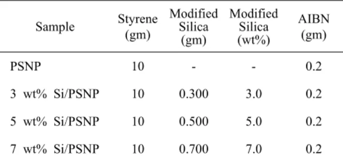
Initially 50 ml of deionized (DI) water, 10 g of styrene, 300 mg of SDS and 2 ml of pentanol were mixed together in 250 ml of beaker. The detailed experimental recipe is given in Table 1. The reactant mixture was then subjected to ultra- sonication for 30 min in ice bath to get the miniemulsion of styrene. In another 250 ml flask 3 - 7 wt% (on the basis of monomer) modified silica (m-silica) was dispersed by ultra- sonication in 50 ml of DI water. The reactant miniemulsified mixture and m-silica was added in the 250 ml three neck flask equipped with magnetic stirrer, nitrogen inlet and condenser.
The reactor was then placed in the oil bath. The polymerization was carried out at 75 ºC. As soon as temperature was reached 75 ºC, the initiator (AIBN) was added into the reaction mixture and continued the polymerization reaction for 4 hr. The ob- tained latex was then washed 3 times with ethanol and water to remove the impurities and surfactant from the reaction mixture. The white precipitate of silica/polystyrene composite nanoparticles (silica/PS) was obtained by vacuum filtration with aluminium oxide membrane filters with a 200 nm pore size (Whatman Inc. UK). The obtained precipitate was dried
Table 1. Recipe for Silica/PS Composite Nanoparticles by Miniemulsion Polymerization
Sample Styrene (gm)
Modified Silica
(gm)
Modified Silica (wt%)
AIBN (gm)
PSNP 10 - - 0.2
3 wt% Si/PSNP 10 0.300 3.0 0.2 5 wt% Si/PSNP 10 0.500 5.0 0.2 7 wt% Si/PSNP 10 0.700 7.0 0.2
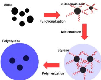
Scheme 1. Schematic illustration showing the multi step proc- ess involved in the formation of silica/PS composite nano- particles by miniemulsion polymerization.
at 60 ºC for 24 hr in vacuum oven. The detail schematic illus- tration is shown in scheme 1.
4. Characterization
The spectroscopic analyses were carried out using fourier transform infrared spectrometer (FTIR; Thermo Nicolet 360 USA). Thermogravimetry/differential thermal analysis (TG/
DTA) was carried out using thermo balance (TGA 2050) from room temperature to 800 °C, with a rate of 10 °C /min in a continuous nitrogen flow. Differential scanning calorimetry (DSC; TA-2910) analysis was carried out from room temper- ature to 600 °C with a rate of 5 °C /min. The morphology of the silica, PS nanoparticles, m-silica and silica/PS composite nanoparticles was examined by a field emission scanning elec- tron microscope (SEM; JSM6700F, JEOL). Furthermore, sam- ples were dispersed in ethanol and drop coated on a copper grid for high resolution transmission electron microscopy (TEM; JEOL 300 kV) to explore the structural properties. The elemental composition of samples were analyze by energy dis- persive X-ray spectrometers (EDS; Oxford INCA Energy) cou- pled to the existing TEM unit.
Ⅲ. Results and Discussion
Surface modification of silica particles was performed with 9-decenoic acid through electrostatic interactions and ester bonding formation. Spectroscopic analysis by FTIR confirms the adsorption of 9-decenoic acid on the silica nanopaticles.
Figure 1 shows the FTIR spectra of 9-decenoic acid (Figure 1a), pure silica nanoparticles (Figure 1b) and m-silica nano-
Figure 1. The FTIR spectrum of (a) silica, (b) 9-decenoic acid, (c) modified silica, (d) pristine PS nanoparticles, and (e) sili- ca/PS composite.
particles (Figure 1c). The 9-decenoic acid showed (Figure 1a) peaks at 3100, 2940, 2740, 1720 and 1470 cm-1 which could be attributed to =C-H, carboxylic -OH stretching, alkane C-H stretching, acid C=O stretching and alkane C-H bending modes respectively. The pristine silica nanoparticles showed (Figure 1b) main peak at 1080 cm-1 which could be attributed to the asymmetrical vibration of Si-O-Si bonds. The silica nano- particles were vacuum dried and therefore do not show any hydroxyl group (-OH) at 3445 cm-1. The m-silica showed (Figure 1c) main peak at 1020 cm-1 which represents Si-O- stretching mode in silica. The peak at 1637 cm-1 represents carboxylate ion (COO-) from 9-decenoic acid. The peaks at 2851 and 2923 cm-1 represents long alkyl chains on silica nanoparticles. The appearance of peak at 3145 cm-1 showed -OH on the surface of silica nanoparticles. When we compare the peaks at 1720 cm-1 (-COOH ) of 9-decenoic acid in Figure 1a with the peak at 1637 cm-1 (COO-) of m-silica in Figure 1c it can be concluded that -COOH reacted with -OH group on the surface of silica to give COO- as a esterification.
The comparison of FTIR spectra for PS nanoparticles (Figure 1d) and silica/PS composite nanoparticles (Figure 1e) gave clear evidence of bonding of silica with polystyrene. The peaks at 1300-1400, 2837 and 2918 cm-1 in Figure 1 d and e showed C-H bending, aliphatic stretching and aromatic stretching in
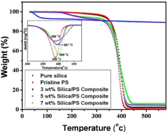
Figure 2. TGA curves of silica, PS nanoparticles and silica/PS composite with difference concentrations of silica.
polystyrene indicating the polymerization of styrene and pres- ence of polystyrene in silica/PS composite. Apart from these peaks in the silica/PS composite the main peak of Si-O was shifted to 1140 cm-1 indicating the bonding of silica with polystyrene.
TGA is widely used for the composition determination of inorganic/organic hybrid particles.22,23 The silica contents in silica/PS composite nanoparticles were determined from weight loss at 500 ºC in the nitrogen environments using TGA.
Figure 2a shows weight loss curve of pristine silica nano- particles, pristine PS nanoparticles and silica/PS composite nanoparticles. The inset of Figure 2a shows the first derivative peaks of TGA curves. In case of silica nanoparticles the first 10% initial weight loss below 100 ºC was attributed to the evaporation of adsorbed moisture in the silica nanoparticles.
Pristine polystyrene started to decompose at 380 ºC and com- pletely degraded above 420 ºC. In all silica/PS nanoparticles composites the weight loss below 200 ºC was attributed to de- composition of lower molecular weight polystyrene and above 200 ºC was attributed to decomposition of higher molecular weight polystyrene and chemisorbed 9-decenoic acid on the silica nanoparticles. In case of all the composite samples the mass remained above 450 ºC was constant and assigned to the silica nanoparticles. It determined that the actual silica con- tent in the 3, 5 and 7 wt% composites were 2.6, 4.3 and 6.0 wt% respectively.
The first derivative of TGA curve (DTA curves shown in inset of Figure 2) clearly exhibits the variation ratio of weight to time (dw/dt) as a function of temperature.
The peaks of DTA exactly indicate the temperature of max- imum reactive velocity. The maximum reactive velocity of DTA curve of pristine polystyrene and silica/PS composites are listed in Table 2. The pure polystyrene reach the maximum reactive velocity at 388 ºC and the value was lowest in all
114℃
113℃
110℃
Figure 3. DSC thermograms of PS and silica/PS composite.
the curves. The temperature at the maximum reactive velocity was increased with increasing amount of silica nanoparticles viz. 389 ºC, 397 ºC and 399 ºC for 3 wt%, 5 wt% and 7 wt%
composites respectively. The temperature of the 7 wt% compo- site increased as by 10 ºC compare to the pure polystyrene polymer, which indicated that the thermal stability of the poly- mer matrix was improved by addition of silica.
The glass transition temperature (Tg) which defines a pseudo second order phase transition is shown in Figure 3. The in- corporation of silica nanoparticles in polystyrene resulted in an increase in Tg. In case of pristine PS nanoparticles Tg was observed at 110 ºC and it was shifted to higher temperature for silica/PS composite nanoparticels. Such type of effect on Tg obtained due to motion restriction of the polymer chain in the nanoparticles-matrix interface.24,25 When the polymer chain present strong interfacial affinity with the filler surface, a region of strongly bonded polymer chain is formed. In this region the polymer chains exhibit different behavior than that in the bulk. The strong packing hinders chain segmental mobility. Thus more energy required to allow the first thermal transition, shifting Tg to higher temperature. In the present case, similar effect might have taken place due to in situ polymerization. The observed increment in Tg with the increas- ing silica nanoparticles suggested good interfacial compati-
Figure 4. SEM images of (a) silica, (b) m-silica, (c) PS nano- particles, and (d) silica/PS composite nanoparticles.
Figure 5. (a) TEM image of PS nanoparticles and (b) histo- gram showing particles size distribution.
bility between the PS and silica nanoparticles. It is worth men- tioning that the observed change in Tg was 4 ºC for 7 wt%
composite (Figure 4c) compare to pristine PS nanoparticles.
The surface morphology and nature of pristine silica, m-sili- ca, pristine PS and silica/PS composite nanoparticles were studied using SEM. Figure 4a shows the SEM image of pris- tine silica nanoparticles. The size of silica nanoparticles was 10-12 nm and therefore could not be resolved in the SEM images. The m-silica also shows (Figure 4b) similar micro- graph. Figure 4c shows the SEM image of pristine PS nano- particles. The synthesized PS particles were uniform nanosized and spherical in nature. The typical SEM image of composite is shown in Figure 4d. The composite nanoparticles are also spherical in nature and nanosized.
Closer inspection of TEM images of pristine PS nano- particles shown in Figure 5a reveals that the resulting particles closed to spherical dots with 55 nm average particle size. The size distribution histogram is plotted in Figure 5b. Various factors are responsible for the particles size and distribution
Figure 6. (a and b) TEM images of silica/PS composite nano- particles.
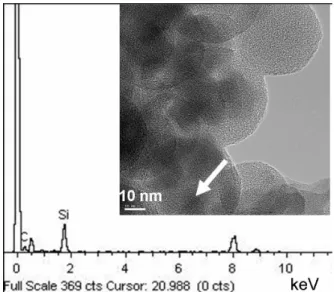
Figure 7. EDS of silica/PS composite nanoparticles. Inset im- age with arrow indicates the scan area.
of emulsion droplets, for example the homogenization method by sonication, the type of hydrophobes, the amount of surfac- tants and so on so forth. Our aim was not to study these effects but to achieve the better compatibility for PS nanoparticles with silica, therefore the stated ratios of chemicals in the ex- perimental part were used throughout the experimentation.
The TEM of silica/PS composite nanoparticles is shown in Figure 6. The purpose of TEM analysis was to study detailed morphology of composite material. In TEM images, the spatial distribution of contrast within the PS nanoparticles can give the presence of silica. The silica is more opaque to electron beam and gave more contrast as compare to PS nanoparticles.
TEM displayed the presence of spherical particles of silica with a diameter more or less corresponding to commercial specification (10 - 20 nm) within the PS nanoparticles which having average diameter of 55 nm.
In order to support the evidences from TEM images EDS has been carried out on different locations within the single
ca/PS composite nanoparticles. Nanosized silica particles mo- dified with 9-decenoic acid can be functionalized by in situ miniemulsion polymerization of styrene monomer. The FTIR spectroscopic analysis gave the evidence of modification of silica surface by esterification, which further attached to the polystyrene.
TGA analysis showed the thermal properties of PS improved with the incorporation of silica in the composite. DSC analysis showed that the Tg of PS was shifted by 4 ºC by incorporating the 7 wt% silica. The morphology of synthesized PS nano- particles and composite revealed from SEM showed the uni- formly distributed spherical nanoparticles. The average PS nanoparticle size was calculated from TEM observations. The PS nanoparticles size distribution histogram showed the aver- age size of PS nanoparticle was 55 nm. TEM observations of composite clearly showed the incorporation of silica nano- particels inside the PS nanoparticles giving more compatible composite. Elemental analysis of composite by EDS confirms the presence of silica nanoparticles within PS nanoparticles.
The results provide us with a new method for an alternative synthetic strategy to fabricate novel materials and provide us a further understanding of the formation mechanism of compo- site materials. In addition, we believe that our method can be easily extended to a variety of organic-inorganic hybrid materials.
Acknowledgement
This work was supported by Korea Materials and Compo- nents Technology Development Program.
References
1. V. Castelvetro and C. de Vita, “Nanostructured Hybrid Materials from Aqueous Polymer Dispersions”, Adv. Colloid.
Interf. Sci., 108‐109, 167 (2004).
2. W. Wu, T. He, J. Chen, X. Zhang, and Y. Chen, “Study on in situ Preparation of Nano Calcium carbonate/PMMA”, Mater. Lett., 60, 2410 (2006).
3. Y. Lu, J. McLellan, and Y. Xia, “Synthesis and Crystalliza- tion of Hybrid Spherical Colloids Composed of Polystyrene Cores and Silica Shells”, Langmuir, 20, 3464 (2004).
R. Pereira,“Polymer–Filler Interactions and Mechanical Properties of a Polyurethane Elastomer”, Polym. Test, 19, 93 (2000).
8. R. C. R. Nunes, R. A. Pereira, J. L. C. Fonseca, and M.
R. Pereira, “X‐ray Studies on Compositions of Polyurethane and Silica”, Polym. Test, 20, 707 (2001).
9. W. Qiu, Luo, Y. F. Chen, Y. Duo, and H. Tan, “Morphology and Size Control of Inorganic Particles in Polyimide Hybrids by using SiO2–TiO2 Mixed oxide”, Polymer, 44, 5821 (2003).
10. P. Viala, E. Bourgeat‐Lami, A . Guyot, P. Legrand, and D.
Lefebvre, “Pigment Encapsulation by Emulsion Polymeriza- tion, Redispersible in Water”, Macromol, 187, 651 (2002).
11. Y. Yang, X. Kong, C. Kan, and C. Sun, “Encapsulation of Calcium Carbonate by Styrene Polymerization”, Polym. Adv.
Technol, 10, 54 (1999).
12. P. Liu, W. M. Liu, and Q. J. Xue, “In situ Radical Transfer Addition Polymerization of Styrene from Silica Nanopar- ticles”, Eur. Polym. J., 40, 267 (2004).
13. I. Tissot, C. Novat, F. Lefebvre, E. Bourgeat‐Lami, “Hybrid Latex Particles Coated with Silica”, Macromolecules, 34, 5737 (2001).
14. J. H. Chen, C. Y. Cheng, W. Y. Chiu, C. F. Lee, and N.Y.
Liang, “Synthesis of ZnO/polystyrene Composites Particles by Pickering Emulsion Polymerization”, Eur. Polym. J., 44, 3271 (2008).
15. R. Y. Hon, B. Feng, X. Cai, G. Liu, H. Z. Li, J. Ding, Y. Zheng, and D. G. Wei, “Double‐Miniemulsion Preparation of Fe3O4/Poly(methyl methacrylate) Magnetic Latex”, J. Appl.
Polym. Sci., 112, 89 (2009).
16. J. Ugelsted, M. S. EI‐Aasser, and J. W. Vanderhoff, “Emul- sion Polymerization Initiation of Polymerization in Emulsion Droplet”, J. Polym. Sci., Polym. Lett., 11, 503 (1973).
17. E. Bourgeat‐Iami, F. Lefebvre, and N. Novat, “Designing Organic/Inorganic Colloids by Heterophase Polymerization”, Macromolecules, 34, 5737 (2001).
18. E. Bourgeat‐Lami, Espiard Ph., A. Guyot, “Poly(ethyl acryl- ate) Latexes Encapsulating Nanoparticles of Silica: Function- alization and Dispersion of Silica”, Polymer, 36, 4385 (1995).
19. J. L. Luna‐Xavier, E. Bourgeat‐Lami, and A. Guyot, “The role of Initiation in the Synthesis of Poly(methyl meth- acrylate) Nanocomposite Latex Particles through Emulsion Polymerization”, Colloid Polym. Sci., 279, 947 (2001).
20. H. Sugimoto, K. Daimatsu, E. Nakanishi, Y. Ogasawara, T.
Yasumura and K. Inomata, “Preparation and Properties of
Poly(methylmethacrylate)–Silica Hybrid materials Incorporat- ing Reactive Silica Nanoparticles”, Polymer, 47, 3754 (2006).
21. C. Kunze, T. Freier, E. Helwig, B. Sandner, D. Reif and A.Wutzler, “Surface Modification of Tricalcium Phosphate for Improvement of the Interfacial Compatibility with Biode- gradable Polymers”, Biomaterials, 24, 967 (2003).
22. R. K. Nagarale, G. S.Gohil, V. K. Shahi, and R. Rangarajan,
“Recent Developments on Ion‐exchange Membranes and Electro‐membrane Processes”, Macromolecules, 37, 10023 (2004).
23. D. Z. Wu, X. G. Ge, Z. C. Zhang, M. Z. Wang, and S. L.
Zhang, “Novel One‐Step Route for Synthesizing CdS/
Polystyrene Nanocomposite Hollow Spheres”, Langmuir, 20, 5192 (2004).
24. P. A. O. Muisener, L. Clayton, J. D. Angelo, and J. P.
Harmona, “Effects of Gamma Radiation on Poly(methyl- methacrylate)/single‐wall Nanotube Composites”, J. Mater.
Res., 17, 2507 (2002).
25. X. Qu, T. Guan, G. Liu, Q. She, and L. Zhang, “Preparation, structural characterization, and properties of Poly(methyl- methacrylate)/Montmoillonite Nanocomposite by Bulk Poly- merization”, J. Appl. Polym. Sci., 97, 348 (2005).