2004; 9(3): 154-161
Modulation of Apoptosis-related Signal Transduction by Celecoxib, a Selective COX-2 Inhibitor in Comparison with Estrogen in
Perimenopausal Mammary Glands
Tae-Kyung Kim, In-Ja Park and Ock Jin Park
Department of Food and Nutrition, Hannam University, Daejeon 306-791, Korea
This investigation was intended to study the expression of cyclooxygenase-2 (COX-2) and mapkinase, and apoptosis related gene expression in normal mammary glands of perimenopausal female rats fed oral celecoxib, a selective COX-2 inhibitor and estrogen.
The expression of pERK1/2 showed the similar patterns as COX-2 by the oral treatment of celecoxib and estrogen. It was found that celecoxib induced up-regulation of bcl-2 in mammary gland buds. The regulation of bax was decreased in celecoxib supplemented rats. The bcl-2/bax ratio was higher in celecoxib supplemented rats. However, bcl-2/bax ratio was highest in celecoxib group. The up-regulation of COX-2 was observed in celecoxib in mammary gland buds. The similar trend was not displayed with mapkinase expression. Compared to estrogen feeding, bcl-2 expression was upregulated in celecoxib and down-regulating effect was observed with bax expression. The up-regulation of bcl-2 was accompanied by the decreased expression of COX-2. The oral administration of celecoxib caused significant reduction in bcl-2/bax ratio compared to the control indicating that there might be more apoptotic activity in celecoxib treatment. However, estrogen, a known stimulator of cell proliferation also showed the apoptotic potential compared to control or celecoxib. The increased apoptotic potential by celecoxib or estrogen resulted from the different patterns in bcl-2 or bax regulation. The lowering of bcl-2/bax ratio by celecoxib was resulted from the increased expression of bas, while the reduction of bcl-2/bax observed with estrogen was from the decreased bcl-2 and the increased bax. These findings suggest that both bcl-2 and bax are involved in the apoptotic control of celecoxib, and bcl-2 is a significant factor in apoptotic control of estrogen.
ꠏꠏꠏꠏꠏꠏꠏꠏꠏꠏꠏꠏꠏꠏꠏꠏꠏꠏꠏꠏꠏꠏꠏꠏꠏꠏꠏꠏꠏꠏꠏꠏꠏꠏꠏꠏꠏꠏꠏꠏꠏꠏꠏꠏꠏꠏꠏꠏꠏꠏꠏꠏꠏꠏꠏꠏꠏꠏꠏꠏꠏꠏꠏꠏꠏꠏꠏꠏꠏꠏꠏꠏꠏ
Key Words: Celecoxib, COX-2, Molecular markers related to apoptosis, Perimeno-
pausal female rat, Mammary gland buds
Corresponding author:Ock Jin Park, Department of Food and Nutrition, Hannam University, 133 Ojeong-dong, Daedeok-gu, Daejeon 306-791, Korea. Tel: +82-42-629-7493, Fax: +82-42-629-7490, E-mail: [email protected]
Received:August 9, 2003, Accepted:September 15, 2003
INTRODUCTION
It has been shown through in vitro and in vivo studies that estrogen might play a central role in the promotion of breast cancer and possibly in the initia- tion process.1,2) In vitro, estrogen exerts direct and indirect proliferative effects on breast cancer cells.3) Estrogen produces alkylation of cellular molecules, and generates active potential carcinogenic metabo- lites such as catechol estrogens and 16 α-hydroxy- estrone.4∼7) It has been also shown that there are strong correlations between estradiol and estrone levels in postmenopausal women and subsequent breast cancer risk.8∼10) Along with the significance of the involvement of estrogen in breast cancer develop- ment, the importance of the modulation of signal transduction pathways for chemoprevention of breast cancer is being recognized.11) It has been observed that COX-2 expression is seen in almost all tumor sites.12) Fifty-six percent of all breast tumors express COX-2 at moderate to high level.13) In rodent breast cancer models, COX-2 is expressed in both car- cinogen-induced and oncogene-induced mammary tu- mors.14,15) Evidence is indicating that the selective COX-2 inhibitors protect against breast cancer. Precli- nical studies have shown that celecoxib reduced tumor incidence and growth of 7,12-dimethylbenzanthra- cene-induced mammary tumors in rats.16∼18) In a mouse model, the COX-2 inhibitor SC236 was effec- tive in inhibiting mammary tumor growth.19) Recent epidemiological data also suggested that aspirin and nonsteroidal anti-inflammatory drugs reduce the risk of breast cancer. The dramatic reduction of breast cancer incidence in women was shown with the supplementation of over-the-counter non-steroidal anti- inflammatory drugs in the Women's Health Initiative study.20) COX-2 inhibitors have been shown to de- crease cell proliferation,21,22) stimulate apoptosis,23) and inhibit angiogenesis.24)
Apoptosis is a form of geneticaly programmed cell death, which plays a key role in regulation of cellula-
rity in a variety of tissue and cell types, and a mechanism by which tissue removes unwanted, aged or damaged cells. Abnormal regulation of apoptosis is related to many disorders including tumor promotion, autoimmune and immunodeficiency disease, and neu- rodegenerative pathologies. One of the major genes responsible for regulating apoptotic cell death is bcl-2 that encodes a 26 kDa protein found in the nuclear envelope, parts of the endoplasmic reticulum, and the outer mitochondrial membrane. Bcl-2 family proteins play a role in regulating apoptosis.25) Overexpression of bcl-2 enhances the survival of several cell types and prevents apoptosis induced by various chemo- therapeutic drugs.26) Whereas, bax represents a pre- apoptotic member of the bcl-2 family, which controls important checkpoints during the apoptotic process.
Overexpression of bax has been shown to accelerate the cell death.27) A number of cellular and animal model experiments indicate that COX-2 may play a role in apoptosis. The overexpression of COX-2 is shown to relate to the inhibition of apoptosis and further the cause of tumorigenesis. It has been also observed that COX-2 is overexpressed in many tumors, and this up-regulation of COX-2 has been shown to promote cancer progression and recurrence. 28∼30) In addition, the selective COX-2 inhibitor such as celecoxib or NS398 has been found to induce apoptosis, which may contribute to their antitumor effects.30,31)
The purpose of this study was to examine the modulation of the expression of bcl-2 and bax, whose gene products are known to in involved the regulation of apoptosis, by celecoxib, a selective COX-2 inhi- bitor in comparison with estrogen in perimenopausal female rats. Also the interaction between apoptosis- related protein expression and COX-2 expression was examined in mammary gland buds. The changes in apoptosis with supplementation of chemopreventive agents might provide the basis for the alteration of apoptosis related proteins in comparison with estrogen.
MATERIALS AND METHODS 1) Chemicals
17β-estradiol, cholesterol, L-cysteine, α-cellulose, choline bitartrate and tert-butylhydroquinone were purchased from Sigma Chemical, St. Louis, USA.
Celecoxib is the product in the form of Celebrex (Korea Searle Co.). Other reagents were all chemical grade and purchased from commercial reagent sup- pliers. Corn starch was supplied by Miwon Co, Seoul, Korea; casein was a product of The New Zealand Dairy Board (Wellington, New Zealand); soybean oil and lard were commercial brands.
2) Animals and feeding regimens
Female Sprague-Dawley rats, forty-eight weeks old, were fed a standard laboratory diet (manufactured by Cheil Feed Co., Seoul, Korea) for one week. Using a randomized complete block design, rats were divided by initial body weight into three groups of nine. Rats were housed individually in an environ- mentally-controlled animal laboratory with a 12-h light:dark cycle. For four weeks, rats were fed one of the three diet regimens (Table 1) and water ad libitum.
Diets were stored at -40oC before use.
3) Tissue Preparation
Rats were fasted for 14 hr before the end of the experiment and anaesthetized with ether. Mammary gland tissues were collected from 4 mammary buds and frozen with liquid nitrogen, and stored at -80oC until analysis.
4) Western Blotting
Collected tissues were lysed in ice-cold 120 ml lysis buffer (150 mM NaCl, 0.5% Triton X-100, 50 mM Tris-HCl, pH 7.4, 20 mM EGTA, 1 mM DTT, 1 mM Na3VO4, protease inhibitor cocktail tablet (Boehringer Mannheim, Mannheim, Germany)) for 40 min. Lysates were centrifuged at 14,800x g for 30 min, and aliqouts of supernatant containing 30 mg
protein were boiled in SDS sample loading buffer for 5 min before electrophoresis on 12% SDS-polya- crylamide gel. After 3 h transfer of SDS-polyacryla- mide gel to PVDF membrane (Amersham Life Sci- ences, Arlington Heights, IL), the blots were blocked with 5% fat-free dry milk-PBST buffer (Phosphate- buffered saline (PBS) containing 0.1% Tween-20) for 2 h at room temperature and then washed in PBST buffer. The membranes were incubated for 1 h at room temperature with 1:1000 dilution of goat bcl-2, bax, COX-2 or ERK polyclonal antibody (Santa Cruz Biotechnology, Santa Cruz, CA, USA) for 2 h. Blots were rinsed with PBST, incubated with 1:5000 dilution of anti-gout-horseradish peroxidase conjugated- secondary antibody and then washed again three times in PBST buffer for 5 min. The transferred proteins
Table 1. The groups of rats and the composition of the experimental diet. The basal diet was high fat (120 g lard/kg diet) and high cholesterol 1g/kg diet1)
ꠧꠧꠧꠧꠧꠧꠧꠧꠧꠧꠧꠧꠧꠧꠧꠧꠧꠧꠧꠧꠧꠧꠧꠧꠧꠧꠧꠧꠧꠧꠧꠧꠧꠧꠧꠧꠧꠧꠧꠧꠧꠧꠧꠧꠧꠧꠧ
Groups
ꠏꠏꠏꠏꠏꠏꠏꠏꠏꠏꠏꠏꠏꠏꠏꠏꠏꠏꠏꠏꠏꠏꠏꠏꠏꠏꠏꠏꠏꠏꠏꠏꠏꠏꠏꠏꠏꠏꠏꠏꠏꠏꠏꠏꠏꠏꠏ
1. Control Basal diet
2. Celecoxib supplemented: celecoxib (500 mg/kg) 3. Estrogen supplemented: 17β-estradiol (500μg/kg) ꠏꠏꠏꠏꠏꠏꠏꠏꠏꠏꠏꠏꠏꠏꠏꠏꠏꠏꠏꠏꠏꠏꠏꠏꠏꠏꠏꠏꠏꠏꠏꠏꠏꠏꠏꠏꠏꠏꠏꠏꠏꠏꠏꠏꠏꠏꠏ 1) High-fat and high-cholesterol diet contains Corn starch 438 g; sucrose 100 g; soybean oil 41 g; lard 120 g;
cholesterol 1 g; casein, 200 g; L-cysteine, 3.0 g; α-cel- lulose, 50 g; choline bitartrate, 2.5 g; tert-butylhydro- quinone, 0.014 g; AIN 93 G salt mix2), 35.0 g; AIN 93 G vitamin mix3) 10.0 g/kg.
2) AIN 93 G salt mix (g/kg): calcium carbonate, 357.0;
potassium phosphate monobasic, 196.0; potassium citrate, 70.78; sodium chloride, 74.0; potassium sulfate, 46.6;
magnesium oxide, 24.4; ferric citrate, 6.08; zinc carbonate, 1.65; manganous carbonate, 0.63; cupric carbonate, 0.3;
potassium iodate, 0.01; sodium selenate, 0.01025; ammo- nium paramolybdate, 0.00795; chromium potassium sul- fate, 0.275; sodium meta-silicate, 1.45; powdered sucrose, 221.2268
3) AIN 93 G vitamin mix (g/kg): nicotinic acid, 3.0; cal- cium pantothenate, 1.6; pyridoxine hydrochloride, 0.7;
thiamin hydrochloride, 0.6; riboflavin, 0.6; D-biotin, 0.02;
folic acid, 0.2; vitamin B12, 0.025; α-tocopherol acetate, 15.0; retinyl acetate, 0.8; vitamin D3, 0.25; vitamin K, 0.075; powdered sucrose, 974.655
were visualized with an enhanced chemiluminescence (ECL) detection kit (Amersham Life Sciences, USA) according to the manufacture's procedure.
5) Measurement of estradiol and leptin
Plasma estradiol was measured by γ-counter (Cobra 5010 II, Packard, USA) using coat-A count estradiol kit (DPC, Diagnostic Products, Co.) and leptin concenrations were measured by by γ-counter (Cobra 5010 Quantum, Packard, USA) using human leptin RIA kit (Finco research, Inc., USA).
6) Statistical analyses
ANOVA was performed to determine whether there were significant differences among the groups (p<
0.05). When ANOVA indicated any significant dif- ference among the means, the Duncan follow-up multiple comparison test was used to determine which means were significantly different.
RESULTS
Fig. 1 shows the estrogen concentrations of rats
grouped 1. control, 2. celecoxib supplemented and 3.
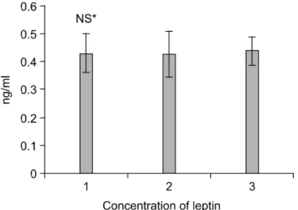
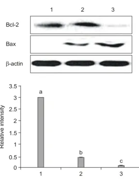
estrogen supplemented. Compared to the control, the estrogen group showed the higher estrogen level, and celecoxib showing the in-between values from control and estrogen. The leptin concentrations were com- pared, and there were no significant differences in their concentrations (Fig. 2). The effects of celecoxib in comparison with estrogen on expressions of COX-2 and molecular markers related to apoptosis in animal model system, bcl-2 and bax expression, the ratio of bcl-2 and bax were examined. In the system of perimenopausal rat mammary gland buds, celecoxib treatment resulted in the down-regulation of COX-2, and the further down-regulation was noticed with estrogen treatment (Fig. 3). The similar trend of pERK1/2 regulation was observed (Fig. 3). Celecoxib induced no changes in bcl-2, but increased bax protein (Fig. 4) compared to control. There were the marked dexpression of bcl-2 expression by estrogen (Fig. 4).
The expression of bax showed the increasing trend in the order of control, celecoxib and estrogen (Fig. 4).
The significant differences in bcl-2-bax-ratio was observed showing control the highest value, the next
Fig. 1. Concentrations of plasma estradiol after celecoxib or estrogen treatments in perimenopausal female rats. Rats were treated with basal diet, celecoxib or estrogen sup- plemented diets for four weeks. Lane 1. Control; lane 2.
celecoxib treated and lane 3. estrogen treated respectively as described in Materials and Methods. Values with different letters in the graph are significantly different (p
<0.05).
Fig. 2. Concentrations of plasma leptin after celecoxib or estrogen treatments in perimenopausal female rats. Rats were treated with basal diet, celecoxib or estrogen sup- plemented diets for four weeks. Lane 1. Control; lane 2.
celecoxib treated and lane 3. estrogen treated respectively as described in Materials and Methods.
*Not significantly different.
as celecoxib and the lowest was estrogen (Fig. 4).
DISCUSSION
The effects of celecoxib or estrogen on cell- signaling protein expression related to apoptosis such as bcl-2 and bax as well as COX-2 were investigated.
The peri-menopausal female rats were supplemented with celecoxib or estrogen. It is known that the arachi- donate cascade generates a series of lipid mediators to regulate various biological events such as cell proliferation, differentiation and inflammation through
COX pathways.32∼34) In perimenopausal female rats, oral supplementation of celecoxib or estrogen showed different COX-2 expressions in mammary gland buds.
Celecoxib clearly down-regulated COX-2 expression, and further down-regulation was observed with estro- gen supplementation. COX-2 expression is a critical part of inflammation and plays a major role in defen- ding against exogenous stimuli,35) whereas its over- expression causes cells to exhibit tumor phenotypical changes.36) Several studies indicated that COX-2 expression is associated with parameters of aggressive breast cancer.12,30,36) Based on the basic research with Fig. 3. Expression of COX-2 (top) and pERK1/2 (bottom)
after celecoxib or estrogen treatments in perimenopausal female rats. Rats were treated with basal diet, celecoxib or estrogen supplemented diets for four weeks. Mammary tissues were collected and lysates were prepared, subjected to electrophoresis on 7% SDS-PAGE, Western- blotted, and visualized with ECL detection kit described in Materials and Methods. Lane 1. Control; lane 2. celecoxib treated and lane 3. estrogen treated respectively as descri- bed in Materials and Methods. Values with different let- ters in the graph are significantly different (p<0.05).
% Vol
1 2 3
a
b
c
COX-2 expression in mammary glands 0
1.2 1
0.8 0.6 0.4 0.2 COX-2
1 2 3
pERK 1/2
β-actin
Fig. 4. Expression of bcl-2 and bax (top) and bcl-2 and bax ratio (bottom) after celecoxib or estrogen treatments in perimenopausal female rats. Rats were treated with basal diet, celecoxib or estrogen supplemented diets for four weeks. Mammary tissues were collected and lysates were prepared, subjected to electrophoresis on 7% SDS- PAGE, Western- blotted, and visualized with ECL detec- tion kit described in Materials and Methods. Lane 1.
Control; lane 2. celecoxib treated and lane 3. estrogen treated respectively as described in Materials and Methods. Values with different letters in the graph are significantly different (p<0.05).
Relative intensity
1 2 3
a
b
c
Bcl-2/Bax expression ratio in mammary glands 0
3.5 3 2.5 2 1.5 1 0.5 Bcl-2
1 2 3
Bax
β-actin
animals clinical trials, a selective COX-2 inhibitor, celecoxib was evaluated on the prevention and treat- ment of breast cancer. Epideiologic evidence suggests the incidence of breast, colon, and lung cancers is inversely related to the use of aspirin and nonsteroidal anti-inflammatory drugs, which are nonspecific inhi- bitors of COX-2 or COX-1.20) In this study, the COX- 2 down-regulatory effect of celecoxib was observed with normal mammary gland buds. Nonetheless, estro- gen, a known potent mitogen for breast cancer cells31) did not showed COX-2 stimulatory effect. Rather estrogen was found to be a highly down-regulating agent of mammary gland buds.
The up-regulated state in antiapoptotic protein bcl-2 expression was observed with celecoxib, and in con- trast there was no up-regulation of bcl-2 by estrogen.
Considering that estrogens are potent mitogen in the mammary gland playing a pivotal role in the develop- ment and progression of mammary carcinoma31), and celecoxib is one of the known breast cancer develop- ment inhibitors,18) these results may be perplexing.
The study of Basu et al. has shown that the oral administration of celecoxib caused increased tumor cell apoptosis which was associated with decreased expression of bcl-2 and increased expression of bax.37) In MCF-7 mammary tumor cells lines, 17β-estradiol inhibits apoptosis by inducing bcl-2 expression. 38) In this study 17β-estradiol induced bcl-2 up-regulation was not observed unlike in pathologic conditions. In this normal tissue, the proapoptotic potential of cele- coxib and estrogen was evident from the pattern of bcl-2 and bax expression. The reason for the dif- ferences in apototic protein expression of celecoxib and estrogen in normal or cancerous states require further investigation. The up-regulation of bcl-2 was accompanied by the decreased expression of COX-2 in celecoxib treated mammary gland buds. In the previous study, the high concentrations of estrogen down-regulated COX-2 in estrogen deficient ani- mals.39) Cellular mechanisms underlying the increased expression of bcl-2 by celecoxib and the marked depression of bcl-2 by estrogen are not clear at pre-
sent. Furthermore, the stimulation of bax by celecoxib and the prominent stimulation of bax by estrogen requires explanation.
In summary, although complete understanding of the molecular mechanism by which celecoxib and estrogen control apoptosis in normal condition of mammary gland buds needs in depth investigation, it was observed that COX-2 expression was clearly down-regulated by celecoxib and estrogen similarly in mammary gland buds, and bcl-2/bax ratio were signi- ficantly reduced by celecoxib and estrogen compared to the control. The lowering of bcl-2/bax by celecoxib and estrogen resulted from the different patterns in bcl-2 and bax expression. The lowering of bcl-2/bax by celecoxib was mainly from the increased expres- sion of bax with unchanged bcl-2, whereas estrogen reduced bcl-2/bax ratio by modulation of both bcl-2 and bax.
ACKNOWLEGEMENT
The present research was supported by Korea Research Foundation Grant (KRF-2003-002-C00289) and by KOSEF grant (F01-2003-000-00081-0).
REFERENCES
1) Wren BG. Do female sex hormones initiate breast cancer? A review of the evidence. Climacteric 2004;
7: 120-128.
2) Pike MC, Spicer DV, Dahmoush L, Press MF. Estro- gens, progestogens, normal breast proliferation and breast cancer risk. Epidemilogic Reviews 1993; 15:
17-35.
3) Rogan EG, Cavalieri EL. Estrogen metabolites, con- jugates, and DNA adducts: possible biomarkers for risk of breast, prostate, and other human cancers. Adv Clin Chem 2004: 38: 135-149.
4) Harvell DM, Strecker TE, Tochacek M, Xie B, Pennington KL, McComb RD, Roy SK, Shull JD. Rat strain-specific actions of 17β-estradiol in the mam- mary gland: correlation between estrogen-induced lobuloalveolar hyperplasia and susceptibility to estro- gen-induced mammary cancers. Proc Natl Acad Sci USA 2000; 97: 2779-2784.
5) Liehr JG, Fang WF, Sirbasku DA, Ari-Ulubelen A.
Carcinogenicity of catechol estrogens in Syrian hams- ters. J Steriod Biochem 1986; 24: 353-356.
6) Li JJ, Li SA. Estrogen carcinogenesis in hamster tissues: role of metabolism. Fed Proc 1987; 46: 1858- 1863.
7) Kohli E, Zahid M, Rogan E, Cavalieri EL. Greater reactivity of estradiol-3,4-quinone versus estradiol-2, 3-quinone with DNA in the formation of depurinating adducts. Proc Am Assoc Cancer Res 2003; 44: 180.
8) Toniolo PG, Levitz M, Zeleniuch-Jacquotte A, Baner- jee S, Koenig KL, Shore RE, Strax P, Pasternack BS.
A prospective study of endogeous estrogens and breast cancer in postmenopausal women. J Natl Cancer Inst 1995; 67: 190-197.
9) Thomas HV, Key TJ, Allen DS, Moore JW, Dowsett M, Fentiman IS, Wang DY. A prospective study of endogenous serum hormone concentrations and breast cancer risk in post-menopausal women on the island of Guernsey. Br J Cancer 1997; 76: 401-405.
10) The Endogenous Hormones and Breast Cancer Colla- borative Group. Endogenous sex hormones and breast cancer risk in post-menopausal women: reanalysis of nine prospective studies. J Natl Cancer Inst 2002; 94:
606-616.
11) Shen Q, Brown PH. Novel agents for the prevention of breast cancer: Targeting transcription factors and signal transduction pathways. J Mammary Gland Biol Neop 2003; 8: 45-73.
12) Howe LR, Subbaramaiah K, Brown AM, Dannenberg AJ. Cyclooxygenase-2: A target for the prevention and treatment of breast cancer. Endocr Relat Cancer 2001: 8: 97-114.
13) Soslow RA, Dannenberg AJ, Rush D, Woerner BM, Khan KN, Masferrer J, Koki A. COX-2 is expressed in human pulmonary, colonic, and mammary tumors.
Cancer 2000; 89: 2637-2645.
14) Robertson FM, Parrett ML, Joarder FS, Ross M, Abou-Issa HM, Alshafie G, Harris RE. Ibuprofen- induced inhibition of cyclooxygenase isoform gene expression and regression of rat mammary carcino- mas. Cancer Lett 1998; 122: 165-175.
15) Nakatsugi S, Ohta T, Kawamori T, Mutoh M, Tani- gawa T, Watanabe K, Sugie S, Sugimura T, Wakaba- yashi K. Chemoprevention by nimesulide, a selective cyclooxygenase-2 inhibitor, of 2-amino-1-methyl-6- phenylimidazo[4,5-b]pyridine (PhIP)-induced mammary gland carcinogenesis in rats. Jpn J Cancer Res 2000;
91: 886-892.
16) Alshafie GAN, Abou-Issa, Seibert K. Chemothera- peutic evaluation of celecoxib, a cyclooxygenase-2 inhibitor, in a rat mammary tumor model. Oncol Rep 2000; 7: 1377-1381.
17) Howe LR, et al. Celecoxib, a selective cyclooxy- genase-2 inhibitor, protect against human epidermal growth factor receptor 2 (HER-2)/neu-induced breast cancer. Cancer Res 2002; 62: 5405-5407.
18) Harris RE, Alshafie GA, Abou-Issa HM, Seibert K.
Chemoprevention of breast cancer in rats by cele- coxib, a cyclooxygenase-2 inhibitor. Cancer Res 2000; 60: 2101-2103.
19) Connolly EM, Harmey JH, O'Grady T, Foley, et al.
Cycloxygenase inhibition reduces tumour growth and metastasis in an orthotopic model of breast cancer. Br J Cancer 2002; 87: 231-237.
20) Rossouw JE, et al. Writing Group for the Women's Health Initiative Investigators. Risks and benefits of estrogen plus progestin in healthy postmenopausal women: principal results from the Women's Health Initiative randomized controlled trial. J Am Med Assoc 2002; 288: 321-333.
21) Shiff SJ, Koutsos LI, Oiai L, et al. Nonsteroidal anti- inflammatory drugs inhibit the cell proliferation of colon adenocarcinoma cells: effects on cell cycle and apoptosis. Exp Cell Res 1996; 222: 179-188.
22) Husain SS, Szabo IL, Pai R, Soreghan B, et al. MAPK (ERK2) kinase-a key target for SAID-induced inhibi- tion of gastric cancer cell proliferation and growth.
Life Sci 2001; 69: 3045-3054.
23) Liu XH, Yao S, Kirschenbaum A, et al. NS 398, a selective cyclooxygenase inhibitor, induces apoptosis and down-regulates bcl-2 expression in LNCaP cells.
Cancer Res 1998; 58: 4245-4249.
24) Tsujii M, Kawano S, Tsuiji S, Sawaoka H, Hori M, DuBois RN. Cyclooxygenase regulates angiogenesis induced by colon cancer cells. Cell 1998; 93: 705- 716.
25) Kroemer G. The proto-oncogene bcl-2 and its role in regulation apoptosis. Nature Med 1997; 3: 614-620.
26) Reed JC. Bcl-2: Prevention of apoptosis as a mech- anism of drug resistance. Hematol Oncol Clin North Am 1995; 9: 451-474.
27) Xu ZW, Friess H, buchler MW, Solioz M. Overex- pression of bax sensitizes human pancreatic cancer cells to apoptosis induced by chemotherapeutic agents.
Cancer Chemother Pharmacol 2002; 49: 504-510.
28) Cao Y, Prescott SM. Many actions of cyclooxy- genase-2 in cellular dynamics and in cancer. J Cell
Physiol 2002: 190: 279-286.
29) Williams CS, Mann M, DuBois RN. The role of cyclooxygenases in inflammation, cancer and develop- ment. Oncogene 1999; 18: 7908-7916.
30) Trifan OC, Hla T. Cyclooxygenase-2 modulates cellu- lar growth and promotes tumorigenesis. J Cell Mol Med 2003; 7: 207-222.
30) Kismet K, Akay MT, Abbasoglu O, Ercan A. Cele- coxib: a potent cyclooxygenase-2 inhibitor in cancer prevention. Cancer Detect Prev 2004; 28: 127-142.
31) Ding H, Han C, Zhu J, Chen CS, D'Ambrosio SM.
Celecoxib derivatives induce apoptosis via the disrup- tion of mitochondrial membrane potential and activa- tion of caspase 9. Int J Cancer 2005; 113: 803-810.
32) Hla T, Ristimaki A, Appleby S, Barriocanal JG.
Cyclooxygenase gene expression in inflammation and angiogenesis. Ann N Y Acad Sci 1993; 696: 197-204.
33) Davies P, Bailey PJ, Goldenberg MM, Ford-Hutchin- son AW. The role of arachidonic acid oxygenation in pain and inflammation. Ann Rev Immunol 1984; 2:
335-357.
34) DuBois RN, Abramson SB, Crofford L, Gupta RA, Simon LS, Van De Putte LB, Lipsky PE. Cyclo-
oxygenase in biology and diesease. FASEB J 1998;
12: 1063-1073.
35) Hinz B, Brune K. Cyclooxygenase-2-10 years later. J Pharmacol Exp Ther 2002; 300: 367-375.
36) Subbaramaiah K, Dannenberg AJ. Cyclooxygenase-2:
a molecular target for cancer prevention and treatment of breast cancer. Endorcr Relat Cancer 2001; 8:
97-114.
37) Basu GD, Pathangey LB, Tinder TL, Lagioia M, Gendler SJ, Mukherjee P. Cyclooxygenase-2 inhibitor induces apoptosis in breast cancer cells in an in vivo model of spontaneous mestastatic breast cancer. Mol Cancer Res 2004; 2: 632-642.
38) Perillo B, Sasso A, Abbondanza C, Palumbo G. 17β- estradiol inhibits apoptosis in MCF-7 cells, inducing bcl-2 expression via two estrogen-responsive elements present in the coding sequence. Mole Cell Biol 2000;
20: 2890-2901.
39) Shin JI, Kim TK, Park OJ. Phytoestrogen and estrogen regulation of cell signal protein expression in human breast cancer cells and animal model system. J Kor Ass Cancer Prev 2003; 8: 73-80.