2004; 9(1): 36-41
INTRODUCTION
During the past decade, the vast evidence has sup- ported the potential benefit of lactic acid bacteria in health promotion as a probiotics. It includes impro- vement of gastrointestinal microfloral ecosystems, reduction of serum cholesterol, stimulation of immu- nological system, removal of oxidative stress, and anticarcinogenic activities. The anticarcinogenicity of LAB has been extensively studied by Burns and
Rowland.1) According to their reports, LAB sup- plementation has been shown to have protective ef- fects against a broad range of events such as induction of DNA damage in the colonic mucosa, formation of aberrant crypt foci, tumor incidence, and alteration of bacterial enzyme activities thereby reducing the levels of enzymes involved in carcinogen formation.2∼4) In gastrointestinal tract, there might be many factors to influence the initiation and promotion of tumor cells, such as change of intestinal cytokine pattern and binding of LAB on the intestinal cells.
Corresponding author:Kun-Young Park, Department of Food Science & Nutrition, Pusan National University, Busan 609-735, Korea.
Tel: +82-51-510-2839, Fax: +82-51-514-3138, E-mail: [email protected] Received:February 17, 2004, Accepted:March 15, 2004
Lactic acid bacteria (LAB) has been emphasized to provide beneficial function to human gastrointestinal (GI) ecosystem as a probiotic. It has not been well-documented that LAB could modulate immune system in GI tract. The purpose of this study is to determine whether adhesion of three LAB (Lactobacillus casei 3260, Lac. casei 3109 and Lac. GG) on HT29 colon adenocarcinoma cells could change cytokine production pattern (Th1/Th2 profiles) in vitro. Our findings revealed that binding of Lac. casei 3260 and Lac.GG to HT-29 cell induced the increased level of Th1 type cytokine, IL-2 and IFN-γ while it decreased Th2 type cytokines, IL-1β, IL-6, and TNF-α, but not IL-4, implicating that binding of LAB on HT-29 normalized Th1/Th2 profile. In conclusion, as a probiotic, LAB may give a good effect on colon cancer by balancing the Th1/Th2 cytokine profile, but it is critically dependent on binding capacity of LAB to tumor/cancer cells and thus the pattern of cytokine production due to LAB adhesion may be bacterial strain-specific.
ꠏꠏꠏꠏꠏꠏꠏꠏꠏꠏꠏꠏꠏꠏꠏꠏꠏꠏꠏꠏꠏꠏꠏꠏꠏꠏꠏꠏꠏꠏꠏꠏꠏꠏꠏꠏꠏꠏꠏꠏꠏꠏꠏꠏꠏꠏꠏꠏꠏꠏꠏꠏꠏꠏꠏꠏꠏꠏꠏꠏꠏꠏꠏꠏꠏꠏꠏꠏꠏꠏꠏꠏꠏ Key Words: Lactic acid bacteria, Adhesion, Colon cancer, Cytokines, Th1/Th2 profile
Adhesion of Lactic Acid Bacteria Can Modulate the Secretion of Cytokines on HT-29 Colon Adenocarcinoma Cells
Jeongmin Lee1, Kwon-Tack Hwang1 and Kun-Young Park2
1Department of Food & Life Sciences, Nambu University, Gwangju 506-824, Korea,
2Department of Food Science & Nutrition, Pusan National University, Busan 609-735, Korea
Cytokines comprise a large family of intracellular communicating molecules that play important roles in immunity, inflammation and repair, as well as general tissue homeostasis. In every tissue, there is a cytokine network where the production of individual cytokines stimulates and inhibits production of, and response to, other cytokines. Therefore, it is well accepted that cytokines are key molecules controlling autocrine or paracrine communications within and between the individual cell types. Because of different patterns of receptor expression, cytokines influence a growth and survival of tumor cells. Wilson and Balkwill have highlightened the evidence for the tumor activity of endogenous TNF, IL-1 and a range of chemokines that are related to chronic inflammation and increased susceptibility to cancers.5) However, over-expression of, or treatment with, the some cytokines can provoke a powerful anti-tumor response.6)
It has been suggested that LAB, as a probiotic, could modulate or stimulate gastrointestinal immune system. One of the important criteria for a good probiotic strain is believed to be its ability to adhere to mucosal surfaces of the human gastrointestinal tract.7) Adhesion of probiotic LAB has been reported to be species specific and temporarily colonize the intestine, which beneficially influence the microbial balance of the host. For a last few years, some strains of LAB such as Lac. acidophilus LA1, Lac. GG, and Lac. casei Shirota have been proved to have an higher ability to adhere to in vitro intestinal cell lines, and their binding efficacy and characteristics have been defined in many studies.8) Despite many studies of LAB binding on colonic cancer cells, there are little reports concerning the cytokine secretion in the intestine. The aim of this study is to investigate that adhesion of three representative LAB on colonic adenocarcinoma cell (HT-29) could modulate the pat- tern of cytokine secretion after lipopolysaccharide (LPS) treatment.
MATERIALS AND METHODS 1) Bacterial Strains, culture, and counts
Lac. casei KCTC 3260, Lac. casei 3109, and Lac.
GG were obtained from the frozen stock culture collection of KCTC (Korean Culture Type Collec- tion). All strains were cultured for 24 hr in deMan- Rogosa-Sharpe (MRS) broth (Oxoid, Hampshire, United Kindom) under aerobic conditions (37oC, 10%
CO2). All strains were serially transferred at least three times prior to use in studies. Bacterial counts were determined by flow cytometry using a FACS Calibur equipped with an air-cooled 488-nm argon-ion laser at 15 mV. Direct counts were enumerated by using Fluoresbrite beads (2.0 um, Polysciences Inc.) as an internal calibration. Viability of bacterial pop- ulations was assessed by using SYTOX green nucleic acid stain (Molecular Probes, S-7020) at 1μM/106∼ 107 bacteria to detect non-viable bacteria. A band pass filter of 525 nm was used to collect the emission for green SYTOX. To support the precise bacterial counts, the colony forming unit (CFU) was deter- mined by plating serial 10-fold dilutions of bacterial suspension with MRS agar plates. For adhesion on HT-29 cells, all strains have been adjusted to 2×10⁹ CFU/ml, respectively.
2) HT-29 cell culture
The HT-29 cell line (ATCC HTB39) was purchased from the American Type Culture Collection (ATCC, Rockville, MD, USA). The cells were cultured in Macoy's 5A (Whitaker, USA) supplemented with 10% heat-inactivated (55oC, 30 min) fetal bovine serum (FBS, Whitaker, USA), 2 mM L-glutamine (Sigma), 100 U/ml penicillin and 100 mg/ml strpto- mycin (BioWhitaker, USA) at 37oC in an atmosphere of 5% CO2. For bacterial adhesion, HT-29 monolayers were prepared on 24-well tissue culture plates with a concentration of 5×10⁵ cells per well to obtain confluence, treated with 3μM methotrexate to induce secretion of mucous, and maintained for 2 weeks prior
to use. The cell culture medium was changed every other day and replaced by fresh non-supplemented Macoy's 5A at least 1 hr before the bacterial adhe- sion.
3) Measurement of cytokines
The production of IL-1β, IL-2, IL-4, IL-6, IFN-γ, and tumor necrosis factor-α (TNF-α) was determined by sandwich ELISA. Mouse anti-human IL-1β, IL-2, IL-4, IL-6, and TNF-α purified antibodies; rabbit anti- human IL-1β, IL-2, IL-4, IL-6, IFN-γ, and TNF-α biotinylated antibodies; and recombinant human IL-1β, IL-2, IL-4, IL-6, IFN-γ, and TNF-α were obtained from Endogen (Cambridge, MA). Briefly, HT-29 cells were treated with 10 ng/ml of LPS to induce Th2 cytokine production and maintained overnight. About 2×10⁹ CFU/ml of bacterial stock in PBS has added into HT-29 monolayer cells and incubated for 6 hours at 37oC in an atmosphere of 5% CO2. After centrifuga- tion of 24 well plate at 800 rpm for 10 min, 500μl of supernatant was carefully transferred to 1.5 ml of eppendorf tube and stored at -80oC until immunoas- say.
4) Statistics
All variables were compared using a one-way anal- ysis of variance (ANOVA), followed by a two-tailed Student's t-test for comparison between any two groups. Differences between two groups were consi- dered significant at p<0.05.
RESULTS AND DISCUSSION 1) Th1 type cytokines
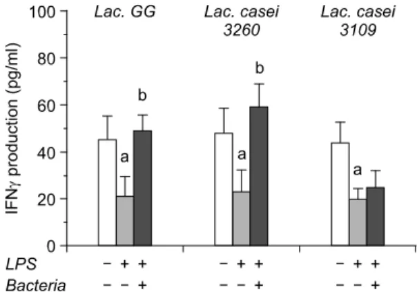
T cells could be split into two main categories, Th1 and Th2, and Th1/Th2 profile is regarded as a critical factor in initiation and promotion of cancer cells. Th1 cell predominantly produces Th1 type cytokines such as IL-2 and IFN-γ that stimulate the growth of T cells and activation of cytotoxic T cell or natural killer (NK) cells. Our data have indicated that LPS treat- ment lowered the secretion of both IL-2 and IFN-γ (Fig. 1, 2). It is not surprised that LPS treatment usually induces Th2 cytokines causing the shift of cytokine pattern from Th1 toward Th2 type. Intere- stingly, binding of LAB has increased IL-2 and IFN-γ level to near normal range. There might be two pos- sibilities to explain the increased level of these cyto-
Fig. 1. Production of IL-2 from HT-29 colon adenocar- cinoma cell after treatment of three different lactic acid bacteria. Data were represented by mean±SD. a: signi- ficantly different (p<0.05) compared to negative control (without treatment of LPS and bacteria), b: significantly different (p<0.05) compared to only LPS treatment.
Fig. 2. Production of IFNγ from HT-29 colon adenocar- cinoma cell after treatment of three different lactic acid bacteria. Data were represented by mean±SD. a: signi- ficantly different (p<0.05) compared to negative control (without treatment of LPS and bacteria), b: significantly different (p<0.05) compared to only LPS treatment.
kines. First, LAB could capture LPS to prevent induc- tion of Th2 cytokines resulting in normalizing the level of Th1 cytokines. Although there is no direct evidence that LPS binds to surface of LAB, we as- sume that there is still a chance because structure of LAB cell wall partially contains LPS with similar composition and thereby interact with exogenous LPS in randomized manner. Haskard et al.9) has reported that AFB1 is bound noncovalently and extracellularly on LAB and suggested that cell wall polysaccharide and peptidoglycan are the two main elements res- ponsible for the binding of mutagens to LAB. This data may indirectly support our data addressing LPS binding to LAB. Secondly, binding of LAB on HT-29 cells may regulate the gene expression, especially Th1 type cytokines in this study. This data could be sup- ported by Servin's review indicating that binding characteristic of LAB on Caco-2 intestinal epithelial cell induced the gene expression for antibiotic pro- teins.10) Among the bacterial strains, it showed the dif- ferent level of IL-2 and IFN-γ production, suggesting that modulation of IL-2 and IFN-γ secretion is strain- specific. This result may be caused by different bin- ding capacity of LAB to either LPS or HT-29 cells.
There are many reports that LAB has a broad range of binding on intestinal cells by approximately 0∼
45%. And the most has agreed that L. GG has higher binding ability to both in vivo and in vitro intestinal cells.11,12) Unpublished data from our laboratory indicated that Lac. casei 3260 has a similar ability on binding to intestinal cell lines as compared to Lac.
GG, but not Lac. casei 3109. The pattern of Th1 cyto- kine secretion was accordance with binding ability of LAB to intestinal cells. Therefore we assume that the binding characteristics could influence cytokine pro- duction in HT-29 cell culture.
2) Th2 type cytokines
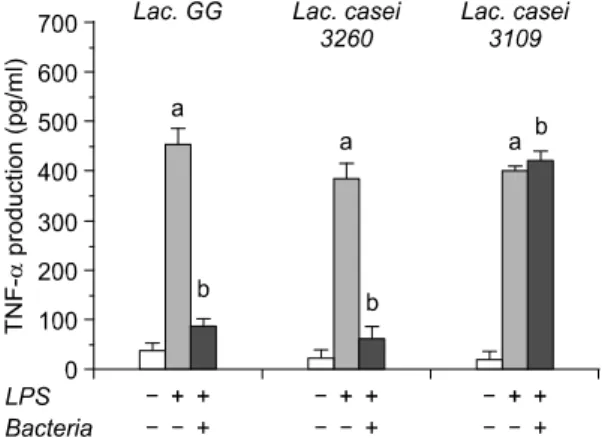
Th2 type cytokines involve IL-1, IL-4, IL-6 and TNF-α that stimulate the growth of B cells and medi- ate the inflammation response. Among them, TNF-α is a major mediator of dramatic cellular change and
dynamic tissue modeling. It can be detected in malig- nant and/or stromal cells in human breast, ovarian and colorectal cancer often in association with IL-1, IL-6 and M-CSF. In breast cancer, infiltrating macrophages are a major source of TNF-α, which may regulate a key angiogenic enzyme (thymidine phosphorylase) in the tumor epithelium.13) In prostate cancer, TNF-α production correlates with loss of androgen respon- siveness and cachexia. And it is also associated with poor prognosis in haematologic malignancies.14) In ovarian cancer, TNF-α mRNA is found in epithelial tumor islands, where there is a correlation with high TNF-α expression and high tumor grade.15) There is also evidence for pro-cancer actions of TNF-α in ani- mal models.16) Treatment of experimental ascitic ovar- ian cancer xenografts promotes adhesion of free floa- ting tumor cells to the peritoneum and solid tumor formation, and over-expression of TNF-α confers invasive properties on some tumor cells. Evidence from animal models suggests that the IL-1, especially IL-1β, may also promote tumor development and spread. In mouse models of metastases, treatment with the IL-1 receptor antagonist significantly decreased tumor development, suggesting that local production of IL-1 aids development of metastases. Moreover, mice deficient in IL-1β were resistant to the develop- ment of metastases.17)
Our data have revealed that IL-1β, IL-6 and TNF-α production significantly reduced after LAB bound to HT-29 cells (Fig. 3, 5, 6). However, IL-4 in this study has not shown any statistical difference, implicating that activation of B cells may be not involved in gastrointestinal ecosystem change caused by LAB adhesion (Fig. 4). Referred as pro-inflammatory cyto- kines, IL-1β, IL-6 and TNF-α intercommunicate each other to induce inflammatory response by media- tion of nuclear factor kappa B (NF-kB). It is so far well-accepted that TNF-α stimulates the translocation of NF-kB, which results in the genetic expression of IL-1β, IL-6 and TNF-α, and further exacerbate in- flammatory reaction in tissue or cells. It is of impor- tance in pointing out that chronic inflammation in-
creases the risk of tumor development.5) Therefore, the data in this study are in accordance with this concept and further represent possible evidence for the asso- ciation of inflammatory cytokine production with tu- mor development.
3) Conclusion
LAB has been emphasized to provide beneficial fun- ction to human gastrointestinal ecosystem. However, it has not been well-documented that LAB could modu- late immune system in GI tract. Thus our study aimed to determine whether adhesion of LAB on HT-29 Fig. 3. Production of IL-1β from HT-29 colon adenocar-
cinoma cell after treatment of three different lactic acid bacteria. Data were represented by mean±SD. a: signifi- cantly different (p<0.05) compared to negative control (without treatment of LPS and bacteria), b: significantly different (p<0.05) compared to only LPS treatment.
Fig. 4. Production of IL-4 from HT-29 colon adenocar- cinoma cell after treatment of three different lactic acid bacteria. Data were represented by mean±SD. a: signifi- cantly different (p<0.05) compared to negative control (without treatment of LPS and bacteria), b: significantly different (p<0.05) compared to only LPS treatment.
Fig. 5. Production of IL-6 from HT-29 colon adenocar- cinoma cell after treatment of three different lactic acid bacteria. Data were represented by mean±SD. a: signifi- cantly different (p<0.05) compared to negative control (without treatment of LPS and bacteria), b: significantly different (p<0.05) compared to only LPS treatment.
Fig. 6. Production of TNF-α from HT-29 colon adenocar- cinoma cell after treatment of three different lactic acid bacteria. Data were represented by mean SD. a: signifi- cantly different (p<0.05) compared to negative control (without treatment of LPS and bacteria), b: significantly different (p<0.05) compared to only LPS treatment.
colon adenocarcinoma cells could change cytokine production pattern (Th1/Th2 profiles) in vitro. LPS has been treated in at least amount (10 ng/ml) to activate TNF-α production and thereby resulted in the shift of cytokine pattern from Th1 to Th2 type. We found that, after binding of LAB on HT- 29 cells, Th1 type cytokines were increased and Th2 type cytokines except IL-4 were decreased, indicating the normaliz- ation of Th1/Th2 profile. In conclusion, as a probiotic, LAB may give a good effect on colon cancer by balancing the Th1/Th2 cytokine profile, but it is criti- cally dependent on binding capacity of LAB to tumor/
cancer cells and thus the pattern of cytokine produc- tion due to LAB adhesion may be bacterial strain- specific.
REFERENCES
1) Burns AJR, Rowland LR. Anti-carcinogenicity of pro- biotics and prebiotics on faecal water-induced DNA damage in human colon adenocarcinoma cells. Mutat Res 2004; 551: 233-243.
2) Pool-Zobel BL, Neudecker C, Domizlaff I, Ji S, Schillinger U, Rumney C, Moretti M, Vilarini I, Scas- sellati-Sforzolini R, Rowland I. Lactobacillus and Bifidobacterium mediated antigenotoxicity in the colon of rats. Nutr Cancer 1996; 26: 365-380.
3) Abdelali H, Cassand P, Bouley D, Narbonne JF. Ef- fects of dairy products on initiation of precursor le- sions of colon cancer rats. Nutr Cancer 1995; 24:
121-132.
4) Gordin BR, Gorbach SL. Effects of Lactobacillus aci- dophilus dietary supplements on 1,2-dimethylhydra- zine dichloride-induced intestinal cancer in rats. Natl Cancer Inst 1980; 64: 263-265.
5) Wilson J, Balkwill F. The role of cytokines in the epithelial cancer microenvironment. Cancer Biol 2002;
12: 113-120.
6) Balkwill F, Mantovani A. Inflammation and cancer:
back to Virchow. Lancet 2001; 357: 539-545.
7) Ouwehand AC, Kirjavainen PV, Shortt C, Salminen S. Probiotics: mechanisms and established effects.
Intern Dairy J 1999; 9: 43-52.
8) Lee YK, Salminen S. The coming of age of probiotics.
Trend Food Sci Techol 1995; 6: 241-245.
9) Haskard C, Binnion C, Ahokas J. Factors affecting the sequestration of aflatoxin by Lactobacillus rhamnosus strain GG. Chemico Biol Interact 2000; 128: 39-49.
10) Servin AL. Adhesion of probiotic strains to the intesti- nal mucosa and interaction with pathogens. Best Pract Res Clin Gastroenterol 2003; 17: 741-754.
11) Rojas M, Ascencio F, Conway PL. Purification and characterization of a surface protein from Lactoba- cillus fermentum 104R that binds to porcine small intestinal mucus and gastric mucin. Appl Environ Mic- robiol 2002; 68: 2330-2336.
12) Rinkinen M, Westermarck E, Salminen S, Ouwehand A. Absence of host specificity for in vitro adhesion of probiotic lactic acid bacteria to intestinal mucus.
Veterin Microbiol 2003; 97: 55-61.
13) Leek RD, Landers R, Fox SB, Harris AL, Lewis CE.
Association of tumour necrosis factor alpha and its receptors with thymidine phosphorylase expression in invasive breast carcinoma. Br J Cancer 1998; 77:
2246-2251.
14) Mizokami A, Gotoh A, Yamada H, Chang C, Matsu- moto T. Tumor necrosis factor-a represses androgen sensitivity in the LNCaP prostate cell cancer line.
Proc Am Assoc Cancer Res 1999; 40: 64.
15) Naylor MS, Foulkes WD, Eccles D, Balkwill FR.
Tumor necrosis factor and its receptors in human ovarian cancer. J Clin Invest 1993; 91: 2194-2206.
16) Roberts RA, Kimber I. Cytokines in non-genotoxic hepatocarcinogenesis. Carcinogenesis 1999; 20: 1397- 1401.
17) Moore R, Owens D, Stamp G, Holdworth H, Burke F, Kollias G, Balkwill FR. Tumor necrosis factor-a deficient mice are resistant to skin carcinogenesis. Nat Med 1999; 5: 828-831.