J Korean Soc Radiol 2018;78(3):163-169 https://doi.org/10.3348/jksr.2018.78.3.163
INTRODUCTION
Solid pseudopapillary neoplasm (SPN) of the pancreas, first reported by Frantz in 1956, is an uncommon but distinct pan- creatic neoplasm with a low malignant potential, accounting for 1–3% of all pancreatic tumors (1). Most SPNs have a ten- dency to affect young women and have excellent prognoses fol- lowing complete surgical resection (2). Although the majority of SPNs have an indolent clinical course and low malignant po- tential, 10% to 15% of cases exhibit aggressive clinical and pathologic features (2). Histologically, most SPNs are composed
of uniform polygonal cells with hemorrhage, necrosis, and characteristically prominent cystic portions, but there is a high- grade malignant histologic variant that is composed of numer- ous nuclear atypia and mitosis, causing a diagnostic dilemma (3). Although there are comprehensive several reports regard- ing imaging features of SPN in literature, there have been few reports specifying detailed description of diffusion weighted MR imaging findings, including mean apparent diffusion coef- ficient (ADC) values and normalized ADC values with respect to differentiating SPN with malignant transformation from be- nign SPN. We herein report a rare case of pancreatic SPN with
Imaging Findings of Pancreatic Solid Pseudopapillary Neoplasm with High-Grade Malignant Transformation: Focusing on Diffusion- Weighted Imaging and Normalized Apparent Diffusion Coefficient Values
고등급 악성전환을 보인 고형 가유두상 종양의 영상소견: 확산강조영상 및 표준화 현성확산계수값
Ka Ram Kang, MD
1, Ok Ran Shin, MD
2, Su Lim Lee, MD
1, Young Mi Ku, MD
1*
Departments of 1Radiology, 2Hospital Pathology, Uijeongbu St. Mary’s Hospital, College of Medicine, The Catholic University of Korea, Uijeongbu, Korea
Solid pseudopapillary neoplasm (SPN) of the pancreas is an uncommon neoplasm characterized by low-grade malignant potential and uncertain histogenesis, and usually afflicts relatively young women. The tumors typically manifest as well-mar- ginated, mixed density lesions consisting of a solid peripheral and central cystic component in conjunction with hemorrhagic foci. SPN with apparent high-grade malignant transformation is a rare histologic variant. Herein, we report a rare case of SPN with high-grade malignant transformation diagnosed in a 75-year-old fe- male, with emphasis on differential diagnoses and role of normalized ADC values.
SPN with malignant transformation should be included in the differential diagnosis in cases of mixed solid and cystic masses with hemorrhagic components, irregular margin and lower normalized apparent diffusion coefficient (ADC) values in an el- derly female patient, as was the case discussed below.
Index terms
Diffusion Magnetic Resonance Imaging Magnetic Resonance Imaging
Pancreatic Neoplasms
Received July 20, 2017 Revised September 13, 2017 Accepted September 18, 2017
*Corresponding author: Young Mi Ku, MD
Department of Radiology, Uijeongbu St. Mary’s Hospital, College of Medicine, The Catholic University of Korea, 271 Cheonbo-ro, Uijeongbu 11765, Korea.
Tel. 82-31-820-3148 Fax. 82-31-846-3080 E-mail: [email protected]
This is an Open Access article distributed under the terms of the Creative Commons Attribution Non-Commercial License (http://creativecommons.org/licenses/by-nc/4.0) which permits unrestricted non-commercial use, distri- bution, and reproduction in any medium, provided the original work is properly cited.
high-grade malignant transformation in an older woman fo- cusing on diffusion weighted MR imaging including normal- ized ADC values.
CASE REPORT
A 75-year-old female patient presented to our internal medi-
D
A B
C
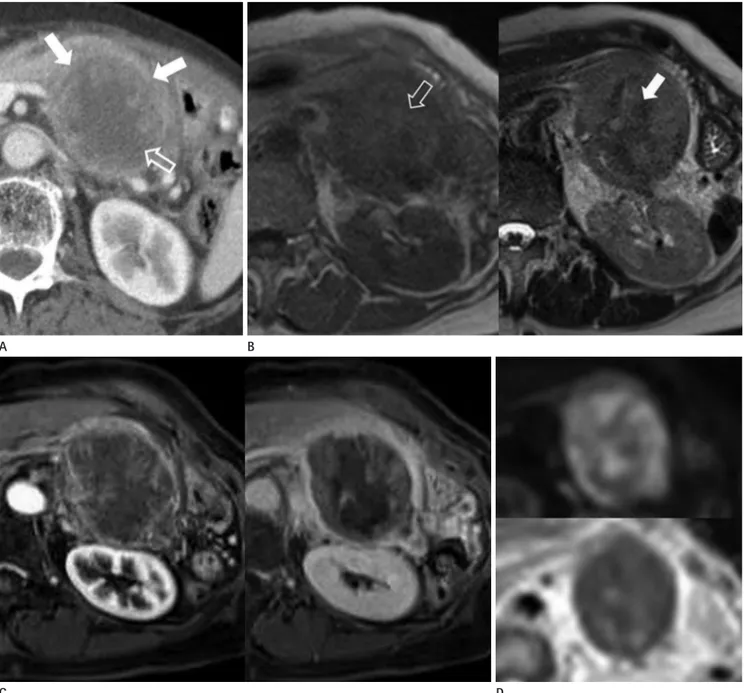
Fig. 1. A 75-year-old female patient presented a pancreatic solid pseudopapillary neoplasm with high-grade malignant transformation.
A. Contrast-enhanced axial computed tomography image demonstrates about 9.2 × 6.6 × 7.0 cm sized relatively well-demarcated lobulated mixed density mass in the tail of the pancreas, composed of enhancing solid component peripherally (solid arrows) and non-enhancing cystic component centrally (open arrow).
B. The MRI shows heterogeneous low to intermediate signal intensity with some central high signal intensity foci (open arrow) on T1-weighted image (left column), heterogeneous intermediate to high signal intensity with internal low signal intensity areas (solid arrow) on T2-weighted im- ages (right column).
C. On the dynamic MRI, the mass shows heterogeneous peripheral enhancement in the arterial phase (left column), and heterogeneous peripher- al gradual enhancement with internal non-enhancing cystic portion in the delayed phase (right column).
D. On diffusion-weighted imaging, the intensity is high, with a high b value (800 sec/mm2) (upper row) and low signal intensity on apparent dif- fusion coefficient mapping (lower row).
MRI = magnetic resonance image
cine department with the chief complaint of worsening upper abdominal discomfort that had persisted for 7 months and weight loss of 10 kg over 3 months. She had a 30-year medical history of diabetes mellitus and two surgical episodes for peri- tonitis 40 years prior.
On admission, physical examination revealed normal body temperature and stable hemodynamics. The lower abdomen was distended with rebound tenderness on palpation. Initial laboratory tests revealed a raised white blood cell count, 15.2 × 10⁹/L, and a high C-reactive protein level of 2.8 mg/dL, consis- tent with an inflammatory reaction. Other biochemical values were normal.
The initial abdominal radiograph revealed normal findings.
Subsequent contrast-enhanced abdominal computed tomogra- phy (CT) examination using a 128-detector-row CT scanner (Definition AS+; Siemens Medical Solutions, Forchheim, Ger- many) was performed to evaluate the cause of abdominal dis- comfort. We obtained axial, coronal, and sagittal reformatted images that were 5 mm in thickness. The CT scan showed an approximately 9.2 × 6.6 × 7.0 cm sized relatively well-demarcat- ed lobulated mixed density lesion with suspicious direct inva- sion to the splenic vessel and gastric lesser curvature, composed of a peripheral solid component and a central cystic component with some high density foci in the pancreatic tail, suggestive of Fig. 1. A 75-year-old female patient presented a pancreatic solid pseudopapillary neoplasm with high-grade malignant transformation.
E. Microscopic findings shows that the tumor cells are radially arranged with papillary configurations composed of delicate and somewhat hya- linized fibrovascular core on the necrotic background (solid arrows) with cellular pleomorphic and numerous mitosis (open arrow) (hematoxylin and eosin stain, × 40).
F. Immunohistochemical staining shows that the tumor cells are positive for alpha-1-antitrypsin (left column) and vimentin (right column) (im- munohistochemical stain, × 200).
E
F
a pancreatic tumor (Fig. 1A). Subsequently, we performed dy- namic magnetic resonance image (MRI) using a 3.0T system (Magnetom Verio; Siemens Medical Solutions, Erlangen, Ger- many) with a body phased-array coil. The MRI showed an en- capsulated mass with heterogeneous low to intermediate signal intensity with some central high signal intensity foci on T1- weighted images and heterogeneous intermediate to high signal intensity with internal low signal intensity areas on T2-weight- ed images (Fig. 1B). This internal high signal intensity on T1- weighted MR images and low signal intensity on T2-weighted MR images were considered as hemorrhagic component within the mass. Dynamic study of the tumor revealed peripheral gradual filling of contrast material with internal a non-enhanc- ing cystic portion (Fig. 1C). On diffusion-weighted imaging, the intensity was high, with a high b value (800 sec/mm2) and low signal intensity on apparent diffusion coefficient mapping (Fig.
1D). By applying the copy and paste function of the picture ar- chiving and communication system (PACS) system (Maroview 5.4; Marotech, Seoul, Korea), region of interests (ROIs) were identically positioned at peripheral solid portion of the pancre- atic mass, normal pancreas and spleen. For the pancreas, 2 ROIs were obtained from the head of the pancreas. For the spleen, 2 ROIs from 3 contiguous slices were measured with a central section obtained through the level of the splenic hilum. The mean and normalized ADC values were calculated using b values of 0, 50, 400, and 800 sec/mm2 from 3 contiguous slices. The mean ADC values of the solid portion, normal pancreas and spleen in our case are 0.932 × 10⁻³ mm2/sec, 1.406 × 10⁻³ mm2/sec and 0.780 × 10⁻³ mm2/sec, respectively. The patient was taken for a distal pancreatectomy with splenectomy. Intraoperatively, the resected mass was measured as 8.0 × 6.0 cm in size. The mass showed central hemorrhagic necrosis, but a whitish glistening nature in the cortical area. Morphologically, the tumor cells were arranged radially around delicate and somewhat hyalin- ized fibrovascular stalks on a necrotic background, with a few hyaline globules, and had pleomorphism and numerous mito- sis, along with deep invasion of the pancreatic parenchyma and capsule, consistent with the histologically malignant features (Fig. 1E). Moreover, the positive rate of Ki-67, a predictive fac- tor for malignancy, was more than 90%. On immune-histochem- ical staining, the tumor cells were positive for vimentin, Alpha 1 antitrypsin (AAT), and CD10 and negative for CD56 and chro-
mogranin-A (Fig. 1F). The patient recovered well and was dis- charged on the 6th postoperative day.
DISCUSSION
SPN is an uncommon exocrine tumor, accounting for 6% of all exocrine tumors in the pancreas (1). The World Health Orga- nization (WHO) classified these tumors as solid pseudopapillary tumors in 1996 and reclassified them as solid pseudopapillary neoplasms (SPNs) in 2010 (1). After the WHO reclassification in 2010, SPN is now classified as an epithelial tumor under the bor- derline subcategory and is pathologically characterized as a rare cystic pancreatic neoplasm (1). The cellular lineage of the tumors is unknown; general theories of histogenesis can be divided into three main groups: pancreatic duct cell origin, acinar cell origin, and primitive cell origin (4). Moreover, some assume that SPNs may originate from genital ridge related cells that are in- corporated into the pancreas during organogenesis on the basis of some similarities between SPN and ovarian surface cells and the proximity between genital ridges and the pancreas anlage during early embryogenesis (4). Others have speculated that sex hormones may play a role in the pathogenesis or growth of SPNs because the tumors have a tendency to affect young wom- en and the growth of an SPN in the pancreas seems to be en- hanced by pregnancy. Patients with malignant transformation of SPN are often older at presentation and have a male predi- lection (4).
SPN has distinctive pathologic features. The smaller tumors are organized in solid sheets and nests of cells. However, large tumors contain a mixture of solid, cystic, and pseudopapillary patterns. Solid areas are contain of necrosis, foamy macrophages, cholesterol granulomas, and calcifications and pseudopapillae are supported by hyalinized fibrovascular stalks (2).
Immunohistochemically, SPNs are typically positive for vi- mentin, neuron-specific enolase (NSE), α-1-antitrypsin, CD10, CD56, progesterone receptors, and β-catenin and negative for chromogranin, epithelial membrane antigen, and cytokeratin (4). Most SPNs have an indolent clinical course, but some of these have malignant potentials, include vascular or perineural invasion, Ki-67 positive, significant cellular pleomorphism, nu- clear atypia, increased mitotic activity and increased β-catenin.
According to the WHO classification scheme, SPNs with clear
criteria for malignancy (vascular and nerve sheath invasion or lymph node and liver metastasis) are designated as solid pseu- dopapillary carcinomas (SPCs) (3). There have been attempts to identify imaging characteristics that aid in differentiating be- nign SPN from malignant SPC. Recent studies demonstrate that hepatic or peritoneal involvement, main pancreatic duct obstruc- tion, infiltration of pancreatic parenchyma, vascular encase- ment, focal discontinuity of the capsule, large tumor size (> 6.0 cm) and pancreatic tail location may suggest malignancy of SPN (5, 6). The case reported in our study is consistent with aforementioned findings.
Typical, classic imaging characteristics are a well-encapsulat- ed heterogenous mass with peripheral capsule containing vary- ing amounts of internal hemorrhagic and cystic degeneration, and necrosis (1). Although imaging features of SPN overlap with those of other pancreatic neoplasms, misdiagnosis is com- mon. Internal hemorrhagic and cystic degeneration is the hall- mark of SPN owing to the fragile vascular network of the tumor.
Therefore the radiologic differential consideration in cases of mixed solid and cystic masses with hemorrhagic components in the pancreas, as in our case, should include cystic neuroen- docrine tumor, acinar cell carcinoma (ACC), and mucinous cyst- adenocarcinoma. First, areas of high signal intensity on T1- weighted images and low or inhomogeneous signal intensity on T2-weighted images, signatures of a hemorrhagic component, may help differentiate SPNs from neuroendocrine tumors, whose cystic components have increased signal intensity on T1- and T2-weighted images (4). Second, ACC of the pancreas typically appears as a well-marginated, exophytic, hypovascular mass which shows less homogenous enhancement than the surrounding pancreas when small, but contains cystic areas owing to necrosis when large. In agreement with clinical mani- festations, ACCs can also cause hyperlipasemia, which may lead to diffuse subcutaneous nodules and, interestingly, polyar- thropathy (7). Third, mucinous cystadenocarcinoma is typically seen as a predominant cystic pancreatic lesion with intracystic enhancing solid components. However, the signal intensity in the cystic portion of the mass in our case is not as high as that of the mucin component on T1- and T2-weighted MR images.
Therefore, our case’s lesions favor a diagnosis of SPN owing to a peripheral gradual enhancement pattern within the tumor and internal hemorrhagic components. In other words, despite the
fact that our patient was an older female and the imaging find- ings were aggressive, SPN should be included in the differential diagnosis in cases of mixed solid and cystic mass with hemor- rhagic components in the pancreas.
In general, the diffusion restriction that is seen primarily in malignant tumors is related to the high degree of cellularity and increased amount of macromolecules. Although there is sub- stantial overlap, the mean ADC value of malignant tumors is significant lower than that of benign lesion. Barral et al. (8) re- ported that SPNs have a lower ADC compared with normal pancreatic parenchyma with a mean ADC of 1.208 × 10⁻³ mm2/ sec (range: 1.05 × 10⁻³ mm2/sec to 2.793 × 10⁻³ mm2/sec) at 1.5 T using b values of 0, 500 and 0, 400, 800 sec/mm2. The mean ADC value of the solid portion in our case (0.932 × 10⁻³ mm2/ sec) is lower than previously reported data (1.208 × 10⁻³ mm2/ sec). However ADC values are influenced by a number of fac- tors including the patient, hardware, acquisition related param- eters and artifacts related to the susceptibility effect (9). To overcome these matters, standardized diffusion-weighted im- aging MR imaging protocols have been made including nor- malization of ADC values using the normal pancreas and spleen (9, 10). Previous report revealed that normalized ADC using the normal pancreas as a reference organ helps characterize focal pancreatic lesions and further discriminate between pancreatic malignant tumor, benign tumor and mass-forming pancreatitis, measuring in 0.933, 1.807, 0.839 in median, respectively (10). In our case, normalized ADC values using the pancreas as a refer- ence organ was 0.662, which is much lower than that of the ma- lignant tumor (0.933) and mass forming pancreatitis (0.893). The reason for decreased ADC value is probably because the mass has histologically high cellularity, pleomorphism and increased mitosis.
According a recent study, the spleen is an ideal reference organ for normalization of ADC value, and is usually used for quanti- tative analysis (9). In our case, normalized ADC value using the spleen as reference organ was calculated to be 1.194, lower than that of reported normalized ADC values of the pancreas (1.208).
Our report suggests that normalized ADC value may have a po- tential role in discriminating between benign and malignant SPN.
However, because we only have a single case review, there is limitation for characterization of benign and malignant pancre-
atic pathologies. Thus, further studies are needed.
In summary, SPN with malignant transformation should be included in the differential diagnosis in cases of mixed solid and cystic masses with hemorrhagic components, irregular margin and lower normalized ADC values in old female patients.
REFERENCES
1. Sunkara S, Williams TR, Myers DT, Kryvenko ON. Solid pseu- dopapillary tumours of the pancreas: spectrum of imaging findings with histopathological correlation. Br J Radiol 2012;
85:e1140-e1144
2. Tang LH, Aydin H, Brennan MF, Klimstra DS. Clinically ag- gressive solid pseudopapillary tumors of the pancreas: a re- port of two cases with components of undifferentiated car- cinoma and a comparative clinicopathologic analysis of 34 conventional cases. Am J Surg Pathol 2005;29:512-519 3. Bosman FT, Carneiro F, Hruban RH, Theise ND. WHO classi-
fication of tumours of the digestive system. Lyon: World Health Organization 2010
4. Coleman KM, Doherty MC, Bigler SA. Solid-pseudopapillary tumor of the pancreas. Radiographics 2003;23:1644-1648 5. Lee JH, Yu JS, Kim H, Kim JK, Kim TH, Kim KW, et al. Solid
pseudopapillary carcinoma of the pancreas: differentia-
tion from benign solid pseudopapillary tumour using CT and MRI. Clin Radiol 2008;63:1006-1014
6. Yin Q, Wang M, Wang C, Wu Z, Yuan F, Chen K, et al. Differ- entiation between benign and malignant solid pseudopap- illary tumor of the pancreas by MDCT. Eur J Radiol 2012;81:
3010-3018
7. Tatli S, Mortele KJ, Levy AD, Glickman JN, Ros PR, Banks PA, et al. CT and MRI features of pure acinar cell carcinoma of the pancreas in adults. AJR Am J Roentgenol 2005;184:
511-519
8. Barral M, Taouli B, Guiu B, Koh DM, Luciani A, Manfredi R, et al. Diffusion-weighted MR imaging of the pancreas: cur- rent status and recommendations. Radiology 2015;274:
45-63
9. Song JS, Hwang SB, Chung GH, Jin GY. Intra-individual, in- ter-vendor comparison of diffusion-weighted MR imaging of upper abdominal organs at 3.0 Tesla with an emphasis on the value of normalization with the spleen. Korean J Radiol 2016;17:209-217
10. Barral M, Sebbag-Sfez D, Hoeffel C, Chaput U, Dohan A, Eveno C, et al. Characterization of focal pancreatic lesions using normalized apparent diffusion coefficient at 1.5-Tes- la: preliminary experience. Diagn Interv Imaging 2013;94:
619-627
고등급 악성전환을 보인 고형 가유두상 종양의 영상소견:
확산강조영상 및 표준화 현성확산계수값
강가람
1· 신옥란
2· 이수림
1· 구영미
1*
췌장의 고형 가유두상 종양은 그 조직 발생에 대해서는 명확히 알려져 있지 않지만, 비교적 낮은 악성도를 보이는 종양으 로 주로 젊은 여성에서 호발한다. 일반적으로 경계가 좋고 주변부는 고형 성분으로, 중심부는 출혈성 병소를 포함하는 낭 성 성분으로 구성된 비균일한 음영을 가지는 병변으로 보인다. 고등급 악성전환을 보이는 고형 가유두상 종양은 흔하지 않은 변이 형태이다. 저자들은 75세 여성에서 고등급 악성전환을 보인 고형 가유두상 종양의 증례를 보고하고, 감별진단 을 하는 데에 있어서 표준화 현성확산계수값의 유용성에 대해 알리고자 한다. 본 증례처럼 고령의 여성에서 명확하지 않 은 경계를 보이며, 주위 조직으로의 침범소견을 보이고 출혈성 병소를 포함하며 낮은 표준화 현성확산계수값을 보이는 고 형성분이 혼합된 경우, 고형 가유두상 종양의 악성전환을 감별에 포함해야 한다.
가톨릭대학교 의과대학 의정부성모병원 1영상의학과, 2병리과