Concurrent Nevus Sebaceus and Nevus Comedonicus
Vol. 26, No. 4, 2014 501
Received May 3, 2011, Revised May 19, 2012, Accepted for publication April 7, 2012
Corresponding author: Weon Ju Lee, Department of Dermatology, Ky- ungpook National University School of Medicine, 130 Dongdeok-ro, Jung-gu, Daegu 700-721, Korea. Tel: 82-53-420-5838, Fax: 82-53-426- 0770, E-mail: weonju@knu.ac.kr
This is an Open Access article distributed under the terms of the Creative Commons Attribution Non-Commercial License (http://
creativecommons.org/licenses/by-nc/3.0) which permits unrestricted non-commercial use, distribution, and reproduction in any medium, provided the original work is properly cited.
Ann Dermatol Vol. 26, No. 4, 2014 http://dx.doi.org/10.5021/ad.2014.26.4.501
CASE REPORT
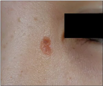
Fig. 1. A yellowish, bean-sized, verrucous plaque on the left me- dial canthus, and multiple, scattered, pea-sized, hyperpigmented macules on the face.
Two Concurrent Facial Epidermal Nevi without Systemic Abnormalities: Nevus Sebaceus and Nevus Comedonicus
Han Jin Jung, Hyun Wuk Cha, Hyun Jung Lim, Seok-Jong Lee, Do Won Kim, Weon Ju Lee
Department of Dermatology, Kyungpook National University School of Medicine, Daegu, Korea
Epidermal nevi (EN) are hamartomatous lesions derived from epidermal components originating from pluripotent cell mutations. They have been categorized according to their predominant component. The existence of >2 types of EN concurrently within a single area or within contiguous areas has been rarely reported. This report describes the case of simultaneous presence of a yellowish plaque on the left medial canthus and an aggregation of closed comedo-like papules on the right side of the cheek of a 15-year-old girl.
(Ann Dermatol 26(4) 501∼504, 2014) -Keywords-
Epidermal nevus, Nevus comedonicus, Nevus sebaceus
INTRODUCTION
Epidermal nevi (EN) are hamartomatous lesions derived from epidermal components originating from pluripotent cell mutations. EN include verrucous epidermal nevus, nevus sebaceus, wooly hair nevus, and nevus comedo- nicus. The incidence of EN has been reported as 1 to 3 per 1,000 live newborns1,2. Their location is variable, following Blaschko lines, and reflecting embryonic migra- tion patterns of the skin1. EN have been classified accor- ding to their predominant component; however, in some
nevi, the predominant tissue may vary with the evolution of the lesion, and different areas of the same lesion may show a variety of components at the same time3. This report describes a 15-year-old girl with concurrent nevus sebaceus (NS) and nevus comedonicus (NC) with no organ involvement.
CASE REPORT
A 15-year-old girl presented with a yellowish plaque on the left medial canthus and a group of closed comedo-like papules on the right side of the cheek. She has not had any developmental problems or a history of seizures. No other family members had similar lesions. The 2 different EN lesions on the patient’s face were noticed during a dermatological examination. The yellowish, asymptoma- tic, bean-sized plaque with a verrucous surface was noted
HJ Jung, et al
502 Ann Dermatol
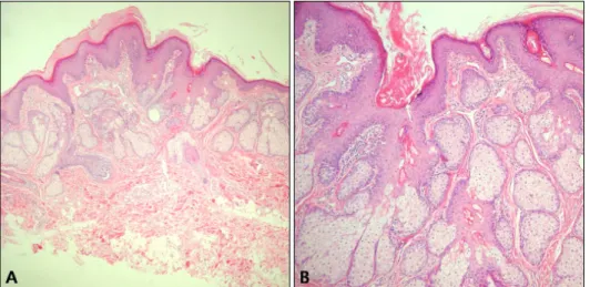
Fig. 3. (A) Marked acanthotic and papillomatous epidermal hyperpla- sia with hyperkeratosis (H&E, ×40).
(B) Excessive number of large seba- ceous glands (H&E, ×100).
Fig. 2. An aggregation of closed comedo-like papules and depressed pinpoint scars on the right side of the cheek.
on the left medial canthus (Fig. 1). It was present at birth.
In addition, an aggregation of closed comedo-like papules and depressed pinpoint craters were found on the right side of the cheek (Fig. 2). It developed at the age of 10 years. She has also had numerous, scattered, pea-sized, hyperpigmented macules on her face for several years (Fig. 1). Her physical examination was normal except for the dermatological findings. Her neurological examination and laboratory examinations, including complete blood cell count, blood chemistry, and urine analysis, were within normal limits. Histopathological examination of the
plaque on the left medial canthus showed acanthotic and papillomatous epidermal hyperplasia and large sebaceous glands located close to the epidermis (Fig. 3). Histopa- thological examination of the papule on the right side of the cheek revealed a dilated follicular infundibulum filled with keratin and incomplete hair follicles, consistent with the clinical diagnosis of NC (Fig. 4). She received laser treatment for NS and comedo extraction for NC in a private clinic before visiting our hospital. However, the NS of the left medial canthus recurred, and depressed craters were left with comedones on the right side of the cheek. The NS was surgically removed from the left medial canthus. The patient declined treatment of the NC and hyperpigmented macules.
DISCUSSION
EN are hamartomatous proliferations of the epithelium, in- cluding keratinocytes, sebocytes, pilosebaceous units, ecc- rine glands, or apocrine glands. Eighty percent of the le- sions appear within the first year of life, with most of the lesions appearing by the age of 14 years. In this patient, NS appeared at birth, and NC was observed at the age of 10 years. The term epidermal nevus syndrome refers to the association of EN with extracutaneous abnormalities4. Abnormalities of the central nervous system, skeletal sys- tem, eyes, and oral cavity were reported in most cases.
We did not detect any other systemic abnormality in our patient. The cutaneous features of EN depend, in part, on the predominant cell type involved, degree of cellular differentiation, location of the body part affected, and age of the patient. EN follow linear patterns known as ‘the lines of Blaschko’. They seem to represent the dorso- ventral migratory pathways of the neuroectoderm during embryogenesis5. The most common sites of involvement
Concurrent Nevus Sebaceus and Nevus Comedonicus
Vol. 26, No. 4, 2014 503 Fig. 4. (A) A wide, deep, epidermal invagination filled with keratin and incomplete hair follicles (H&E,
×100). (B) Well-defined keratin-fi- lled cyst in the mid-dermis (H&E,
×100).
are the head and neck. In the present case, NS and NC were localized on the face following the lines of Blaschko.
NS is relatively common, representing approximately one- half of all EN6. NS is linear, hairless, yellowish-brown, waxy, and flat at birth. It becomes plaque-like with a ve- rrucous surface owing to the hormonal changes during puberty, and usually affects the scalp, neck, and face7. NC is a rare, sporadic epidermal nevus, characterized by an aggregation of dilated hair follicles filled with keratin plugs resembling comedones. NC was first described as an entity by Kofmann8 in 1895, and it usually has a linear or zosteriform configuration. NC appears on the face, chest, trunk, or abdomen. It may develop at any time from birth to middle age but is usually present by the second decade. NC lesions follow a noninflammatory or inflam- matory course and do not resolve spontaneously. The inflammatory course may result in cyst formation with a recurrent infection, fistulae, abscesses, and scarring9. Our case showed the typical clinical findings of noninfla- mmatory NC.
The NC in our case needed to be clinically differentiated from comedonal acne. Consistent with NC, our case sh- owed the presence of comedones, which on extraction left a crater on the skin surface. In addition, it followed the lines of Blaschko, and it was confined to one side of the face. Besides the typical grouping of comedones, the age of onset and persistence made it easy to distinguish this condition from comedonal acne10. Histopathological exami- nation was used to differentiate NC from comedonal acne.
Unlike comedonal acne, the pilosebaceous units in NC were poorly formed. A dilated pore of Winer can some- times be confused with NC in a histopathological exami- nation. However, this condition is usually seen in the elderly and can be clinically differentiated.
Vidaurri-de la Cruz et al.1 evaluated 443 patients with EN.
They found NS in 168 (38%) patients, and NC in 5 (1%) patients. They did not detect ≥2 EN together in their series. Köse et al.11 described a 22-year-old man with 3 different EN (NS, Becker’s nevus, and NC). The patient had NS on his neck, NC on the right side of the spine, and Becker’s nevus on the left shoulder. Kim and Kang12 also reported on a 21-year-old man with NC and epidermal nevus at the same site. He presented with comedo-like papules on the right buttock and thigh that had been present since infancy, and at age 14 years, he noticed the epidermal nevus lesion surrounding the comedones.
Although some cases of NC associated with other EN, including verrucous linear nevus, have been reported, NC accompanying NS is very rare13. Our patient showed NC on the right side of the cheek and NS on the left medial canthus.
NS and NC are considered components in the spectrum of androgen-sensitive disorders11. It is well known that NS increases in size at puberty, revealing a correlation with the androgen hormone. Although there are no data about the androgen receptor, NC shows acneiform eruption pro- moted by androgen stimulation.
Facial hyperpigmented macules cannot be categorized as a particular type of EN. However, the macules of our pa- tient were in close proximity to the NS and the NC. Me- lanocytic nevus is considered one of the cutaneous ab- normalities in EN14. Moreover, considering that phacoma- tosis pigmentokeratotica is characterized by the presence of an NS and a contralateral or ipsilateral speckled len- tiginous nevus, hyperpigmented macules may be asso- ciated with NS or NC15.
We report a rare case of 2 different EN, including NS and NC, with no systemic involvement.
HJ Jung, et al
504 Ann Dermatol
REFERENCES
1. Vidaurri-de la Cruz H, Tamayo-Sánchez L, Durán-McKinster C, de la Luz Orozco-Covarrubias M, Ruiz-Maldonado R.
Epidermal nevus syndromes: clinical findings in 35 patients.
Pediatr Dermatol 2004;21:432-439.
2. Solomon LM, Esterly NB. Epidermal and other congenital organoid nevi. Curr Probl Pediatr 1975;6:1-56.
3. Rogers M, McCrossin I, Commens C. Epidermal nevi and the epidermal nevus syndrome. A review of 131 cases. J Am Acad Dermatol 1989;20:476-488.
4. Hodge JA, Ray MC, Flynn KJ. The epidermal nevus syn- drome. Int J Dermatol 1991;30:91-98.
5. Moss C, Larkins S, Stacey M, Blight A, Farndon PA, Davison EV. Epidermal mosaicism and Blaschko's lines. J Med Genet 1993;30:752-755.
6. Rogers M. Epidermal nevi and the epidermal nevus syndro- mes: a review of 233 cases. Pediatr Dermatol 1992;9:342- 344.
7. Perez-Munoz MA, Hernandez Garcia MJ, Ríos JJ, Camacho F.
Sebaceous naevi: a clinicopathological study. J Eur Acad
Dermatol Venereol 2002;16:319-324.
8. Kofmann S. Ein Fall von seltener Lokalisation und Verbrei- tung von Komedon. Arch Dermatol Syphilol 1895;32:177- 178.
9. Beck MH, Dave VK. Extensive nevus comedonicus. Arch Dermatol 1980;116:1048-1050.
10. Decherd JW, Mills O, Leyden JJ. Naevus comedonicus--treat- ment with retinoic acid. Br J Dermatol 1972;86:528-529.
11. Köse O, Calişkan E, Kurumlu Z. Three different epidermal naevi with no organ involvement: sebaceous naevus, naevus comedonicus and Becker's naevus. Acta Derm Venereol 2008;88:67-69.
12. Kim SC, Kang WH. Nevus comedonicus associated with epidermal nevus. J Am Acad Dermatol 1989;21:1085-1088.
13. Domonkos AN, Arnold HL, Odom RB. Andrew’s disease of the skin. 7th ed. Philadelphia: W.B. Saunders Co., 1982:795.
14. Hill VA, Felix RH, Mortimer PS, Harper JI. Phacomatosis pigmentokeratotica. J R Soc Med 2003;96:30-31.
15. Happle R, Hoffmann R, Restano L, Caputo R, Tadini G.
Phacomatosis pigmentokeratotica: a melanocytic-epidermal twin nevus syndrome. Am J Med Genet 1996;65:363-365.