2020 Korean Society for Surgery of the Hand, Ko- rean Society for Microsurgery, and Korean Society for Surgery of the Peripheral Nerve. All Rights re- served.
This is an open-access article distributed under the terms of the Creative Commons Attribution Non-Commercial license (http://creativecommons.
org/licenses/by-nc/4.0/), which permits unrestricted non-commercial use, distribution, and reproduction in any medium, provided the original work is prop- erly cited.
Resection of biliary tract malignancies may require resection of the hepatic vascula- ture. While immediate revascularization of the liver is necessary, reconstruction is dif- ficult when the original vessels are unavailable. We document a case in which a seg- ment of the common hepatic artery was excised during tumor resection and the re- maining proximal vessel displayed intima dissection. A greater saphenous vein was placed as a bridge between the remaining left hepatic artery and gastroduodenal ar- tery for successful revascularization.
Keywords: Hepatic artery, Saphenous vein, Autograft
대복재정맥편을 이용한 좌간동맥 재관류 증례
최종윤1, 정이룸1, 최장연1, 김기환2, 정성노1, 서보미1
1가톨릭대학교 의정부성모병원 성형외과, 2가톨릭대학교 의정부성모병원 일반외과
Greater Saphenous Vein Graft
Revascularization of the Left Hepatic Artery after Resection of Intrahepatic
Cholangiocarcinoma with Common Hepatic Artery Resection
Jong Yun Choi
1, Ee Room Jung
1, Jangyoun Choi
1, Ki Hwan Kim
2, Sung-No Jung
1, Bommie Florence Seo
11Department of Plastic and Reconstructive Surgery, Uijeongbu St. Mary’s Hospital, College of Medicine, The Catholic University of Korea, Uijeongbu, Korea
2Department of General Surgery, Uijeongbu St. Mary’s Hospital, College of Medicine, The Catholic University of Korea, Uijeongbu, Korea
pISSN 2586-3290 · eISSN 2586-3533 Arch Hand Microsurg 2020;25(2):161-165 https://doi.org/10.12790/ahm.19.0064
Case Report
Received: November 7, 2019 Revised: April 17, 2020 Accepted: April 21, 2020 Corresponding author:
Bommie Florence Seo Department of Plastic and
Reconstructive Surgery, Uijeongbu St.
Mary’s Hospital, College of Medicine, The Catholic University of Korea, 271 Cheonbo-ro, Uijeongbu 11765, Korea Tel: +82-31-820-3074
Fax: +82-31-874-0301
E-mail: bommiefseo@catholic.ac.kr ORCID:
https://orcid.org/0000-0002-6907-5962
INTRODUCTION
In hilar biliary tract malignancies, achieving negative surgical margins is greatly challenging due to the proximity of the liver and hepatic artery (HA) vasculature.
Sufficient resection of the tumor may compromise arterial flow to the liver, and necessitate fabrication of the HA. Arterial reconstruction is also complicated by the factor of individual anatomical variations [1]. There have been studies report- ing successful HA reconstruction using intra- or extra-abdominal vascular grafts during liver transplantation [2-5]. However, when there is an extensive vascular defect that is difficult to overcome with intra-abdominal methods, extra-abdomi- nal grafts must be utilized. Here we report a case of left HA revascularization us- ing an interpositional greater saphenous vein (GSV) graft bridge from the gastro- duodenal artery after resection of intrahepatic cholangiocarcinoma.
CASE REPORT
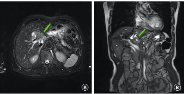
An 80-year-old male presenting with abdominal discomfort was diagnosed with cholangiocarcinoma. Magnetic resonance imaging revealed a 2.5-cm sized mass with delayed enhance- ment in the caudate lobe, with hilar involvement abutting the portal vein (Fig. 1). During open liver hemihepatectomy and cholecystectomy a 3-cm sized mass involving the caudate lobe, hepatic bifurcation, and perihilar lymph node enlargement was exposed. In the process of en bloc tumor resection around 2 cm of the left HA was excised, and the resulting arterial defect re- quired reconstruction. Because the proximal stump of the proximal common HA revealed intimal dissection, attempts at in situ anastomosis between both margins of the defect were unsuccessful. Our team evaluated the gastroduodenal artery for a turnover arterial graft, however, much of the gastroduodenal artery also revealed intimal dissection, and after trimming to a healthy margin, the proximal stump was too short to be con- nected to the hepatic vasculature. A bridging vein graft from the GSV was planned. A portion of the left GSV 10 cm in length was harvested from the lower leg with loupes, and inter- posed to prevent obstruction at valves. Under microscope vi- sion, after trimming to 3 cm, an end-to-end anastomosis was done between the left HA and the GSV graft using 8-0 ETHILON sutures first (Ethicon, Somerville, NJ, USA). Addi- tional trimming to a length of 2.7 cm was done, and the proxi- mal end of the GSV graft was anastomosed in the same manner
to the gastroduodenal artery (Fig. 2). With removal of the ves- sel clamps, fresh bleeding increased at the cut surface of the hepatectomy. Empty-and-refill test and ultrasound Doppler were done and showed successful anastomosis.
Informed consent was obtained.
DISCUSSION
Radical surgical resection until a negative margin is secured provides a higher chance for cure and event free survival in ad- vanced biliary cancer. However, the vascular anatomy in the hi- lar area is very complex, consisting of major vessels that directly supply vital organs, including the HA. Therefore vascular inva- sion of the biliary tumor is one of the main limitation factors that result in irresectability. Sufficient resection of the tumor along with the HA may compromise hepatic perfusion, and re- quire HA reconstruction. Although Hu et al. [2] reported that HA resection during surgical excision of hilar cholangiocarci- noma may not require HA reconstruction in highly selected cases, most other studies have noted that failure to reconstruct the HA may lead to serious complications including hepatone- crosis or hepatic abscesses [2,6]. Thus, the surgeon must be equipped with the ability to fabricate the deficient HA.
When the arterial defect is smaller than 1 cm, the remaining HA can be dissected and advanced from the surrounding tis- sue. Anatomical variations of the HA vasculature should be
Fig. 1. Magnetic resonance imaging revealed a 2.5-cm sized mass (arrow) with delayed enhancement in the caudate lobe of the liver abutting the portal vein, leading to suspicion of mass forming cholangiocarcinoma: (A) axial view and (B) coronal view.
A B
contemplated. While the majority (80%) of cases are reported to have normal (Michels type I) HA anatomy, the remaining 20% may express one of numerous variations (Michels type II, replaced left HA from the left gastric artery; Michels type III, replaced right HA from the superior mesenteric artery; Michels type IV, replaced right HA and left HA; Michels type V, acces- sory left HA; Michels type VI, accessory right HA; Michels type VII, accessory right HA and left HA; Michels type VIII, re- placed right HA or left HA with other HA being accessory one;
Michels type IX, hepatic trunk as a branch of the superior mes- enteric artery; and Michels type X, the common HA from the left gastric artery). A replaced right HA or left HA may be found, while accessory right and left HA occur in 0.8%-8% of patients [1,7,8]. Larger defects may be reconstructed using in- tra- or extra-abdominal vessels. Intra-abdominal vascular re- construction requires knowledge on the various proximal ar- teries that may be utilized within the area. Extra-anatomic transposition that origin from the celiac trunk such as the right gastroepiploic artery, the gastroduodenal artery, the left gastric artery, or the splenic artery may be used for reconstruction.
Defects larger than 2 cm will warrant a vessel graft. Intra-ab- dominal arterial grafts from the gastroduodenal artery, inferior mesenteric artery, iliac artery, splenic artery, and branches of the celiac trunk may be harvested [3-5].
When manipulation of the abdominal vasculature is unsafe and unwanted, the surgeon may opt to use an extra-abdominal or ectopic autologous graft. Reports on the use of the radial ar- tery and the brachiocephalic artery for intraabdominal vascular reconstruction have been reported [9,10]. However, harvesting a major artery is inevitably accompanied by donor morbidity, and may be limited in availability. The GSV graft is a reliable
autologous vessel graft donor, with less anatomical variation and minimal donor morbidity. It is a readily available option that is swiftly and easily harvested with a diameter that is simi- lar to the hepatic vasculature. Bhatti et al. [5] have compared the outcomes of GSV grafts to arterial reconstruction using na- tive HAs, and found that patency was excellent, though GSV grafts did have a higher rate of complications.
Although not optimal, the GSV graft is a reliable option that may be utilized in cases where the native hepatic vasculature has been sacrificed, or when the surrounding arteries are diffi- cult to use. Much care should be taken to preserve the normal anatomy, however this should not be a restricting factor for tu- mor resection. To ensure radical resection of hilar malignancies of the biliary tract, the surgeon should be equipped with a solu- tion for revascularization of the HA.
CONFLICTS OF INTEREST
The authors have nothing to disclose.
REFERENCES
1. Fonseca-Neto OC, Lima HC, Rabelo P, Melo PS, Amorim AG, Lacerda CM. Anatomic variations of hepatic artery: a study in 479 liver transplantations. Arq Bras Cir Dig. 2017;
30:35-7.
2. Hu HJ, Jin YW, Zhou RX, et al. Hepatic artery resection for bismuth type III and IV hilar cholangiocarcinoma: is recon- struction always required? J Gastrointest Surg. 2018;22:1204- 12.
3. Ozer A, Aktas H, Eren N, Karakayali H, Emiroglu R. Hepatic arterial reconstruction using right gastroepiploic artery in liv- ing donor liver transplantation. Transplant Proc. 2018;
50:3559-61.
4. Del Gaudio M, Grazi GL, Ercolani G, et al. Outcome of hepat- ic artery reconstruction in liver transplantation with an iliac arterial interposition graft. Clin Transplant. 2005;19:399-405.
5. Bhatti AB, Dar FS, Qureshi AI, Haider S, Khan NA. Saphe- nous vein conduits for hepatic arterial reconstruction in living donor liver transplantation. Langenbecks Arch Surg. 2019;
404:293-300.
6. Wang WL, Tang XF, Yao MY, et al. Safety and efficiency of left hemihepatectomy combined with hepatic artery resection for hilar cholangiocarcinoma with artery infiltration: report of 2 cases. Can J Surg. 2008;51:305-7.
7. Noussios G, Dimitriou I, Chatzis I, Katsourakis A. The main anatomic variations of the hepatic artery and their importance Fig. 2. A bridging vein graft from the greater saphenous vein (GSV)
was done between the left hepatic artery and gastroduodenal artery.
in surgical practice: review of the literature. J Clin Med. Res.
2017;9:248-52.
8. Michels NA. Newer anatomy of the liver and its variant blood supply and collateral circulation. Am J Surg. 1966;112:337-47.
9. Lin TS, Yang JC, Chen CL. Hepatic artery reconstruction us- ing radial artery interposition graft in living donor liver trans-
plantation. Transpl Int. 2013;26:e28-30.
10. Javerliat I, Pichon A, Glorion M, Coscas R, Goeau-Brisson- niere O, Coggia M. A new technique for intra-abdominal ar- teries revascularization via extra-anatomic bypass from the brachiocephalic artery with a videoscopic retrosternal tunnel.
J Vasc Surg. 2015;62:256-8.
간내 담관암 및 총간동맥 절제술 후 대복재정맥편을 이용한 좌간동맥 재관류 증례
최종윤1, 정이룸1, 최장연1, 김기환2, 정성노1, 서보미1
1가톨릭대학교 의정부성모병원 성형외과, 2가톨릭대학교 의정부성모병원 일반외과
담관암의 수술적 절제 시, 통상 간내 혈관에 대한 절제를 같이 시행하는 경우가 많다. 절제 후 간의 즉각적인 재관류가 필수적이지만, 기존의 혈관이 손상되었을 경우, 이에 대한 재건에 어려움이 있다. 본 증례의 경우 종양 절제 시 총간동맥의 일부가 같이 절제되었으며, 잔존하는 혈관내피조직 또한 손상되어 있었다. 이에 저자는 대복재정맥편을 이용하여 잔존하는 좌간동맥과 위십이지장동맥 사이의 성공적인 재관류를 회복하였기에 이를 보고하고자 한다.
색인단어: 간동맥, 대복재정맥, 자가이식
접수일 2019년 11월 7일 수정일 2020년 4월 17일 게재확정일 2020년 4월 21일 교신저자 서보미
11765, 경기도 의정부시 천보로 271, 가톨릭대학교 의정부성모병원 성형외과 TEL 031-820-3074 FAX 031-874-0301 E-mail bommiefseo@catholic.ac.kr ORCID https://orcid.org/0000-0002-6907-5962