Proteomic Identification of Proteins Interacting with a Dual Specificity Protein Phosphatase, VHZ
Jae-Hoon Kim
1,2,* and Dae-Gwin Jeong
31Faculty of Biotechnology, College of Applied Life Science, Cheju National University, Jeju 690-756, Korea
2Gene & Material Bank for Citrus Breeding, Cheju National University, Jeju 690-756, Korea
3Systemic Proteomic Research Center, Korea Research Institute of Bioscience and Biotechnology, Daejeon 305-806, Korea
Received April 17, 2007; Accepted May 9, 2007
Identification of Dual-specificity protein phosphatase (DSP) substrates is essential in revealing physiological roles of DSPs. We isolated VHZ-interacting proteins from extracts of 293T cells overexpressing a VHZ (C95S, D65A) mutant known to be substrate- trapping mutant. Analysis of specific proteins bound to VHZ by 2D gel electrophoresis and mass spectroscopy revealed that these proteins contained Chaperonin containing TCP1, Type II phosphatidylinositol phosphate kinase γ, Intraflagellar transport 80 homolog, and Kinesin superfamily protein 1B. VHZ- interacting proteins showed that VHZ is involved in many important cellular signal pathways such as protein folding, molecular transportation, and tumor suppression.
Key words: dual specificity protein phosphatase, interacting protein, proteomic identification, VHZ
A family of dual-specificity protein phosphatases (DSPs) dephosphorylates protein substrates at both the tyrosine and serine/threonine residues [Alonso et al., 2004c], regulating the phosphorylation levels of several proteins that are essential for the physiological processes involved in cell survival, proliferation, and differentiation [Yu et al., 2007; Rahmouni et al., 2006; Sakaue et al., 2004; Wu and Bunnett, 2005]. The mammalian genome encodes dozens of DSPs, and all of the catalytic domains of DSPs contain a consensus signature motif HCXXGXXR and have good alignments in the overall structures among them [Denu and Dixon, 1998]. However, each DSP has significant differences in the conformation of many residues and the surface charges, and presumably shows an individual function in the recognition of substrates and targeting to defined subcellular locations [Kim et al., 2007]. Of these DSPs, the Vaccina H1-related phosphatase VHR represents a prototype of DSPs that lacks the MAP kinase-binding (MKB) domain. Without the MKB domain, VHR binds to the activated MAP kinases and downregulates the MAP kinase signaling [Alonso et al., 2001].
Recently, several interesting Vaccina H1-related phosphatases including VHX, VHY, and VHZ were cloned and characterized. VHX specifically activates c- Jun N-terminal kinase (JNK) pathway in HEK 293T cells [Chen et al., 2002]. In jurkat T cells, VHX suppressed T cell antigen receptor-induced activation of extracellular signal-regulated kinase 2 (Erk2) [Alonso et al., 2002].
Transient expression of VHX in COS7 cells deactivated p38 and JNK, but not Erk [Aoyama et al., 2001].
Therefore, function of VHX may differ in different cell types. VHY has an N-terminal myristoylation recognition sequence and is expressed at high levels in the testis [Alonso et al., 2004b].
VHZ was found to be expressed in most tissues such as heart, spleen, prostate, testis, colon, and small intestine [Alonso et al., 2004c; Takagaki et al., 2004]. However, to date, little is known of the physiological substrate of VHZ. VHZ was also predicted to have a different surface charge from VHR, suggesting that the substrate of VHZ may differ from that of VHR that dephosphorylates MAP kinase [Alonso et al., 2004c]. In fact, overexpression of VHZ enhances the activation of JNK and p38 in COS-7 cells by activating their respective upstream kinases [Takagaki et al., 2004]. To understand fully the function of VHZ, it is necessary to identify direct substrates of VHZ. In the case of protein tyrosine phosphatases (PTPs),
*Corresponding author Phone: + 82-64-754-3358 E-mail: [email protected]
there are some reports that the alternation of both active Cys to Ser and/or general acid Asp to Ala resulted in an enhanced substrate-trapping mutant [Flint et al., 1997;
Wu et al., 2006; Sun et al., 1993; Shiozaki and Russell et
al., 1995].
Here, we constructed a trapping mutant of VHZ (C95S, D65A) and expressed the protein in 293T cells to identify substrates of VHZ in vivo. Proteomic approach identifies some VHZ-interacting proteins involved in the signal pathways such as protein folding, molecular transportation, and tumor suppression.
Materials and Methods
Construction of expression plasmid. A full-length cDNA molecule encoding VHZ was obtained from human kidney Quick-Clone cDNA (Clonetech) by polymerase chin reaction (PCR). The obtained PCR product was inserted into the NheI-XhoI sites of the mammalian expression plasmid pcDNA3.1/Zeo (+)-FH that is constructed from pcDNA3.1/Zeo (+) (Invitrogen) to tag the expressing protein flag and Hisx6 sequence in its N-terminus. VHZ (C95S, D65A) mutant was prepared using the site-directed method and was confirmed by DNA sequence analysis.
Cell culture and cell line. 293T cells were grown in Dulbecco’s modified Eagle’s medium (DMEM) (Gibco) supplemented with 10% fetal bovine serum and antibiotics at 37oC, in a 5% CO2 incubator. To obtain cell lines that express VHZ stably, pcDNA3.1/Zeo (+)-F-VHZ (C95S, D65A) was introduced into 293T cells using the LipofectAMINE methods (Invitrogen). Several transfectants were selected in the complete medium containing Zeocin (0.2 mg/mL) and the protein expression was confirmed by western blotting.
Pull-down analysis. 293T cells were lysed with a buffer (50mM Tris-HCl, pH 7.4, 150mM NaCl, 1% triton X-100, 1mM DTT, 1 mM EDTA, 10µg/mL leupeptin, 10µg/mL aprotinin, 1 mM PMSF). The His-tagged VHZ (C95S, D65A) protein was pull-downed by Ni-NTA His-Bind Resin (Qiagen). After three times washing, the pull-downed proteins were used in 2-Dimentional electrophoresis analysis.
2-Dimentional electrophoresis and protein analysis.
Isoelectric focusing (IEF) was performed using a Multiphor II electrophoresis unit (Amersham Bioscience) equipped with 13cm IPG strips (pH 3-10). The protein spots in gels were stained with CBB G-250 and excised using a clean scalpel. After in-gel protein digestion with trypsin, the resulting tryptic peptides were analyzed by MALDI-TOF (Applied Biosystem). The proteins were confirmed using a peptide matching method based on the theoretical peptide masses of proteins in NCBI database.
Results
We prepared 293T cell lines expressing flag-tagged VHZ (C95S, D65A) stably. 293T cells were transfected with a mammalian expression vector pcDNA3.1/Zeo (+)- F-VHZ (C95S, D65A), which was constructed to express VHZ (C95S, D65A) proteins. Several transfectants were selected and designated as 293T-VHR (C95S, d65A).
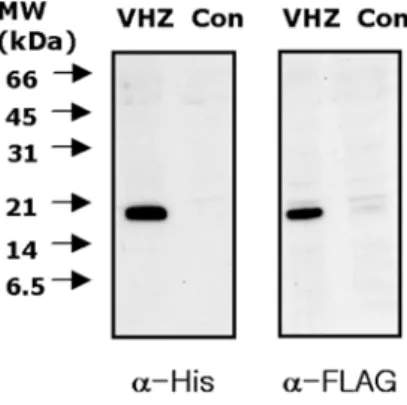
Expression of VHZ (C95S, D65A) was confirmed by immunoblotting the cell lysates with anti-His and anti- flag antibodies, respectively. Compared to the parental 293T cells, 293T-VHZ (C95S, D65A) cells showed a protein band of predicted molecular weight (Fig. 1).
Parental and 293T-VHZ (C95S, D65A) cells were lysed, and VHZ (C95S, D65A) proteins were pull- downed with Ni-NTA bead. The pull-downed proteins were visualized by Coomassie bule staning of the 2-D gels. Several specific protein spots appeared in the both gels in similar intensities (Fig. 2), and those proteins might represent proteins interacting with His-tag or beads. We also found some specific protein spots in the lysate of 293T-VHZ (C95S, D65A), representing potential VHZ-binding proteins.
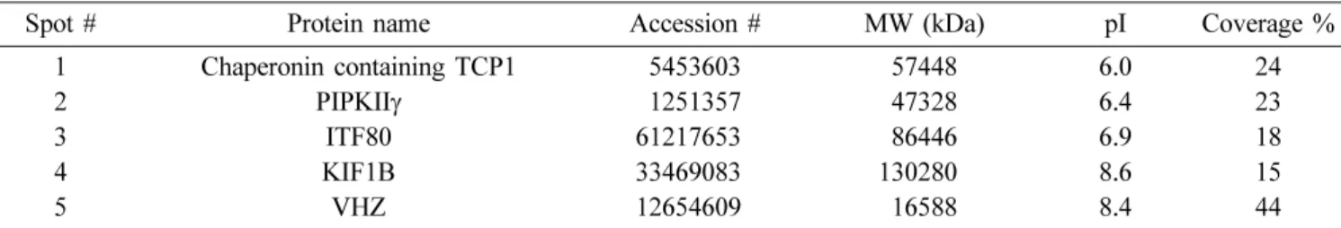
Two-D images of the potential VHZ-interacting proteins characterized by MALDI-TOF Mass spectroscopy are shown in Fig. 3 and listed in Table 1. Chaperonin containing TCP1, which is known to be involved in protein folding upon ATP hydrolysis [Kubota et al., 1994], was found to interact with VHR. Type II phosphatidylinositol phosphate kinase (PIPKII) phosphorylates phosphatidylinositol-5-bisphosphate (Ptdins-5-P) at D-4
Fig. 1. Expression of VHZ in 293 T cells. HEK 293 T cells were transfected with a plasmid expressing double- tagged VHZ (C95S, D65A) and selected for stable expres- sion of VHZ. Whole-cell lysates of the selected HEK 293 T cells and parental cells were resolved by 15% SDS/
PAGE and transferred to a nitrocellulose membrane. To confirm the expression of VHZ, the membrane was immu- noblotted with anti-His and anti-flag antibodies, respec- tively. Positions of the molecular mass markers (kDa) are shown on the left.
position, synthesizing Ptdins-4,5-P2, a key phospholipid in the production of second messengers such as diacylglycerol and inositol 1, 4, 5-triphosphate [Itoh et
al., 1998]. We also detected PIPKIIγ in the precipitant of the cells expressing VHZ (C95S, D65A), suggesting VHZ is involved in the phosphoinositide signaling pathway.
Intraflagellar transport 80 homolog (ITF80) was also included in the pull-downed proteins. Although its cellular functions are not clear, ITF80 has the WD domain repeat that is often found in proteins playing crucial roles in the signal transduction such as transcriptional regulation, RNA processing, and cell cycle progression [Smith et al., 1999]. KIF1B is a member of the kinesin superfamily proteins that transport protein complexes and mRNAs to specific destinations within the cell [Miki et al., 2001], and is shown to be a candidate for tumor suppressor gene of neuroblastoma cells [Yang et
al., 2001]. Compared to the parental HEK 293T cells, HEK 293T cells expressing VHZ (C95S, D65A) only showed a strong specific protein spot that is expected to VHZ at the site designated as 5 (Figs. 2 and 3). This protein was identified as VHZ by MALDI-TOF Mass spectroscopy analysis, showing that the pull-downed proteins contained VHZ and VHZ-interacting proteins.
Fig. 2. Coomassie-stained 2-D SDS PAGE gels of proteins with Ni-NTA beads. Ni-NTA beads were used to pull-down proteins from whole cell lysates. The proteins bound to the beads were subjected to 13 strip, and SDS-PAGE was per- formed on a 12% gel. Left panel, pull-down proteins from the lysate of parental HEK 293T cells, Right panel, pull-down proteins from the lysate of HEK 293T cells expressing VHZ (C95S, D65A). The numbers represent protein spots character- ized by MALDI-TOF analysis.
Fig. 3. Insets from Fig. 2 showing the images of altered patterns. Enlarged images of the VHZ-interacting pro- teins from HEK 293T cells. Arrows indicate protein spots that were selected and analyzed.
Table 1. Identification of pull-downed proteins by MALDI-TOF analysis of HEK293T cells expressing VHZ (C95S, D65A)
Spot # Protein name Accession # MW (kDa) pI Coverage %
1 Chaperonin containing TCP1 5453603 57448 6.0 24
2 PIPKIIγ 1251357 47328 6.4 23
3 ITF80 612176530 86446 6.9 18
4 KIF1B 334690830 1302800 8.6 15
5 VHZ 126546090 16588 8.4 44
Discussion
VHZ consists of a minimal essential core of the phosphatase having only 150 amino acid residues and is a phylogenetically well-conserved enzyme with a probable ortholog from mammals to the Archea T. kodakaraensis
[Alonso et al., 2004a], suggesting that basic function of VHZ in cell physiology. In some respect, VHZ is similar to VHR, a prototype of atypical DSPs that is well characterized structurally and biologically. Although VHR is a member of the dual specificity protein phosphatases, it has a relatively deep active site pocket and prefers pY to pT in the dephosphorylation reactions [yuvaniyama et al., 1996]. Similarly, recombinant VHZ was shown to dephosphorylate Tyr(P)-MBP 103 times rapidly than and Ser/Thr (P)-MBP [Takagaki et al., 2004]. Similar to VHR, VHZ also has no MKB domain.
However, VHZ is known to be different from VHR in its cellular functions. VHR shows inhibitory role in CD28- induced Erk and JNK activations, but not in p38 activation [Alonso et al., 2001]. However, VHZ enhances the activation of JNK and p38, and do not affect the phosphorylation level of Erk. Both Inactive and active VHZs were similar in the activation of JNK and p38 in COS-7 cells [Takagaki et al., 2004]. Therefore, the real substrate of VHZ may not be Erk, JNK, and p38 in vivo.
VHZ was found to interact with Chaperonin contataining TCP1, suggesting that VHZ is involved in the protein folding. However, previous studies showed that VHZ also interacts with the overexpressed proteins [Guo et al., 2005; Andersen et al., 2005]. Thus, it cannot be rule out that Chaperonin contataining TCP1 may help the overexpressed VHZ to be folded correctly in cells.
PIPKIIγ is one of the substrate candidates for VHZ in
vivo. Similar to VHZ, PIPKIIγ could be detected in almost all tissues including heart, brain, spleen, kidney, and testis. Upon extracellular stimuli such as EGF, PDGF, and lysophosphatidic acid, PIPKIIγ can be phosphorylated [Itoh et al., 1998]. This implies that the level of phosphorylation may regulate PIPKIIγ activity, and the resulting Ptdins-4,5-P2 can function as a second messengers in the cellular pathway leading to Map kinase. VHZ can also participate in the transcriptional regulation and RNA processing by interacting with ITF 80 and molecular transportation by KIF1B. It now remains to be determined how VHZ regulates the interacting proteins mentioned above. This is under further investigation in our laboratory.
Acknowledgments. This work was supported by Korea Research Foundation Grant (KRF-2004-002-C00127).
References
Alonso A, Saxena M, Williams S, and Mustelin T (2001) Inhibitory role for dual specificity phosphatase VHR in T cell antigen receptor and CD28-induced Erk and Jnk activation. J Biol Chem276, 4766-4771.
Alonso A, Merlo JJ, Na S, Kholod N, Jaroszewski L, Khari- tonenkov A, Williams S, Godzik A, Posada JD, and Mustelin T (2002) Inhibition of T cell antigen receptor signaling by VHR-related MKPX (VHX), a new dual specificity phosphatase related to VH1 related (VHR). J Biol Chem.277, 5524-5528.
Alonso A, Burkhalter S, Sasin J, Tautz L, Bogetz J, Huynh H, Bremer MC, Holsinger LJ, Godzik A, and Mustelin T (2004a) The minimal essential core of a cysteine- based protein-tyrosine phosphatase revealed by a novel 16-kDa VH1-like phosphatase, VHZ. J Biol Chem 279, 35768-35774.
Alonso A, Narisawa S, Bogetz J, Tautz L, Hadzic R, Huynh H, Williams S, Gjorloff-Wingren A, Bremer MC, Hols- inger LJ, Millan JL, and Mustelin T (2004b) VHY, a novel myristoylated testis-restricted dual specificity pro- tein phosphatase related to VHX. J Biol Chem 279, 32586-32591.
Alonso A, Sasin J, Bottini N, Friedberg I, Osterman A, Godzik A, Hunter T, Dixon J, and Mustelin T. (2004c) Protein tyrosine phosphatases in the human genome. Cell
117, 699-711.
Andersen JS, Lam YW, Leung AK, Ong SE, Lyon CE, Lamond AI, and Mann M (2005) Nucleolar proteome dynamics. Nature 433, 77-83.
Aoyama K, Nagata M, Oshima K, Matsuda T, and Aoki N (2001) Molecular cloning and characterization of a novel dual specificity phosphatase, LMW-DSP2, that lacks the cdc25 homology domain. J Biol Chem 276, 27575- 27583.
Chen AJ, Zhou G, Juan T, Colicos SM, Cannon JP, Cabri- era-Hansen M, Meyer C F, Jurecic R, Copeland NG, Gil- bert DJ, Jenkins NA, Fletcher F, Tan TH, and Belmont JW (2002) The dual specificity JKAP specifically acti- vates the c-Jun N-terminal kinase pathway. J Biol Chem
277, 36592-36601.
Denu JM and Dixon JE (1998) Protein tyrosine phos- phatases: mechanisms of catalysis and regulation. Curr Opin Chem Biol2, 633-641.
Flint AJ, Tiganis T, Barford D, and Tonks NK (1997) Development of “substrate-trapping” mutants to identify physiological substrates of protein tyrosine phos- phatases. Proc Natl Acad Sci USA 94, 1680-1685.
Guo D, Han J, Adam BL, Colburn NH, Wang MH, Dong Z, Eizirik DL, She JX, and Wang CY (2005) Proteomic analysis of SUMO4 substrates in HEK293 cells under serum starvation-induced stress. Biochem Biophys Res Commun 337, 1308-1318.
Itoh T, Ijuin T, and Takenawa T (1998) A novel phosphati- dylinositol-5-phosphate 4-kinase (phosphatidylinositol-
phosphate kinase IIgamma) is phosphorylated in the endoplasmic reticulum in response to mitogenic signals.
J Biol Chem 273, 20292-20299.
Kim SJ, Jeong DG, Yoon TS, Son JH, Cho SK, Ryu SE, and Kim JH (2007) Crystal structure of human TMDP, a testis-specific dual specificity protein phosphatase: impli- cations for substrate specificity. Proteins66, 239-245.
Kubota H, Hynes G, Carne A, Ashworth A, and Willison K (1994) Identification of six Tcp-1-related genes encod- ing divergent subunits of the TCP-1-containing chapero- nin. Curr Biol4, 89-99.
Miki H, Setou M, Kaneshiro K, and Hirokawa N (2001) All kinesin superfamily protein, KIF, genes in mouse and human. Proc Natl Acad Sci USA 98, 7004-7011.
Rahmouni S, Cerignoli F, Alonso A, Tsutji T, Henkens R, Zhu C, Louis-dit-Sully C, Moutschen M, Jiang W, and Mustelin T (2006) Loss of the VHR dual-specific phos- phatase causes cell-cycle arrest and senescence. Nat Cell Biol8, 524-531.
Sakaue H, Ogawa W, Nakamura T, Mori T, Nakamura K, and Kasuga M (2004) Role of MAPK phosphatase-1 (MKP-1) in adipocyte differentiation. J Biol Chem 279, 39951-39957.
Shiozaki K and Russell P (1995) Cell-cycle control linked to extracellular environment by MAP kinase pathway in fission yeast. Nature378, 739-743
Smith TF, Gaitatzes C, Saxena K, and Neer EJ (1999) The WD repeat: a common architecture for diverse func- tions. Trends Biochem Sci 24, 181-185.
Sun H, Charles CH, Lau LF and Tonks NK (1993) MKP-1
(3CH134), an immediate early gene product, is a dual specificity phosphatase that dephosphorylates MAP kinase in vivo. Cell75, 487-493.
Takagaki K, Satoh T, Tanuma N, Masuda K, Takekawa M, Shima H, and Kikuchi K (2004) Characterization of a novel low-molecular-mass dual-specificity phosphatase-3 (LDP-3) that enhances activation of JNK and p38. Bio- chem J383, 447-455.
Wu JJ and Bennett AM (2005) Essential role for mitogen- activated protein (MAP) kinase phosphatase-1 in stress- responsive MAP kinase and cell survival signaling. J Biol Chem.280, 16461-16466.
Wu J, Katrekar A, Honigberg LA, Smith AM, Conn MT, Tang J, Jeffery D, Mortara K, Sampang J, Williams SR, Buggy J, and Clark JM (2006) Identification of sub- strates of human protein-tyrosine phosphatase PTPN22. J Biol Chem 281, 11002-11010.
Yang HW, Chen YZ, Takita J, Soeda E, Piao HY, and Hayashi Y (2001) Genomic structure and mutational analysis of the human KIF1B gene which is homozy- gously deleted in neuroblastoma at chromosome 1p36.2.
Oncogene20, 5075-5083.
Yu W, Imoto I, Inoue J, Onda M, Emi M, and Inazawa JA (2007) novel amplification target, DUSP26, promotes anaplastic thyroid cancer cell growth by inhibiting p38 MAPK activity. Oncogene26, 1178-1187.
Yuvaniyama J, Denu JM, Dixon JE, and Saper MA (1996) Crystal structure of the dual specificity protein phos- phatase VHR. Science272, 1328-1331.