Risk Factor Analysis for C5 Palsy after Double-Door Laminoplasty for Cervical
Spondylotic Myelopathy
Satoshi Baba, Ko Ikuta, Hiroko Ikeuchi, Makoto Shiraki, Norihiro Komiya, Takahiro Kitamura, Hideyuki Senba, Satoshi Shidahara
Department of Orthopaedic Surgery, Karatsu Red Cross Hospital, Karatsu, Japan
Study Design: A retrospective comparative study.
Purpose: To clarify the risk factors related to the development of postoperative C5 palsy through radiological studies after cervical double-door laminoplasty (DDL).
Overview of Literature: Although postoperative C5 palsy is generally considered to be the result of damage to the nerve root or segmental spinal cord, the associated pathology remains controversial.
Methods: A consecutive case series of 47 patients with cervical spondylotic myelopathy treated by DDL at our institution between April 2008 and April 2015 were reviewed. Postoperative C5 palsy occurred in 5 of 47 cases after DDL. We investigated 9 radiologic factors that have been reported to be risk factors for C5 palsy in various studies, and statistically examined these between the two groups of palsy and the non-palsy patients.
Results: We found a significant difference between patients with and without postoperative C5 palsy with regards to the posterior shift of spinal cord at C4/5 (p=0.008). The logistic regression analyses revealed posterior shift of the spinal cord at C4/5 (odds ratio, 12.066; p=0.029; 95% confidence interval, 1.295–112.378). For the other radiologic factors, there were no statistically significant dif- ferences between the two groups.
Conclusions: In the present study, we showed a significant difference in the posterior shift of the spinal cord at C4/5 between the palsy and the non-palsy groups, indicating that the “tethering phenomenon” was likely a greater risk factor for postoperative C5 palsy.
Keywords: C5 palsy; Double door laminoplasty; Cervical spondylotic myelopathy
Copyright Ⓒ 2016 by Korean Society of Spine Surgery
This is an Open Access article distributed under the terms of the Creative Commons Attribution Non-Commercial License (http://creativecommons.org/licenses/by-nc/3.0/) which permits unrestricted non-commercial use, distribution, and reproduction in any medium, provided the original work is properly cited.
Asian Spine Journal • pISSN 1976-1902 eISSN 1976-7846 • www.asianspinejournal.org
Received Aug 10, 2015; Revised Nov 04, 2015; Accepted Nov 04, 2015 Corresponding author: Satoshi Baba
Department of Orthopaedic Surgery, Karatsu Red Cross Hospital, 1-5-1 Futago Karatsu, Saga 847-8588, Japan Tel: +81-95-572-5111, Fax: +81-95-573-9530, E-mail: [email protected]
ASJ A SJ
Introduction
Cervical laminoplasty is a reliable surgical procedure in the treatment of cervical spondylotic myelopathy (CSM).
This operation has been widely performed in place of laminectomy for multilevel posterior decompression of the spinal cord and has been considered as a relatively safe
operation with low risk of complications. Among various types of laminoplasty, double door laminoplasty (DDL) is widely accepted due to its simpler and safer technical requirements [1]. However, postoperative upper extremity palsy, especially C5 palsy, is a relatively frequent compli- cation after laminoplasty. The incidence of C5 palsy has been reported to be 4.6% but varies from 0% to 30% [2].
Although postoperative C5 palsy is generally considered to be the result of the damage to the nerve root or seg- mental spinal cord, the exact mechanisms underlying its pathology remain controversial, particularly as to whether the nerve is damaged during or after surgery [2]. Generally, the postoperative C5 palsy is defined as muscle paresis of deltoids and biceps corresponding to the C5 segment.
However, Tamiya et al. [3] reported that there is also a case for ensuing pain from the C6 segment area, and par- ticipation of C6 segment as the cause for C5 palsy. Thus, we also investigated the possibility of C6 segment area be- ing related to the C5 palsy.
In the past, there have been several C5 palsy-related papers, which concluded that operations involving “open door laminoplasty” and “ossification of posterior longitu- dinal ligament (OPLL)” carried risk factors [1]. However, there has not been much report on patients who under- went DDL for CSM, and the risk factor for C5 palsy as the risks, in general, have been reported to be relatively low.
Therefore, we examined the patients who underwent the DDL for CSM patient in terms of risks for C5 palsy.
The objective of this study was to review the radiologi- cal findings in patients after DDL for CSM and the occur- rence of associated adverse effects from the operation.
Materials and Methods
A consecutive case series of 76 patients with CSM treated by DDL at our institution between April 2008 and April 2015 were retrospectively reviewed. Exclusion criteria were as follows: (1) preoperatively, patients with Grade 3 or less weakness of the deltoid or biceps in a manual muscle test (MMT); (2) patients with previous cervical
surgery; (3) patients who needed spinal fusion; and (4) patients who could not be examined in all radiological factors described later.
A total of 29 patients were excluded and therefore 47 patients (35 men and 12 women) were enrolled in the study. The mean age of the patients was 70.6 years (range, 37–85 years).
Postoperative C5 palsy was defined as deterioration of muscle strength by one or more grades on the manual muscle testing of the deltoid and biceps after surgery, and in absence of other neurologic symptoms. Even if the palsy developed bilaterally, or was considered to include C5 and other multi-segments, it was counted one C5 palsy case.
There were a total of 5 patients (3 men and 2 women, average age 69.2 years) with C5 palsy, and clinical details of each patient are shown in Table 1. The MMT score of the deltoid at the onset of palsy was between 1 and 3, and that of the biceps brachii was between 1 and 5. Paralysis was noted after a mean of 2.6 days after surgery (range, 8 hours–5 days), and one patient experienced right side paralysis, 2 patients on the left side, and in 2 patients pa- ralysis was on both sides. Upper extremity pain appeared upon the development of the palsy in 3 patients. Of the 5 patients, one patient required additional foraminotomy because there was a possibility of kinking intervertebral foramen from postoperative CT images; other patients, however, were treated conservatively. All of the 5 patients fully recovered within 11 months. We categorized these 5 patients as the P-group (as having palsy), and the remain- ing 42 patients (32 men and 10 women, average age 70.7 years) were categorized as the N-group (no palsy).
According to patient demographics, there were no sig- nificant differences between the two groups (Table 2). We Table 1. Demographic and clinical data of patients of P-group
No Age (yr) Sex Procedure Palsy
side Deterioration of deltoid
and biceps (MMT) Onset
(pstoperative day) Treatment Recovery (mo)
1 77 Female DDL (C4–6) Right Deltoid: 5–2, biceps: 5–4 3 Conservative Complete (7.5)
2 72 Female DDL (C4–5)+
foraminotomy (left C4/5)
Left Deltoid: 5–1, biceps: 5–1 1 Reforaminotomy
(left C4/5) Complete (11)
3 56 Male DDL (C3–6) Left Deltoid: 5–2, biceps: 5–4
R: deltoid: 5–3, biceps: 5–5
5 Conservative Complete (2)
4 82 Male DDL (C4–7) Both L: deltoid: 5–3, biceps: 5–5
R: deltoid: 5–1, biceps: 5–1 4 Conservative Complete (3) 5 60 Male DDL (C3–6) Both L: deltoid: 5–2, biceps: 5–2 0 (8 hr after surgery) Conservative Complete (11) MMT, manual muscle test; DDL, double-door laminoplasty; R, right side; L, left side
then statistically performed a comparison between the P- group and the N-group, and also determined the associ- ated risk factors occurring with the onset of postoperative palsy.
1. Radiological examination
We investigated 9 factors that have been reported to be risk factors for C5 palsy in various studies. We investi- gated cervical alignment [1,4], anterior protrusion of the superior articular process [1,4,5], width of the interver- tebral foramen [4,5], and number of the compressed seg- ment of the cord [4] as preoperative risk factors. We also considered vertebral arch opening angle [6], hinge angle
[6], position of the gutters [2], value of posterior shift of the spinal cord [1,2,4,5], and appearance of a high inten- sity area in the cord [2,4] as preoperativ and postoperative risk factors.
With preoperative X-ray images, cervical alignment was measured from C2–C7 sagittal angle (Fig. 1). Using pre- operative computed tomography (CT) images, the width of the intervertebral foramen at C4/5 and C5/6 levels, and the anterior protrusion of the superior articular process of C5 and C6 were measured according to the method described by Sunago et al. (Fig. 2) [7]. The width of the intervertebral foramen and the anterior protrusion of the Table 2. Comparison of demographic data between two groups
Parameters P-group N-group p-value
No. of patients 5 42
Mean age (yr) 69.2±9.66 70.7±10.87 0.629
Sex 0.433
Male 3 32
Female 2 10
No. of opened laminae 3.4±0.8 4.17±00.78 0.066
Fig. 1. C2–7 sagittal angle was measured as illustrated.
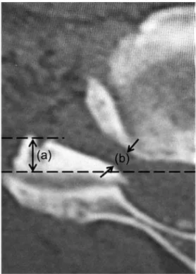
Fig. 2. The position of the superior articular process was measured by the method of Sunago et al. as illustrated (a). The black arrows (b) indicate the width of the inter- vertebral foramen of C4–5 at the narrowest point.
superior articular process were both measured at their narrowest and the most prominent points, respectively.
Using postoperative CT images, the vertebral arch opening angle, the hinge angle, and the position of the gutters were measured. The vertebral arch opening angle was measured at C4, C5, and C6 by the method in Fig. 3, and the hinge angle was measured at C4, C5 and C6 by the method in Fig. 4. The position of the gutter was mea- sured the distance to the gutter from the vertebral body center line portion at C4, C5, and C6 by the method in Fig. 4. With the CT images, the P-group was evaluated by measuring the palsy side, and the N-group was evaluated by measuring both sides.
Using preoperative magnetic resonance imaging (MRI), the number and the position of the compressed segments of the cord were evaluated. Using postoperative MRI, the value of posterior shift of the spinal cord, and appearance of a high intensity area in the cord were evaluated. The
posterior shift of spinal cord at level C4/5 and C5/6 was measured from the posterior edge of the compressive mass to the midpoint of the spinal cord in the mid-sagittal plane of T2-weighted MRI (Fig. 5). The midpoint of the spinal cord was selected because it is believed that this measure- ment is less influenced by postoperative spinal cord ex- pansion and thus provides a more accurate determination of spinal cord displacement. Appearance of a high signal intensity lesion in the cord at the C3–5 level was evaluated using T2-weighted sagittal and axial MRI and any new high intensity lesion was defined as positive (Fig. 5).
2. Statistical analysis
The comparison of continuous variables between two groups was Mann-Whitney U-test or chi-square test. In addition, we conducted logistic regression analysis in each
Fig. 3. The vertebral arch opening angle was measured by (A°)–(a°).
Fig. 4. The hinge angle (theta) was measured as illustrated. The position of the gutter was the distance to the gutter from the ver- tebral body centerline portion as illustrated (c).
Fig. 5. Posterior shift of the spinal cord at the level of C4/5 and C5/6 was measured from the posterior com- pressive mass to the preoperative and postoperative midpoint of the spinal cord on T2-weighted midsagittal magnetic resonance imaging (grey arrows). The preop- erative value was then subtracted from the postopera- tive value for the measurement of the spinal cord shift.
The appearance of new high signal intensity lesion in the cord at the level of C3–C5 was evaluated before and after surgery as illustrated (black arrow).
parameter. In the logistic regression model, P-group and N-group were treated as dependent variables.
All p-value less than 0.05 were considered statistically significant. Logistic regression analysis was performed with SPSS software ver. 21.0 (SPSS Inc., Chicago, IL, USA). Each parameter was measured 2 times by the same person and the median was adopted.
Results
Details of the radiological parameters of the P-group are
shown in Table 3, and comparison of radiological param- eters for the P- and N-groups is shown in Table 4.
1. Comparison of radiological parameters
On radiographic studies, the C2–7 sagittal angle had a mean of 15.4°±6.0° in the P-group and 17.9°±10.7° in the N-group (p=0.849), and there was no significant differ- ence.
On CT images evaluations, there were no significant differences between the two groups with regard to the Table 3. Radiological parameters of the P-group
Parameters No. 1 No. 2 No. 3 No. 4 No. 5
X-ray
SA (°) 17 19 21 4 16
Computed tomography APSAP
C4/5 (mm) 5.7 1.6 3.4 R: 6.3, L: 4.3 R: 5.6, L: 2.8
C5/6 (mm) WIF
C4/5 (mm) 2.4 3.5 3.1 R: 3.0, L: 3.0 R: 3.0, L: 3.0
C5/6 (mm) 3.6 2.5 1.8 R: 3.8, L: 2.6 R: 2.9, L: 3.6
VAOA
C4 (°) 8 24 20 R: 20, L: 12 R: 18, L: 20
C5 (°) 22 24 18 R: 33, L:14 R: 14, L: 30
C6 (°) 20 - 18 R: 31, L: 6 R: 20, L: 28
HA
C4 (°) 147 145 141 R: 136, L: 142 R: 121, L: 147
C5 (°) 129 133 125 R: 118, L: 145 R: 134, L: 141
C6 (°) 122 - 122 R: 138, L: 141 R: 138, L: 141
PG
C4 (mm) 9.0 9.0 10.0 R: 9.3, L: 9.3 R: 10.6, L: 8.9
C5 (mm) 11.2 9.6 11.4 R: 9.4, L: 10.6 R: 8.8, L:,10.0
C6 (mm) 10.4 - 10.0 R: 9.0, L: 10.8 R: 10.0, L: 8.4
Magnetic resonance imaging
No. of compression 3 2 4 4 3
VPS
C4/5 (mm) 3.8 3.1 3.6 1.9 5.5
C5/6 (mm) 3.2 2.3 2.5 1.9 5.4
T2-high lesion None None None None None
SA, the C2–7 sagittal angle; APSAP, anterior protrusion of the superior articular process; WIF, width of the intervertebral foramen; VAOA, vertebral arch opening angle; HG, hinge angle; PG, position of the gutter; No. compression, number of the compressed segment of the cord; VPS, value of posterior shift of the spinal cord; T2-high lesion, appearance of intramedullary T-2 high signal intensity lesion; R, right side; L, left side.
anterior protrusion of the superior articular process at C4/5 and C5/6 (p=0.846 and 0.783, respectively) along with the width of the intervertebral foramen at C4/5 and C5/6 (p=0.946 and 0.917, respectively), the vertebral arch opening angle at C4, C5 and C6 (p=0.793, 0.293, and 0.413, respectively), the hinge angle at C4, C5, and C6 (p=0.064, 0.858, and 0.783, respectively) and the position of the gutters at C4, C5 and C6 (p=0.098, 0.536, and 0.770, respectively).
On MRI evaluations, there was significant difference between the two groups with regards to the amount of the posterior shift of the spinal cord at C4/5 (p=0.008).
The posterior shift of the spinal cord at C5/6 presented
no statistical significant difference (p=0.261) between the groups. The number of the compressed segments of the cord (p=0.824), and the appearance of an intramedul- lary T2-weighted high signal lesion after surgery at C3–5 (p=0.537) revealed no significant differences between the two groups. Thus, with the 9 radiological factors, there was only significant difference of the posterior shift of the spinal cord at C4/5 between the two groups (p=0.008 as above).
2. Risk factor analysis
Logistic regression analyses revealed posterior shift of the Table 4. Comparison of radiological parameters between the two groups
Parameters P-group N-group p-value
SA (°) 18.0±8.1 17.8±10.5 0.849
APSAP
C4/5 (mm) 4.24±1.60 4.20±1.29 0.846
C5/6 (mm) 3.94±1.70 3.92±1.19 0.783
WIF
C4/5 (mm) 3.00±0.30 2.94±0.75 0.958
C5/6 (mm) 2.97±0.68 3.02±0.83 0.917
VAOA
C4 (°) 17.43±5.10 18.83±6.22 0.793
C5 (°) 22.14±6.90 18.81±5.31 0.293
C6 (°) 20.50±7.9 18.79±6.34 0.413
HA
C4 (°) 138.71±7.98 134.62±7.48 0.064
C5 (°) 131.86±9.17 131.25±7.73 0.858
C6 (°) 128.00±9.54 129.01±7.50 0.783
PG
C4 (mm) 9.44±0.58 9.93±1.44 0.098
C5 (mm) 9.86±0.95 10.09±1.32 0.536
C6 (mm) 9.77±0.82 9.63±1.45 0.770
No. of compression 3.2±0.75 3.36±0.89 0.824
VPS
C4/5 (mm) 3.62±1.15 2.17±0.72 0.008
C5/6 (mm) 3.08±1.23 2.29±0.90 0.261
T2-high lesion Positive: 0, negative: 5 Positive: 3, negative: 39 0.537
Values are presented as mean ± standard deviation.
P-group was evaluated by measuring the palsy side. N-group was evaluated by measuring the both sides.
SA, the C2-7 sagittal angle; APSAP, anterior protrusion of the superior articular process; WIF, width of the intervertebral foramen; VAOA, vertebral arch opening angle; HA, hinge angle; PG, position of the gutter; No. of compression, number of the compressed segment of the cord; VPS, value of posterior shift of the spinal cord; T2-high lesion, appearance of intramedullary T-2 high signal intensity lesion.
spinal cord at C4/5 (odds ratio, 12.066; p-value=0.029;
95% confidence interval, 1.295–112.378) was significantly involved in C5 palsy, whereas other radiologic examina- tions did not significantly affect the occurrence of postop- erative C5 palsy (Table 5).
Odds ratio of posterior shift of the spinal cord at C4/5 was 1 or more, and it became a risk factor for C5 palsy in this study. For the T2-high lesion, because there were too few cases of any new high intensity lesions, a statistical analysis was not possible.
Discussion
In 1961, Stoops and King [8] and Scoville [9] reported postoperative paresis of the upper extremity as a neu- rologic complication following cervical laminectomy.
Although the occurrence of C5 palsy after laminoplasty has been reported in many studies, its detailed mecha- nism remains poorly understood. There have been several hypotheses regarding the etiology of C5 palsy, including direct injury to nerve root during surgery, tethering of the nerve root, segmental spinal cord disorder, and reperfu- Table 5. Logistic regression analysis for the risk factors of postoperative C5 palsy
Parameters Odds raio p-value 95% Confidence interval
X-ray
SA (°) 0.974 0.615 0.881–1.078
Computed tomography APSAP
C4/5 (mm) 1.022 0.941 0.571–1.830
C5/6 (mm) 1.018 0.955 0.547–1.896
WIF
C4/5 0.952 0.827 0.612–1.480
C5/6 0.928 0.877 0.360–2.390
VAOA
C4 (°) 0.962 0.562 0.845–1.096
C5 (°) 1.108 0.134 0.969–1.268
C6 (°) 1.042 0.532 0.916–1.185
HA
C4 (°) 1.093 0.089 0.986–1.211
C5 (°) 1.010 0.843 0.914–1.116
C6 (°) 0.983 0.755 0.883–1.094
PG
C4 (mm) 0.789 0.376 0.467–1.333
C5 (mm) 1.031 0.920 0.571–1.862
C6 (mm) 1.070 0.825 0.587–1.950
Magnetic resonance imaging
No. of compression 0.809 0.707 0.269–2.436
VPS
C4/5 (mm) 12.066 0.029 1.295–112.378
C5/6 (mm) 2.224 0.106 0.845–5.854
T2-high lesion 0 0.999 0
SA, the C2-7 sagittal angle; APSAP, anterior protrusion of the superior articular process; WIF, width of the intervertebral foramen; VAOA, vertebral arch opening angle; HA, hinge angle; PG, position of the gutter; No. of compression, number of the compressed segment of the cord; VPS, value of posterior shift of the spinal cord; T2-high lesion, appearance of intramedullary T-2 high signal intensity lesion.
sion injury to the spinal cord, but none of these hypoth- eses have been completely established.
1. Direct injury to nerve root during surgery
Tamiya et al. [3] reported that direct injury or heat injury has become a cause of damage during surgery. However, Sasai et al. [10] suggested that additional microcervi- cal foraminotomy could prevent postoperative C5 palsy, although during foraminotomy high-speed drills come much closer to nerve root than any other procedure, so that frictional heat reaching the roots must be the high- est. It is inconsistent with injury due to heat if the injury is preventable by a procedure that generates even more heat.
Hirabayashi et al. [11] along with Iwamoto et al. [12]
reported that if damage occurs during surgery by direct injury, C5 palsy becomes immediately evident after sur- gery. However, none of our cases immediately developed C5 palsy after surgery, so we thought the possibility of damage during surgery, as the cause of palsy, was low in our study. There were, however, only 5 cases of C5 palsy in our study, so we could not definitively conclude that di- rect injury to nerve root during surgery did not induce C5 palsy in our study. We thought that future study should examine more cases.
2. Tethering of the nerve root
Nerve root traction may be caused by posterior shift of the spinal cord after laminoplasty, and the so-called “teth- ering effect” has been considered as one of the more ac- ceptable pathologic mechanisms of C5 palsy. We showed a significant difference between the two groups with regards to the posterior shift of spinal cord at C4/5. The extent of posterior shift of the spinal cord after laminoplasty was measured by various methods and the reports indicate a 1 to 3 mm displacement [13,14]. Yamashita et al. [15]
reported that the posterior shift averaged 5 mm at C4 or C5 in patients with C5 palsy; this was significantly greater than in patients without clinical signs of palsy. In this study, we indicated that the posterior shift of the spinal cord averaged 3.6 mm at C4/5 in patients with C5 palsy.
There was a significant difference in the posterior shift between the two groups with almost the same results as those of the previous studies.
Radiological examinations, however, may some prob- lems with respect to the time of imagings performed post-
operatively. Shiozaki et al. [16] reported that the posterior shift of the spinal cord at 24 hours postoperation had a tendency to shift more posteriorly than that observed at 2 weeks after cervical laminoplasty. In this study, the P- group had their MRI recorded immediately upon occur- rence of their palsy. Most of the N-group, however, had the MRI more than one month after their surgery. There is a possibility that difference of time in taking the MRI caused a significant difference in the posterior shift of the spinal cord between the two groups. It is difficult to sim- ply compare this data with those of previous reports, be- cause many of reports with the exception of Shiozaki et al.
[16] do not mention the time of the MRI postoperation.
Posterior shift of the spinal cord at C4/5 in this study was the risk factor of C5 palsy by logistic regression analysis.
But for the reasons mentioned above, this risk factor has this confounding factor in intrepretation. However we thought that the posterior shift of the spinal cord at C4/5 is likely to be the largest factor of C5 palsy from this study and that in a future study, a greater number of cases post- operation with matched timing of their MRI should be examined.
There are other causes of nerve root lesion that have been thought to play a part in C5 palsy; these include kinking by the superior articular process, pressuring and impinging the nerve root by an osteophyte, excessive teth- ering on the nerve roots to one side caused by deviation of the vertebral arch opening angle, and excessive stress on the nerve roots caused by edge of too steep a hinge, and kinking caused by excessively lateral bony gutter [2,4,6].
For preoperative risk factors, there are the anterior pro- trusion of the superior articular process and the width of the intervertebral foramen that are thought to play a role [17].
Most posterior shifts occurred at the C5 vertebral level because C5 is the apex of cervical lordosis. In addition, the superior articular process of C5 protrudes in a more an- terior direction and as the root of C5 is shorter compared with other levels, a posterior shift might create a tension on the C5 nerve root, causing C5 palsy. However, there was no significant difference between the two groups with regards to the anterior protrusion of the superior articular process in this study.
Preexisting foraminal stenosis has been suggested to be associated with C5 palsy in several studies. Imagama et al. [17] presented radiographic evidence of nerve root impingement from CT in a retrospective study of 1,858
patients who underwent a cervical laminoplasty. They found that the width of the C5 intervertebral foramen was significantly smaller, and the anterior protrusion of C5 superior articular process was significantly greater in patients with C5 palsy. In this study, there was no signifi- cant difference between the two groups with regards to the width of the intervertebral foramen. Some researchers reported that patients who have narrow foramen should have prophylactic foraminotomy to prevent C5 palsy.
Komagata et al. [18] reported that prophylactic bilateral partial foraminotomy could reduce the incidence of C5 palsy after open-door laminoplasty from 4.0% to 0.6%.
In contrast, Tamiya et al. [3] reported that foraminotomy was not necessary to prevent dysfunction of the cervical nerve root. Thus, whether foraminotomy is beneficial as a prophylactic measure on the nerve root is not yet clear.
Foraminotomy may be preventative when the cause of C5 palsy is tethering of the nerve root. However, we didn’t perform foraminotomy to prevent C5 palsy in all cases when we performed the laminoplasty. We only investigat- ed respective risk factors in this study. But we also thought that the risk factors might also increase when performing additional procedures. As a future study, it may be neces- sary to consider the case of overlapping risk factors.
For additional postoperative risk factors, there were the vertebral arch opening angle, the hinge angle, and the po- sition of the gutters to consider. Matsunaga et al. [6] have reported that the occurrence of C5 palsy was more com- mon in the cases where the vertebral arch opening angle was large and the hinge angle was acute [5]. They reported that excessive tethering to one side of the nerve root by the vertebral arch opening angle deviation and excessive stress on the nerve root by the edge of the sharp hinge were causes of C5 palsy. As a precautionary measure against C5 palsy, Matsunaga et al. proposed that the vertebral arch opening angle should be 15°–20° and the hinge angle should not be too acute. The authors also reported that this insight during surgery could reduce the incidence of C5 palsy from 35.3% to 16.4%. In our study, there were no sig- nificant differences between the two groups with regards to the vertebral arch opening angle and the hinge angle.
Creating the bony gutter too laterally or too medi- ally might also result in kinking of the nerve root after posterior shifting of the cord. Some researchers recom- mend making the gutter on the medial aspect of the zygapophyseal joint to prevent C5 palsy [19,20], whereas others reported that the incidence of C5 palsy did not cor-
relate with the position of the bony gutter [13,21]. In this study, there was no significant difference between the two groups with regards to the position of the gutter.
3. Segmental spinal cord disorder and reperfusion injury Some researchers have reported that reperfusion injury occurs when the spinal nerve is depressurized. Chiba et al. [22] and Hasegawa et al. [23] proposed that local reperfusion injury in the spinal cord could cause the de- terioration of gray matter and could lead to postoperative segmental motor palsy. The lesions identified as T2 high- signal intensity zone in the spinal cord newly arose or increased in intensity after surgery. Kaneko et al. [24] also suggested the association of gray matter lesion with the palsy as a high signal change on T2-weighted MRI in spi- nal cord at the relevant segment was shown in all patients who developed postoperative palsy. However, Seichi et al.
[25] reported that the expansion of intramedullary high intensity lesion on T2-weighted MRI was only minimally associated with postoperative segmental motor palsy. In our study, none of C5 palsy cases had new high signal intensity lesions in the spinal cord at the level of C3–C5 after surgery; therefore, this measure could not predict C5 palsy occurrence.
4. Involvement of C6 segment
In the present study, we showed a significant difference of the posterior shift of the spinal cord at C4/5 between palsy group and non-palsy group. If the shift of the spinal cord at C4/5 is large, there is a possibility that there is a tethering effect also to C6 ventral root. But in this study, an involvement of the C6 segment was not recognized with various radiological examinations (Table 4). In ad- dition, the strength of the deltoid muscle was not lower than the biceps muscle (Table 1) for all cases. Thus, we could not establish an involvement of C6 segment in the C5 palsy cases.
The limitations of this study were the difference in the average time postoperation for the MRI between the P- and N-groups and the relatively small number of research cases studied.
Conclusions
In the present study, the large amount of postoperative
posterior shift of the spinal cord at the C4/5 level was the only factor that correlated with occurrence of postopera- tive C5 palsy. From this study, it may be reasonable that the tethering phenomenon might be the most significant contributing cause to C5 palsy. Since the present study had some limitations, additional studies with a greater number of cases of DDL for CSM may better address the causes of postoperative C5 palsy in this population.
Conflict of Interest
No potential conflict of interest relevant to this article was reported.
References
1. Kaneyama S, Sumi M, Kanatani T, et al. Prospective study and multivariate analysis of the incidence of C5 palsy after cervical laminoplasty. Spine (Phila Pa 1976) 2010;35:E1553-8.
2. Sakaura H, Hosono N, Mukai Y, Ishii T, Yoshikawa H. C5 palsy after decompression surgery for cervical myelopathy: review of the literature. Spine (Phila Pa 1976) 2003;28:2447-51.
3. Tamiya A, Hanakita J, Nakanishi K, et al. Analysis of the postoperative palsy of upper extremities of the cases undergone spinous process-splitting lamino- plasty without foraminotomy. Spinal Surg 2005;19:
321-8.
4. Wu FL, Sun Y, Pan SF, Zhang L, Liu ZJ. Risk factors associated with upper extremity palsy after expansive open-door laminoplasty for cervical myelopathy.
Spine J 2014;14:909-15.
5. Gu Y, Cao P, Gao R, et al. Incidence and risk factors of C5 palsy following posterior cervical decompres- sion: a systematic review. PLoS One 2014;9:e101933.
6. Matsunaga H, Inada M, Takeuchi M, et al. Pathogen- esis and prevention of C5 palsy after cervical lami- noplasty. Cent Jpn J Orthop Surg Traumatol 2007;
50:135-6.
7. Sunago K, Nakahara Y, Mitsuyama H. Impairment of postoperative cervical nerve root for cervical com- pressive myelopathy. Cent Jpn J Orthop Surg Trau- matol 1991;34:227-8.
8. Stoops WL, King RB. Neural complications of cervi- cal spondylosis: their response to laminectomy and foramenotomy. J Neurosurg 1962;19:986-99.
9. Scoville WB. Cervical spondylosis treated by bilateral facetectomy and laminectomy. J Neurosurg 1961;
18:423-8.
10. Sasai K, Saito T, Akagi S, Kato I, Ohnari H, Iida H.
Preventing C5 palsy after laminoplasty. Spine (Phila Pa 1976) 2003;28:1972-7.
11. Hirabayashi K, Satomi K, Ichimura M, et al. Com- plications of expansive open-door laminoplasty for ossification of the posterior longitudinal ligament in the cervical spine (in Japanese). Rinsho Seikei Geka 1988;23:509-15.
12. Iwamoto Y, Fujimura Y, Nishi Y, et al. Neurological complications in early stage after expansive open- door laminoplasty (in Japanese). Bessatsu Seikeigeka 1996;29:200-4.
13. Morishita K, Kasai Y, Takegami K. Disturbance of shoulder elevation after cervical laminoplasty. Cen- tral Japan J Orthop Surg Traumatol 1999;42:97-8.
14. Sodeyama T, Goto S, Mochizuki M, Takahashi J, Moriya H. Effect of decompression enlargement laminoplasty for posterior shifting of the spinal cord.
Spine (Phila Pa 1976) 1999;24:1527-31.
15. Yamashita T, Yokogusu K, Yokozawa H, et al. C5 nerve palsy after cervical laminoplasty: an analysis of three cases (in Japanese). Seikei Geka 1996:1365-9.
16. Shiozaki T, Otsuka H, Nakata Y, et al. Spinal cord shift on magnetic resonance imaging at 24 hours after cervical laminoplasty. Spine (Phila Pa 1976) 2009; 34:274-9.
17. Imagama S, Matsuyama Y, Yukawa Y, et al. C5 palsy after cervical laminoplasty: a multicentre study. J Bone Joint Surg Br 2010;92:393-400.
18. Komagata M, Nishiyama M, Endo K, Ikegami H, Tanaka S, Imakiire A. Prophylaxis of C5 palsy after cervical expansive laminoplasty by bilateral partial foraminotomy. Spine J 2004;4:650-5.
19. Uematsu Y, Tokuhashi Y, Matsuzaki H. Radiculopa- thy after laminoplasty of the cervical spine. Spine (Phila Pa 1976) 1998;23:2057-62.
20. Uematsu Y, Tokuhashi Y, Matsuzaki H, et al. Clinical features of postoperative C5 palsy after laminoplasty (in Japanese). J Jpn Spine Res Soc 2002;13:232.
21. Minoda Y, Nakamura H, Konishi T, et al. Radicu- lopathy after bilateral open laminoplasty with split- ting of the spinous processes of the cervical spine (in Japanese). J Jpn Spine Res Soc 2001;12:272.
22. Chiba K, Toyama Y, Matsumoto M, Maruiwa H,
Watanabe M, Hirabayashi K. Segmental motor pa- ralysis after expansive open-door laminoplasty. Spine (Phila Pa 1976) 2002;27:2108-15.
23. Hasegawa K, Homma T, Chiba Y. Upper extremity palsy following cervical decompression surgery re- sults from a transient spinal cord lesion. Spine (Phila Pa 1976) 2007;32:E197-202.
24. Kaneko K, Hashiguchi A, Kato Y, Kojima T, Ima- jyo Y, Taguchi T. Investigation of motor dominant
C5 paralysis after laminoplasty from the results of evoked spinal cord responses. J Spinal Disord Tech 2006;19:358-61.
25. Seichi A, Takeshita K, Kawaguchi H, Nakajima S, Akune T, Nakamura K. Postoperative expansion of intramedullary high-intensity areas on T2-weighted magnetic resonance imaging after cervical lamino- plasty. Spine (Phila Pa 1976) 2004;29:1478-82.