Open Access
Risk Factor Analysis for Secondary Malignancy in Dexrazoxane-Treated Pediatric Cancer Patients
Original Article
Purpose
Dexrazoxane has been used as an effective cardioprotector against anthracycline cardiotox- icity. This study intended to analyze cardioprotective efficacy and secondary malignancy development, and elucidate risk factors for secondary malignancies in dexrazoxane-treated pediatric patients.
Materials and Methods
Data was collected from 15 hospitals in Korea. Patients who received any anthracyclines, and completed treatment without stem cell transplantation were included. For efficacy eval- uation, the incidence of cardiac events and cardiac event-free survival rates were compared.
Data about risk factors of secondary malignancies were collected.
Results
Data of total 1,453 cases were analyzed; dexrazoxane with every anthracyclines group (D group, 1,035 patients) and no dexrazoxane group (non-D group, 418 patients). Incidence of the reported cardiac events was not statistically different between two groups; however, the cardiac event-free survival rate of patients with more than 400 mg/m
2of anthracyclines was significantly higher in D group (91.2% vs. 80.1%, p=0.04). The 6-year cumulative inci- dence of secondary malignancy was not different between both groups after considering follow-up duration difference (non-D, 0.52%±0.37%; D, 0.60%±0.28%; p=0.55). The most influential risk factor for secondary malignancy was the duration of anthracycline adminis- tration according to multivariate analysis.
Conclusion
Dexrazoxane had an efficacy in lowering cardiac event-free survival rates in patients with higher cumulative anthracyclines. As a result of multivariate analysis for assessing risk fac- tors of secondary malignancy, the occurrence of secondary malignancy was not related to dexrazoxane administration.
Key words
Dexrazoxane, Childhood, Cancer, Anthracyclines, Risk factors, Second neoplasm
Hyery Kim, MD, PhD
1,2Hyoung Jin Kang, MD, PhD
1Kyung Duk Park, MD, PhD
1Kyung-Nam Koh, MD, PhD
2Ho Joon Im, MD, PhD
2Jong Jin Seo, MD, PhD
2Jae Wook Lee, MD, PhD
3Nack-Gyun Chung, MD, PhD
3Bin Cho, MD, PhD
3Hack Ki Kim, MD, PhD
3Jae Min Lee, MD, PhD
4Jeong Ok Hah, MD, PhD
4,5Jun Ah Lee, MD, PhD
6Young Ho Lee, MD, PhD
7Sang Kyu Park, MD, PhD
8Hee Jo Baek, MD, PhD
9Hoon Kook, MD, PhD
9Ji Yoon Kim, MD, PhD
10,11Heung Sik Kim, MD, PhD
10Hwang Min Kim, MD, PhD
12Hee Won Chueh, MD, PhD
13Meerim Park, MD, PhD
14Hoi Soo Yoon, MD, PhD
15Mee Jeong Lee, MD, PhD
16Hyoung Soo Choi, MD, PhD
17Hyo Seop Ahn, MD, PhD
1,17Yoshifumi Kawano, MD, PhD
18Ji Won Park, MS
19Seokyung Hahn, PhD
19Hee Young Shin, MD, PhD
1*A list author’s aliations appears at the end of the paper.
+ + + + + + + + + + + + + + + + + + + + + + + + + + + + + + + + + + + + + + + + + + + + + + + + + + + + + + + + + + + + + + + + + + + + + + + + + + + + + + + + + + + + + + + + + + + + + + + + + + + + + + + + + + + + + + + + + + + + + + + + + + + + + + + + + + + + + + + + + + + + + + + + + + + + + + + + + + + + + + + + + + + + + + + + + + + + + + + + + + + + + + + + + + + + + + + + + + + + + + + + + + + + + + + + + + + + + + + + + + + + + + + + + + + + + + + + + + + + + + + + + + + + + + + + + + + + + + + + + + + + + + + + + + + + + + + + + + + + + + + + + + + + + + + + + + + + + + + + + + + +
Correspondence: Hee Young Shin, MD, PhD Division of Hematology/Oncology, Department of Pediatrics, Cancer Research Institute, Seoul National University College of Medicine, 103 Daehak-ro, Jongno-gu, Seoul 03080, Korea
Tel: 82-2-2072-2917 Fax: 82-2-743-3455 E-mail: [email protected]
Received September 26, 2017
Accepted May 11, 2018
Published Online May 14, 2018
Introduction
Anthracycline plays an important role in chemotherapy of various cancer types, including breast cancer, leukemia, and sarcoma so that about 50% of pediatric cancer patients are receiving anthracycline based chemotherapy [1]. However, cardiotoxicity is well known side effect of anthracycline, and leaves permanent cardiac damage in certain patients. The likelihood of anthracycline-related cardiotoxicity is reported to be up to 57%, and mortality rate due to these heart prob- lems is reported as 8.2 times higher than that of the normal people [2]. In particular, the risk of late onset cardiotoxicity is reported to be higher in patients younger at the time of anthracycline administration, and received higher total cumulative anthracycline dose.
Because it is important to prevent late anthracycline induced cardiac event due to irreversibility, various attempts to prevent cardiac toxicity of anthracyclines were tried, such as changing administration methods or co-infusion of candi- date cardio-protective agents. However, those methods were not effective in preventing cardiac toxicity only except dexra- zoxane (Cardioxane) [1].
Dexrazoxane has been widely used to prevent cardiac tox- icity after anthracycline administration including pediatric cancer patients, and showed clinically significant cardio-pro- tective effects [3,4]. However, in a previous prospective study of Hodgkin’s disease, there was a claim that dexrazoxane might have increased the incidence of myelodysplastic syn- drome (MDS) and secondary cancers [5].
However, on the contrary to the report with Hodgkin’s disease, following studies with various diseases and large numbers of patients showed that there was no difference in the occurrence of secondary malignancies (SMN) between patients administered dexrazoxane and the others [6-8]. In addition, there was no previous report about risk factor analysis including multivariate factors influencing the devel- opment of SMN. It is needed to verify whether SMN occurs in patients who received dexrazoxane, and whether other risk factors are associated with SMN. This study intended to analyze cardioprotective efficacy and SMN development related to dexrazoxane, and find out risk factors for SMN in Korean patients who received dexrazoxane.
Materials and Methods
1. Study population
This nationwide study was conducted in 15 institutions in Korea. The subjects for this study were selected from all pediatric cancer patients whose treatment was ended at the time of study initiation (November 2012). Among those, patients who had received anthracycline during their chemo- therapy and did not receive hematopoietic stem cell trans- plantation were finally selected for the analysis. The patients were divided into two groups: (1) patients who used dexra- zoxane with anthracyclines, and (2) patients who did not use the dexrazoxane. For the comparison with control group, patients who had received dexrazoxane with every dose of anthracyclines during their treatment regardless of the pre- ceding risk factors for cardiotoxicity were included, and those who were administered dexrazoxane intermittently or only during selected schedules were excluded.
2. Data collection
Various clinical and laboratory data were collected, which included patients’ demographics, diagnosis, chemotherapy history, dexrazoxane related data (total dose, date of first and last administration), any cardiac event, survival status and other late effects at the last follow up. Data reported to be risk factors of SMN were also collected including any con- genital condition or hereditary syndrome, granulocyte colony-stimulating factor (G-CSF) administration and the details of radiation therapy. Irradiation on the thorax- involved fields (thorax, lung, craniospinal, mediastinum, etc.) might influence late cardiac function, thus radiation in those locations was considered as an independent factor for the analysis of late cardiac function. Cumulative doses of var- ious chemotherapeutic agents including anthracyclines (daunorubicin, doxorubicin, idarubicin, epirubicin, and mitoxantrone), etoposide and cyclophosphamide were cal- culated. All anthracycline doses were converted to doxoru- bicin equivalent doses with the formula using doxorubicin equivalent dose conversion [9].
All the information was obtained using the electronic case
report form developed by the Medical Research Collaborat-
ing Center (MRCC) in Seoul National University of Medi-
cine, and was inputted by a designated researcher at each
hospital. Data-management experts at MRCC performed
quality-control checks to ensure accuracy of the recorded
data, and audits were conducted thoroughly.
3. Statistical analysis
Baseline and demographic characteristics were summa- rized by standard descriptive statistics. Cumulative doses of chemotherapeutic agents and data related to dexrazoxane were summarized by descriptive statistics. For continuous data, basic statistical values were obtained, and T-test for parametric data and Wilcoxon rank sum test for nonpara- metric data were conducted. In case of categorical data, the frequencies and percentages were obtained, and Pearson's chi-square test or Fisher exact test were conducted for com- parison between groups.
Events were defined as any relapse, death, or SMN. Car- diac events were defined as any cardiac abnormalities detected during and after the administration of anthracy- clines including sudden cardiac death, and early cardiac tox- icity. We defined early cardiotoxicity as which developed during treatment or first year after treatment completion, whereas late cardiotoxicity as which developed at least one year after therapy in cancer survivors [10,11]. Probabilities of event-free survival (EFS) and overall survival (OS) were estimated by Kaplan-Meier method, and survival differences were analyzed with log-rank test. Cox proportional hazard regression model was made after being adjusted for baseline and other factors which might influence on the outcome vari- ables. Cardiac EFS was separately calculated for four groups which were divided according to each quartile values of the cumulative doses of anthracyclines and dexrazoxane.
The incidence of SMN and death in the two groups and unadjusted odds ratios were estimated with 95% confidence intervals. Considering the competing risks for death or relapse, cumulative incidence curves were calculated, and Fine and Gray models were used for the analysis of the dif- ference between two groups. In addition, competing risk analysis with Fine and Gray model was conducted for the evaluation of risk factors for SMN. The correction factors for competing risk analysis were age, sex, diagnosis, G-CSF administration, hereditary or genetic syndrome, cumulative doses of chemotherapeutics, irradiation history, duration of anthracycline administration, duration between first anthra- cycline and last echocardiography, and total duration of chemotherapy.
All the statistical analysis was performed by biostatistics specialists at the MRCC in Seoul National University of Med- icine, and SAS ver. 9.4 (SAS Institute Inc., Cary, NC) was used as a statistics program.
4. Ethical statement
This study has been approved by the Institutional Review Board of Seoul National University Hospital (H-1212-107- 453), and each participating hospital with a waiver of
informed consent.
Results
1. Characteristics of the patients
Data of total 1,788 patients from 15 institutions were col- lected, and data of total 1,453 patients were analyzed for the study (S1 Fig., S2 Table). The date of initial diagnosis of all patients were ranged from August 1996 to September 2012.
Dexrazoxane group (D group) included 1,035 patients, and non-dexrazoxane group (non-D group) included 418 pati- ents. Median age at diagnosis was 5 years in non-D group, and 6 years in D group (Table 1). Age and sex proportions were not statistically different between two groups, but fol- low-up duration was longer in non-D group, as median 124.3 months comparing with 59.4 months in D group (p < 0.01).
Diagnosis of patients was various as shown in Table 1. In D group, there were acute lymphoblastic leukemia (26.5%), non-Hodgkin's lymphoma (16.0%), and osteosarcoma (12.2%) in the order of proportion, and acute lymphoblastic leukemia (42.6%), non-Hodgkin's lymphoma (18.7%) and neuroblas- toma (6.5%) in non-D group. The distribution of diagnosis was statistically different between two groups (p < 0.01).
We collected data of hereditary syndrome or genetic dis- ease in all subjects (Table 1). There were 12 patients who were diagnosed with hereditary syndrome or genetic dis- eases; two (0.5%) patients in non-D group and 10 (1.0%) patients in D group, respectively.
2. Chemotherapy and dexrazoxane
Cumulative doses of chemotherapeutic agents are shown in Table 2. The median cumulative doses of etoposide were 1,830 mg/m
2in non-D group and 1.82 g/m
2in D group (p=0.52). Cumulative doses of cyclophosphamide were sig- nificantly higher in the D group, as median cumulative doses were 4,030 mg/m
2in D group and 2,570 mg/m
2in non-D group (p < 0.01). The data about anthracyclines doses was collected in 1,443 patients, and the dose of total cumulative anthracyclines was significantly higher in D group, as 210 mg/m
2in D group and 150 mg/m
2in non-D group, respec- tively (p < 0.01). Among anthracyclines, cumulative doses of doxorubicin and idarubicin were significantly higher in the D group.
The median duration of entire chemotherapy was 652 days
in non-D group and 366 days in D group, and total duration
of chemotherapy was significantly longer in non-D group
(p < 0.01) (Table 2), and the median total duration of anthra-
cycline administration was 161 days in non-D group and 168 days in D group (p=0.90). The median age of first anthracy- cline administration was 5 years in non-D group and 6 years in D group, respectively (p=0.73). The median dose of dexra- zoxane was 2,390 mg/m
2, and the median total duration of dexrazoxane administration was 167 days (range, 1 to 2,974 days).
Routine G-CSF administration during neutropenia was more frequent in D group (86.0%) than in non-D group (71.2%) (p < 0.01), and more patients in non-D group (32.1%) received radiotherapy (p < 0.01) (Table 2). The total dose of
radiation was significantly higher in D group (p=0.01); how- ever, radiation fields were various and the number of patients who received radiation therapy on thorax-related fields was not significantly different between two groups (p=0.48).
3. Efficacy of dexrazoxane
Cardiac events in 1,452 patients until the time of data col- lection were collected. A total of 75 cardiac events occurred, and there were 16 cases (3.8%) in non-D group, and 59 cases Table 1. Clinical characteristics of all patients
Characteristic Non-dexrazoxane Dexrazoxane
p-value
(n=418) (n=1,035)
Age at diagnosis (yr) 5 (0-26) 6 (0-21) 0.10
Sex
Female 174 (41.6) 411 (39.7) 0.50
Male 244 (58.4) 624 (60.3)
Follow-up duration (mo) 124.3 (0-209.2) 59.4 (0-182.1) < 0.01
Diagnosis
Acute lymphoblastic leukemia 178 (42.6) 274 (26.5) < 0.01
Acute myeloid leukemia 23 (5.5) 61 (5.9)
Acute biphenotypic leukemia 9 (2.2) 8 (0.8)
Ewing/Primitive neuroectodermal tumor 7 (1.7) 44 (4.3)
Extracranial germ cell tumor 5 (1.2) 26 (2.5)
Hepatic tumor 18 (4.3) 39 (3.8)
Hodgkin lymphoma 12 (2.9) 34 (3.3)
Leukemia other 2 (0.5) 4 (0.4)
Neuroblastoma 27 (6.5) 78 (7.5)
Non-Hodgkin's lymphoma 78 (18.7) 166 (16.0)
Non-rhabdomyosarcoma soft-tissue sarcomas 8 (1.9) 34 (3.3)
Osteosarcoma 20 (4.8) 126 (12.2)
Other solid tumor 5 (1.2) 14 (1.4)
Renal tumor 17 (4.1) 30 (2.9)
Retinoblastoma 5 (1.2) 75 (7.2)
Rhabdomyosarcoma 4 (1.0) 22 (2.1)
Hereditary syndrome or genetic disease
No 416 (99.5) 1,025 (99.0) 0.53
Yes 2 (0.5) 10 (1.0)
13q deletion syndrome 0 ( 2 (20.0)
Beckwith-Wiedemann syndrome 0 ( 1 (10.0)
Marfans syndrome 1 (50.0) 0 (
Neurofibromatosis 0 ( 2 (20.0)
Noonan syndrome 0 ( 2 (20.0)
Rothmund Thompson syndrome 0 ( 1 (10.0)
Tuberous sclerosis 1 (50.0) 0 (
WAGR 11P13 deletion syndrome 0 ( 1 (10.0)
Williams syndrome 0 ( 1 (10.0)
Values are presented as median (range) or number (%).
Non-dexrazoxane (n=418) Dexrazoxane (n=1,035)
p-value No. (%) Median (range) No. (%) Median (range)
Cumulative doses of chemotherapeutics (mg/m
2)
Etoposide 122 1,830 (120-6,340) 447 1,820 (40-15,430) 0.52
Cyclophosphmide 297 2,570 (310-35,370) 718 4,030 (4-74,110) < 0.01
Total anthracyclines
a)411
b)150 (15.4-665.1) 1,032
c)210 (17-753.9) < 0.01
Daunomycin (daunorubicin) 164 75 (18-347) 304 80 (18.4-392) 0.20
Doxorubicin (adriamycin) 364 120 (15-691) 909 180 (17.3-665) < 0.01
Idarubicin 27 50 (6-113) 80 60 (10.7-248) 0.03
Epirubicin 1 208 ( 13 100 (31.1-1,091) 1.00
Mitoxantrone 15 40 (16-100) 35 40 (12-55) 0.73
Total duration of chemotherapy (day)
d)- 652 (0-2,655) - 366 (1-3,444) < 0.01
Total duration of anthracycline (day)
d)- 161 (0-1,889) - 168 (0-2,974) 0.90
Age at first anthracycline (yr) - 5 (0-26) - 6 (0-21) 0.73
Total dose of dexrazoxane (mg/m
2) - - - 2,390 (125-36,989) < 0.01
Total duration of dexrazoxane (day)
d)- - - 167 (1-2,974) < 0.01
G-CSF administration during neutropenia 297 (71.2) - 890 (86.0) - < 0.01
Radiotherapy 134 (32.1) - 201 (19.4) - < 0.01
Radiotherapy on thorax 9 (2.2) - 29 (2.8) - 0.48
Radiation dose (Gy) - 18 (2-63) - 22.5 (0.45-147) 0.01
Table 2. Cumulative doses of chemotherapeutics and details of treatment
G-CSF, granulocyte colony-stimulating factor.
a)Cumulative dose of total anthracyclines (mg/m
2)=daunorubicin (mg/m
2)
0.833+doxorubicin (mg/m
2)+idarubicin (mg/m
2)5+epirubicin (mg/m
2)0.67+mitoxantrone (mg/m
2)4,
b)Dose data of 7 patients were not available,
c)Dose data of 3 patients were not available,
d)Total duration of the drug=date of last adminis- tration–date of first administration.
Fig. 1. Cardiac event-free survival (EFS) of all patients (A), patients who received more than 400 mg/m
2of total cumulative anthracyclines (B). (A) The cardiac EFS rate was 95.4% in non-dexrazoxane group (non-D group) (n=416), and 93.4% in dexra- zoxane group (D group) (n=1,034) (p=0.09), (B) but there was significant difference in cardiac EFS between non-D group (n=32) and D group of patients (n=172) who received more than 400 mg/m
2, as 80.1% and 91.2% (p=0.04).
Ca rd ia c ev en t-f re e su rv iv al ra te
1.0
0.5 0.6 0.7 0.9
0
Time (mo) 60
40
20 80 100 120 140 160 180 200
A
0.8
Dexrazoxane (n=1,034) Non-dexrazoxane (n=416)
p=0.09 Ca rd ia c ev en t-f re e su rv iv al ra te 1.0
0 0.2 0.4 0.8
0
Time (mo) 60
40
20 80 100 120 140 160 180 200
B
0.6
Dexrazoxane (n=172)
Non-dexrazoxane (n=32)
p=0.04
P ri m ar y tr ea tm en t T im e to A ge a t C u m u la ti ve d os es o f se co n d ar y N o. S ex in it ia l P ri m ar y R T R T f ie ld G -C S F ch em ot h er ap eu ti cs m al ig n an cy S ec on d ar y S u rv iv al s ta tu s d ia gn os is d ia gn os is E to p os id e C yc lo p h os p h am id e fr om i n it ia l m al ig n an cy (y r) (g /m
2) (g /m
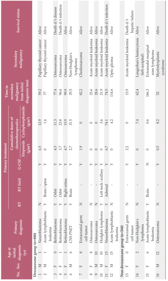
2) d ia gn os is ( m o) D ex ra zo xa n e gr ou p ( n = 88 0) 1 M 1 N eu ro bl as to m a N - Y 6. 5 12 .9 59 .2 P ap il la ry th yr oi d c an ce r A li ve 2 M 1 A cu te ly m p h ob la st ic Y B ra in / sp in e Y 0 1. 6 77 P ap il la ry th yr oi d c an ce r A li ve le u ke m ia 3 F 0 R et in ob la st om a Y O rb it Y 4. 5 11 .3 77 .4 O st eo sa rc om a D ea th d / t d is ea se 4 F 1 R et in ob la st om a Y O rb it Y 3. 7 23 .8 96 .6 O st eo sa rc om a D ea th d / t i n fe ct io n 5 F 1 R et in ob la st om a Y R ig h t r et in a Y 4. 7 15 .9 99 .9 O st eo sa rc om a A li ve 6 M 6 C N S P N E T Y B ra in Y 2. 7 31 .5 29 .5 N on -H od gk in 's A li ve ly m p h om a 7 F 8 E xt ra cr an ia l g er m N - Y 1. 9 0 82 .2 C h od ro sa rc om a A li ve ce ll tu m or 8 F 8 O st eo sa rc om a N - Y 0 0 25 .6 A cu te m ye lo id le u ke m ia A li ve 9 M 10 O st eo sa rc om a N - Y 0 0 28 .6 A cu te m ye lo id le u ke m ia A li ve 10 M 10 H od gk in ly m p h om a Y L ef t n ec k/ ax il la ry Y 0 3. 6 21 .9 A cu te m ye lo id le u ke m ia A li ve 11 F 13 N eu ro bl as to m a Y A d re n al Y 6. 7 74 .1 74 .5 A cu te m ye lo id le u ke m ia D ea th d / t i n fe ct io n 12 M 15 A cu te ly m p h ob la st ic N - Y 0 4. 2 11 4. 6 O p ti c gl io m a A li ve le u ke m ia N on -d ex ra zo xa n e gr ou p ( n = 34 4) 13 F 0 E xt ra cr an ia l g er m N - Y 2. 2 0 15 .9 A cu te m ye lo id le u ke m ia D ea th d / t ce ll tu m or h ep at ic f ai lu re 14 F 2 N on -H od gk in 's N - Y 0 7. 4 62 .4 L an ge rh an 's h is ti oc yt os is A li ve ly m p h om a (l ef t o cc ip it al ) 15 F 6 A cu te ly m p h ob la st ic Y B ra in N 0 0. 6 16 6. 3 E xt ra n od al m ar gi n al A li ve le u ke m ia zo n e ly m p h om a 16 M 12 O st eo sa rc om a N - Y 0. 5 8. 2 52 M ye lo d ys p la st ic A li ve sy n d ro m e
T ab le 3 . C h ar ac te ri st ic s of p at ie n ts w it h s ec on d ar y m al ig n an ci es R T , r ad ia ti on ; G -C SF , g ra nu lo cy te c ol on y- st im u la ti ng fa ct or ; M , m al e; N , n o; Y , y es ; F , f em al e; d / t, d u e to ; C N S P N E T , c en tr al n er vo u s sy st em p ri m it iv e ne u ro ec - to d er m al tu m or .
(5.7%) in D group (p=0.24) (S3 Table). Among all cardiac events, asymptomatic ventricular dysfunction in echocardio- graphy was the most common cardiac manifestation (3 cases in non-D group, 26 cases in D group). According to the time of cardiotoxicity, early cardiotoxicity occurred in 13 cases of non-D group, and 52 cases of D group.
The cardiac EFS rate was 95.4% in non-D group, and 93.4%
in D group. There was no significant difference in EFS after considering the corrections factors (p=0.09) (Fig. 1A).
According to the total anthracyclines cumulative doses, there were 204 patients who received more than 400 mg/m
2of anthracyclines (32 patients in non-D group, 172 patients in D group). There were 6 cardiac events in non-D group, and 12 events in D group (S4 Table). When analyzing the cardiac EFS rates according to the total cumulative anthracycline doses, there was significant difference between non-D group and D group of patients who received more than 400 mg/m
2, as 80.1% and 91.2% (p=0.04) (Fig. 1B).
4. Secondary malignancy and survival
Data of SMN was available in 1,224 patients (non-D group 344 patients and D group 880 patients), and there were 4 and 12 patients who developed SMN or MDS in non-D group and D group, respectively (Table 3). The median latency from initial diagnosis to SMN was 5.7 years (range, 1.3 to 13.9 years). The most common SMN was acute myeloid leukemia
(AML), as total five patients developed AML, and osteosar- coma was developed in three patients. The follow up dura- tion of both groups were significantly different, so the cumu- lative incidence of both groups were compared at the same time points. The 6-year cumulative incidence rate of SMN in all patients was 0.67±0.24%. The rates of SMN were 0.52%±
0.37% in non-D group and 0.60%±0.28% in D group, respec- tively (Fig. 2A), and the cumulative incidence rate after con- sidering correction factors was not statistically different between two groups (p=0.55). When patients with Hodgkin lymphoma were analyzed (non-D group 12 patients, and D group 34 patients), the 6-year cumulative incidence rate of SMN was 0% in non-D group and 3.1%±2.9% in D group, and the difference between two groups was not significant (p=0.56) (Fig. 2B).
The OS in D group was 87.4%, and that in non-D group was 86.3% (Fig. 3A). The EFS was 80.3% in D group and 80.4% in non-D group (Fig. 3B). The survival rates were not significantly different between two groups (OS, p=0.06; EFS, p=0.64).
5. Risk factor for secondary malignancy
Risk factors affecting the occurrence of SMN after consid- ering relapse and death as competing risks were analyzed with Fine and Gray model, and the results are shown in Table 4. According to the univariate analysis with significant Fig. 2. The 6-year cumulative incidence rate of secondary malignancy in all patients (n=1,224) (A), patients with Hodgkin disease (n=46) (B). (A) The 6-year cumulative incidence rate of secondary malignancies (SMN) in all patients was 0.67±0.24%.
The rates of SMN were 0.52%±0.37% in non-dexrazoxane group (non-D group) (n=880) and 0.60%±0.28% in dexrazoxane group (D group) (n=344) (p=0.55). (B) When patients with Hodgkin lymphoma were analyzed, the rates of SMN was 0% in non-D group (n=12) and 3.1%±2.9% in D group (n=34) (p=0.56).
Cu m ul at iv e in ci de nc e of s ec on da ry m al ig na nc
y 1.0
0 0.2 0.4 0.8
0
Time (mo)
50 100 150 200
A
0.6
Dexrazoxane (n=880) Non-dexrazoxane (n=344) p=0.55
Cu m ul at iv e in ci de nc e of s ec on da ry m al ig na nc
y 1.0
0 0.2 0.4 0.8
0
Time (mo)
50 100 150
B
0.6
Dexrazoxane (n=34)
Non-dexrazoxane (n=12)
p=0.56
level of 5%, irradiation history, higher cumulative doses of etoposide, cyclophosphamide and anthracyclines, longer duration of anthracycline, shorter time after last anthracy- cline, and longer duration of chemotherapy were related with statistically significant higher hazard ratio of the occur- rence of SMN. The result of multivariate analysis showed that the incidence of SMN was not increased due to dexra- zoxane, but increased approximately 1.05 times in response
to every 1-month prolongation of anthracycline administra- tion (p < 0.01) (Table 4). Whereas, the incidence of SMN was reduced by about 0.99 times as every month after last anthra- cycline went by (p < 0.01).
Factor Univariate analysis Multivariate analysis
Unadjusted HR (95% CI) p-value Adjusted HR (95% CI) p-value
Dexrazoxane group vs. non-dexrazoxane group 3.46 (0.92-13.07) 0.07 - -
Age (yr) 0.97 (0.85-1.09) 0.55 - -
Sex (female vs. male) 2.30 (0.82-6.47) 0.11 - -
Syndrome or genetic disease (yes vs. no) 3.14 (0.03-23.39) 0.50 - -
G-CSF (yes vs. no) 5.05 (1.03-91.25) 0.09 - -
Previous radiation (yes vs. no) 3.12 (1.15-8.45) 0.03 - -
Cumulative dose of etoposide (mg/m
2) 1.31 (1.16-1.47) < 0.01 - -
Cumulative dose of cyclophosphamide (mg/m
2) 1.08 (1.06-1.10) < 0.01 - -
Cumulative dose of total anthracyclines (mg/m
2) 2.88 (1.82-4.58) < 0.01 - -
Total duration of anthracycline (mo) 1.06 (1.05-1.07) < 0.01 1.05 (1.03-1.06) < 0.01 Time after last anthracycline (mo) 0.98 (0.97-0.99) < 0.01 0.99 (0.98-0.99) < 0.01
Total duration of chemotherapy (mo) 1.05 (1.04-1.07) < 0.01 - -
Table 4. Analysis of the risk factors for secondary malignancy
HR, hazard ratio; CI, cumulative incidence.
Fig. 3. Survival rates of all patients. (A) Overall survival rate of 1,035 patients in dexrazoxane group (D group) was 87.4%, and that of 418 patients in non-dexrazoxane group (non-D group) was 86.3% (p=0.06). (B) Event-free survival rate was 80.3%
in D group (n=1,035) and 80.4% in non-D group (n=418) (p=0.64).
Ov er al l s ur vi va l r at e
1.0
0.5 0.6 0.7 0.9
0
Time (mo) 60
40
20 80 100 120 140 160 180 200
A
0.8
Dexrazoxane (n=1,035) Non-dexrazoxane (n=418) p=0.06
Ev en t-f re e su rv iv al ra te
1.0
0 0.2 0.4 0.8
0
Time (mo) 60
40
20 80 100 120 140 160 180 200
B
0.6
Dexrazoxane (n=1,035)
Non-dexrazoxane (n=418)
p=0.64
Discussion
In this study, multivariate analysis for assessing risk fac- tors of SMN in pediatric cancer patients who received dexra- zoxane was conducted, and there was no increased incidence or risk of SMN in those patients.
The mechanism of anthracycline’s early cardiotoxicity is known to be related to free radical injury, contributing to the formation of reactive oxygen species and leading to the apop- tosis of cardiomyocyte and intracellular damage [12]. On the other hand, the mechanism of delayed cardiotoxicity in the long-term survivors is multifactorial including myocardial mitochondria related apoptosis which results in metabolic remodeling of heart [13]. However, delayed cardiotoxicity presents as overt clinical manifestations such as heart failure only in extreme cases, and only slowly progressing ventric- ular abnormalities are detected in many cases [13].
For assessing cardiotoxicity in pediatric cancer patients, cardiac event has been counted as one of the markers for car- diotoxicity [3,14]. A cumulative dose of anthracycline has been known as the most significant risk factor for cardiac dysfunction. The incidence of heart failure was approxi- mately 3.0% in patients receiving a cumulative dose of dox- orubicin of 400 mg/m
2, 7.5% at doses of 550 mg/m
2, and 18.0% at doses of 700 mg/m
2[15]. In our study, although there was no significant difference in EFS between two groups and the median follow up duration was significantly longer in non-D group, patients who received more than 400 mg/m
2of cumulative anthracyclines and dexrazoxane showed a slightly higher cardiac EFS than the other patients without dexrazoxane. These results might be related to the relatively low incidence of clinical cardiologic events in patients with less than 400 mg/m
2of anthracycline, thus the protective effect would be apparent only in patients receiving higher amounts of anthracyclines. Longer follow-up would be needed considering late cardiotoxicity especially in patients with short treatment off period.
Despite of the cardioprotective effects of dexrazoxane, dexrazoxane was reported to be related with increased risk of SMN. Tebbi et al. [5] reported the results of the Pediatric Oncology Group study with Hodgkin lymphoma, in which patients were randomly assigned to receive dexrazoxane to evaluate its cardiopulmonary protective effect. The authors found an increased incidence of SMN after a median follow- up time of 4.8 years, especially AML or MDS, in the dexra- zoxane-received patients.
However, many refutations against the Tebbi’s report have been followed. Since in that article, risk factors of SMN such as cumulative doses of etoposide, cyclophosphamide, and doxorubicin or G-CSF administration were not considered as confounding factors in the analysis. Also, many studies in
other patient groups have not shown the same results [6-8, 13].
In our study, multiple risk factors known to affect SMN development were collected. In the multivariate analysis, longer duration of anthracycline, and shorter time after last anthracycline were related with statistically significant higher hazard ratio of the occurrence of SMN. There was no significant risk factor related to AML or MDS, specifically.
There was no data related to the association of SMN and duration of certain chemotherapeutic agents. Considering the final adjusted hazard ratios in our study were relatively low as 1.05 and 0.99, our final multivariate factors could not be the most powerful risk factors for SMN. Further analysis could elucidate more influential risks factors for SMN in these patients group.
Risk factors for SMN are known to be multifactorial.
Chemotherapy agents known to be carcinogenic are the alky- lating agents, topoisomerase II agents, and anthracyclines [16]. In addition, the cumulative dose, the schedule of chemo- therapy administration, and the use of multiple drugs all sig- nificantly impact the risk of developing SMN [16].
In our study, patient No. 2 in Table 3 developed papillary thyroid cancer within radiation fields. Although there were cases where the irradiated fields and location of SMN were not related, radiation itself has been reported as one of the risk factors for SMN in childhood cancer patients [17-19]. Sec- ondary osteosarcoma was developed in three retinoblastoma patients. The risk of secondary bone tumors in retinoblas- toma survivors is higher in patients with bilateral disease or germline mutations in RB gene. In addition, radiation ther- apy could also increase the risk of secondary osteosarcoma in retinoblastoma patients [20].
When patients with Hodgkin disease were analyzed sepa- rately, dexrazoxane was not a significant risk factor for SMN and the cumulative incidence of SMN was not significantly different regardless of dexrazoxane. In the study of the Childhood, Adolescent and Young Adult Cancer Survivors Research Program of British Columbia which analyzed long term results of 442 Hodgkin lymphoma survivors, radiation therapy (hazard ratio, 2.7), and female sex (hazard ratio, 1.8) were significant risk factors for SMN in multivariate analysis [21]. To analyze risk factors for SMN in Hodgkin disease, many factors other than dexrazoxane should be considered.
This study has several limitations. First, there would be
possibility of selection bias due to retrospective design
although data management and analysis were conducted by
medical statistics specialists. Second, the follow up duration
was variable, and there were patients with insufficient fol-
low-up duration for late complication analysis. Especially,
the follow up duration of D group was significantly shorter
than non-D group so that cardiac events or SMN results
might be changed after longer follow-up. Third, there could
be other risk factors for SMN development such as cancer predisposing genetic factors and other alkylating agents including ifosfamide. In addition, specific medical conditions which might cause cardiac events other than anthracycline could not be considered because of the limitation of retro- spective design.
In conclusion, dexrazoxane had an efficacy in the preven- tion of lowering cardiac EFS rates in patients with higher cumulative anthracyclines. According to the multivariate analysis, the occurrence of SMN was not related to dexrazox- ane. Given the efficacy of dexrazoxane as a cardioprotectant, dexrazoxane could be used in anthracycline-containing pediatric regimens.
Electronic Supplementary Material
Supplementary materials are available at Cancer Research and Treatment website (https://www.e-crt.org).
Conflicts of Interest
Conflict of interest relevant to this article was not reported.
Acknowledgments
This research was supported by a grant (12172MFDS231) from the Ministry of Food and Drug Safety, Republic of Korea.
Author Details
1
Department of Pediatrics, Cancer Research Institute, Seoul National University College of Medicine, Seoul,
2Department of Pediatrics, Asan Medical Center, University of Ulsan College of Medicine, Seoul,
3Department of Pediatrics, College of Medicine, The Catholic University of Korea, Seoul,
4Department of Pediatrics, Yeungnam University Medical Center, Yeungnam University College of Med- icine, Daegu,
5Department of Pediatrics, Daegu Fatima Hospital, Daegu,
6Department of Pediatrics, Korean Cancer Center Hospital, Seoul,
7Department of Pediatrics, Hanyang University Medical Cen- ter, Hanyang University College of Medicine, Seoul,
8Department of Pediatrics, Ulsan University Hospital, University of Ulsan College of Medicine, Ulsan,
9Department of Pediatrics, Chonnam National University Hwasun Hospital, Chonnam National University School of Medicine, Hwasun,
10Department of Pediatrics, Keimyung Uni- versity Dongsan Medical Center, Keimyung University School of Medicine, Daegu,
11Department of Pediatrics, School of Medicine, Kyungpook National University, Daegu,
12Department of Pediatrics, Wonju Severance Christian Hospital, Yonsei University Wonju Col- lege of Medicine, Wonju,
13Department of Pediatrics, Dong-A Uni- versity Hospital, Dong-A University College of Medicine, Busan,
14
Department of Pediatrics, Chungbuk National University Hospital, Chungbuk National University College of Medicine, Cheongju,
15