ISSN 0378-6471 (Print)⋅ISSN 2092-9374 (Online)
http://dx.doi.org/10.3341/jkos.2015.56.9.1365
Original Article
안내염 배양에서 소아용 혈액배양병의 유용성
The Effectiveness of Pediatric Blood Culture Bottle in Endophthalmitis
김경호1,2⋅권한조3⋅박성후3,4⋅변익수1,2⋅이지은3,4⋅엄부섭5⋅신경화6
Kyong Ho Kim, MD1,2, Han Jo Kwon, MD3, Sung Who Park, MD3,4, Ik Soo Byon, MD1,2, Ji Eun Lee, MD, PhD3,4, Boo Sup Oum, MD, PhD5, Kyung Hwa Shin, MD6
부산대학교 의학전문대학원 양산부산대학교병원 안과학교실1, 부산대학교 의학전문대학원 양산부산대학교병원 의생명융합연구소2, 부산대학교 의학전문대학원 부산대학교병원 안과학교실3, 부산대학교 의학전문대학원 안과학교실4, 정근안과병원5,
부산대학교 의학전문대학원 부산대학교병원 진단검사의학과학교실6
Department of Ophthalmology, Pusan National University Yangsan Hospital, Pusan National University School of Medicine1, Yangsan, Korea Research Institute for Convergence of Biomedical Science and Technology, Pusan National University Yangsan Hospital, Pusan National
University School of Medicine2, Yangsan, Korea
Department of Ophthalmology, Pusan National University Hospital, Pusan National University School of Medicine3, Busan, Korea Department of Ophthalmology, Pusan National University School of Medicine4, Yangsan, Korea
Junggun Eye Hospital5, Busan, Korea
Department of Laboratory Medicine, Pusan National University Hospital, Pusan National University School of Medicine6, Busan, Korea
Purpose: To investigate the effectiveness of the pediatric blood culture bottle for vitreous sample culture in endophthalmitis patients.
Methods: All consecutive cases with clinically suspected endophthalmitis treated and cultured in our institution between January 2009 and June 2013 were included in the study. Vitreous samples were obtained by vitreous needle aspiration (tap), anterior chamber aspiration, or mechanized vitreous biopsy (vitrectomy). The samples obtained using the conventional method until August 2011 were classified as group I. Since August 2011, the BacT/Alert PF pediatric blood culture bottle (bioMérieux, Marcy l’Etoile, France) was used for culture in group II. We investigated age, gender, biopsy method, cause of infection, use of anti- biotics, bacterial culture, and culture positive rate.
Results: Thirty-three cases were included in group I and 17 cases in group II. There was no significant difference in age, gender, sampling technique, cause of infection, and use of antibiotics between the 2 groups. The culture positive rate in group II (60.7%) was significantly higher than group I (33.3%, p = 0.032). In group II, Enterococcus feacalis was the most common pathogen (8 eyes). In group I, Streptococcus pneumoniae and Pseudomonas aeruginosa were confirmed in 3 cases.
Conclusions: The pediatric blood culture bottle can be used successfully in the examination of clinically suspected endophthalmitis.
The method showed higher culture positive rate compared with the conventional method. This technique is simple and maintain- ing a supply of fresh agar media is not necessary.
J Korean Ophthalmol Soc 2015;56(9):1365-1370
Key Words: Culture, Endophthalmitis, Pediatric culture media
■Received: 2015. 4. 10. ■ Revised: 2015. 5. 31.
■Accepted: 2015. 7. 21.
■Address reprint requests to Sung Who Park, MD
Department of Ophthalmology, Pusan National University Hospital, #179 Gudeok-ro, Seo-gu, Busan 49241, Korea Tel: 82-51-240-7326, Fax: 82-51-242-7341
E-mail: [email protected]
* This study was presented as a narration at the 110th Annual Meeting of the Korean Ophthalmological Society 2013.
ⓒ2015 The Korean Ophthalmological Society
This is an Open Access article distributed under the terms of the Creative Commons Attribution Non-Commercial License (http://creativecommons.org/licenses/by-nc/3.0/) which permits unrestricted non-commercial use, distribution, and reproduction in any medium, provided the original work is properly cited.
안내염(endophthalmitis)은 방수, 유리체 등의 안내 조직 에 세균이나 진균 등이 감염된 상태로, 드물지만 발병하면 시력을 위협하는 심각한 질환이다.1 감염 경로에 따라 술후 안내염, 외상후 안내염, 내인성 안내염으로 구분할 수 있 다.2 술후 안내염의 빈도는 안내수술의 0.05-0.7%이며3-5 외 상성 안내염은 천공외상의 3.3-17%6,7에서 발생한다. 내인 성 안내염의 경우는 전체 안내염의 2-6%로 당뇨나 간농양
Figure 1. Conventional culture media. Blood agar plate (A),
chocolate agar (B), thioglycollate broth (C), sabouraud dex- trose agar (D), and slide glasses for Gram stain and KOH test (E).Figure 2. BacT/Alert® PF (bioMérieux,
Marcy l’Etoile, France). A widely used broth culture medium for blood and other body fluids in pediatric practice.It is suitable for bacteria as well as fungi.
이 가장 흔한 기저 질환으로 알려져 있다.8
안내염은 조기 치료가 중요하기에 대부분의 경우 배양 결과를 확인하기 전 광범위 항균제를 투입하는 것을 원칙 으로 한다.1 하지만 균 동정은 감염 원인을 추정함에 있어 중요할 뿐 아니라 오진 가능성을 줄여주고, 치료 효과가 충 분하지 않아 항생제를 변경하거나 치료 방법을 변경하는 결정을 할 때 중요한 정보를 제공한다.
안내염에서 원인균을 확인하기 위해서 전통적으로 검체를 혈액한천배지(blood agar plate), 초콜렛한천배지(chocolate agar plate) 등에 접종하여 배양한다. 배양 양성률은 26-70%
로 다양하며,9-12 검사자의 숙련도에 영향을 받고 검체의 보 관이 까다롭기 때문에 양성률의 편차가 큰 것으로 생각된다.
반면 소아용 혈액배양병을 이용한 안내염 균 배양 양성률은 Yospaiboon et al13이 보고한 52%부터 Joondeph et al14의 결 과인 69%까지 양성률의 편차가 크지 않다. 이는 검체 보관 방법이나 접종 및 검사자의 숙련도에 영향을 덜 받기에 일정 수준 이상의 양성률을 보이는 것으로 생각된다.
저자들은 2011년 8월에 안내염 원인균 배양 방법을 전통 적 배양 방법에서 소아용 혈액배양병을 사용한 배양으로 변경하였다. 이에 변경 전후의 배양 결과를 비교하고자 하 였다.
대상과 방법
2009년 1월부터 2013년 6월 사이 본원에서 안내염이 의 심되어 배양검사를 시행한 환자의 의무기록을 후향적으로 분석하였다. 유리체천자, 전방천자 혹은 유리체절제술을 통 해 검체 채취를 시행한 경우를 포함하였고, 안구적출술 중 검체를 채취하거나 각막궤양 부위에서 검체를 채취한 경우 는 제외하였다. 안내염이 의심되어 검체를 채취하였더라도 이후 경과 관찰에서 임상의가 안내염이 아닌 것으로 판단 한 경우는 제외하였다. 유리체절제술을 시행한 경우 관류액 을 잠근 상태에서 전방수를 채취한 후 유리체 검체를 채취하 였으며, 유리체절제술 없이 유리체 내 항생제 주사를 시행한 경우는 전방수를 채취한 후 26게이지 바늘을 이용하여 유리 체 검체를 채취하였고, 무효천자(dry tap)된 경우는 대상에서 제외하였다. 전방천자와 유리체 천자는 0.1 mL 이상, 유리체 절제술은 0.5 mL 이상 채취하는 것을 원칙으로 하였다.
2011년 8월 이전 시행된 경우 I군으로 하였고, 세균 감염 을 확인하기 위하여 채취된 검체는 즉시 그람염색에 필요 한 슬라이드를 제작하고, 혈액한천배지, 초콜렛한천배지, 그리고 타이오글리콜레이트 배지(thioglycolate broth)에 접 종하였다. 진균 감염 여부를 확인하기 위해 KOH 표본도말 검사 및 사브로덱스트로스한천배지(Sabouraud dextrose agar) 에 접종하였으며(Fig. 1) 접종한 배지를 적합한 환경에서 배양하기 위해 즉시 미생물검사실로 보내는 것을 원칙으로
Table 1. Baseline characteristics of this study between the conventional method group and pediatric blood culture bottle method
groupConventional method Pediatric blood culture bottle p-value
Period January 2009 to August 2011 August 2011 to June 2013
Number (n, %) 33 (54.1) 28 (45.9)
Sex (male/female) 13/20 11/17 0.601*
Age (years) 65.6 ± 14.1 70.4 ± 11.0 0.153†
Sampling technique 0.082*
Vitrectomy (n, %) 17 (51.5) 22 (78.6)
Vitreous and anterior chamber tapping (n, %) 16 (48.5) 6 (21.4)
Cause of endophthalmitis 0.317‡
Postoperative (n, %) 23 (69.7) 17 (60.7)
Trauma (n, %) 6 (18.2) 2 (7.1)
Endogenous (n, %) 4 (12.1) 9 (32.1)
Values are presented as mean ± SD unless otherwise indicated.
*Chi-square test; †Mann-Whitney test; ‡Fisherexacttest.
Figure 3. Culture positive rate between two groups. Graphs
shows that culture positive rate of pediatric blood culture bot- tle group is higher than that of conventional method group (p= 0.032).
하였다. 본원 미생물검사실에 접수된 즉시 그람염색과 KOH 표본도말검사를 시행하였고, 세균을 검출하기 위하여 혈액한천배지, 초콜렛한천배지는 35°C, 5% CO2 환경에서 최소 48시간 배양 후, 타이오글리콜레이트 배지는 최소 5 일간 배양하여 균이 자라지 않으면, 배양 음성으로 판정하 였다. 진균을 검출하기 위한 사브로덱스트로스한천배지는 30°C에서 최소 14일 배양 후 균이 자라지 않으면 배양 음 성으로 판정하였다.
2011년 8월 이후에 시행한 경우를 II군으로 하였고, 채취 한 검체는 모두 즉시 소아용 혈액배양병인 BacT/Alert PF (bioMérieux, Marcy l’Etoile, France, Fig. 2)에 접종하였고, 1-3일 내 미생물검사실로 보냈다. 혈액배양병은 실시간 감 시 자동혈액배양장비인 BacT/Alert 3D (bioMérieux)를 이 용하여 최소 5일간 배양 후 균이 자라지 않으면, 배양 음성 으로 판정하였다. 두 방법 모두 배양이 양성인 검체는 통상 적인 방법으로 균주를 동정하였다.
대상 환자들의 나이, 성별, 검체 채취 방법, 감염경로, 항 생제 사용 여부, 배양률 및 균 종류를 조사하였고, 두 군의 차이를 비교하기 위해 Chi-square test 혹은 Fisher exact test를 사용하여 p<0.05일 때 통계적 의의가 있는 것으로 판 단하였다.
결 과
I군 33명(54.1%), II군 28명(45.9%)이 포함되었다. I군의 나이는 65.6 ± 14.1세, 남자 13명(39.4%), 여자 20명(60.6%) 이었고, II군은 나이 70.4 ± 11.0세, 남자 11명(39.3%), 여자 17명(60.7%)이었다. 두 군 간 나이와 성별의 유의한 차이는 없었다(p=0.601, p=0.153, Table 1).
I군 중 17안(51.5%)은 유리체절제술을 통한 검체 채취였 고, 16안(48.5%)은 유리체천자 및 전방천자를 통한 검체 채취
였다. II군의 경우 22안(78.6%)은 유리체절제술, 6안(21.4%) 은 유리체천자 및 전방천자를 통한 검체 채취가 이루어져 II군에서 유리체절제술의 비율이 높았으나 통계학적 의미 는 없었다(p=0.082, Table 1). 전체 대상 중 유리체절제술로 검체를 채취한 39안 중 21안(53.8%)과 유리체천자로 채취 한 22안 중 7안(31.8%)에서 균이 배양되어 검체 채취 방법 에 따른 배양 양성률의 통계학적 차이는 없었다(p=0.116).
I군에서는 전신 혹은 점안 항생제를 사용했던 한 경우가 25안(75.8%), II군은 21안(75%)으로 두 군 간 차이는 없었 고(p=0.65, Table 1), 두 군 모두에서 술 전 점안 혹은 전신 항생제 사용과 균 배양률은 의미 있는 상관관계를 보이지 않았다(I군: p=0.503, II군: p=0.774). 검체 채취 전 유리체 내 항생제를 주입한 경우는 I군의 1안이 유일하였으며, 균 은 배양되지 않았다.
안내염 감염 경로는 I군에서 술 후 감염 23안(69.7%), 외 상 6안(18.2%), 내인성 감염 4안(12.1%)이었고, II군에서 술 후 감염 17안(60.7%), 외상 2안(7.1%), 내인성 감염 9안
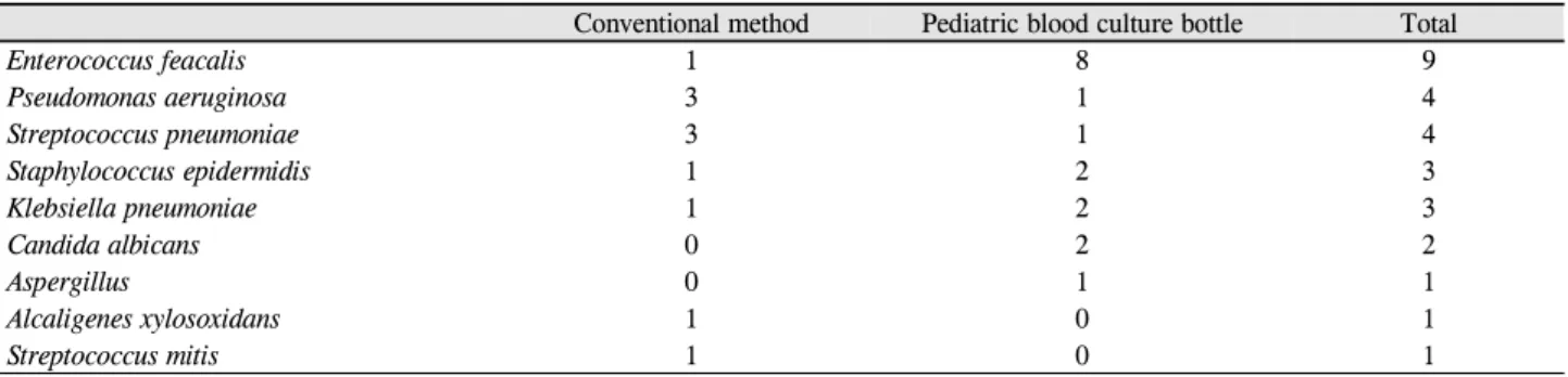
Table 2. Frequency of organism according to the result of culture
Conventional method Pediatric blood culture bottle Total
Enterococcus feacalis 1 8 9
Pseudomonas aeruginosa 3 1 4
Streptococcus pneumoniae 3 1 4
Staphylococcus epidermidis 1 2 3
Klebsiella pneumoniae 1 2 3
Candida albicans 0 2 2
Aspergillus 0 1 1
Alcaligenes xylosoxidans 1 0 1
Streptococcus mitis 1 0 1
(32.1%)이었으며, 두 군 간의 유의한 차이는 없었다(p=0.317, Table 1). 안내염 감염 경로에 따른 배양률의 차이 여부를 분석해 본 결과, 술 후 감염의 경우 40안 중 18안(45%), 외 상 8안 중 3안(37.5%), 내인성 감염 13안 중 7안(53.8%)에 서 균 배양 양성이었고, 감염 경로에 따른 배양률의 차이는 없었다(p=0.755).
배양 양성률은 I군에서 33.3% (11안), II군에서 60.7%
(17안)로 II군에서 높았다(p=0.032, Fig. 3). I군에서 배양된 11안 중 Streptococcus pneumoniae, Pseudomonas aeruginosa 가 각 3안에서 배양되었고, Enterococcus feacalis, Staphylococcus
epidermidis, Klebsiella pneumoniae, Alcaligenes xylosoxidans, Streptococcus mitis가 각 1안에서 배양되었다. 반면 II군에
서 배양된 17안 중에서는 Enterococcus feacalis가 8안으로 가장 많았고, Staphylococcus epidermidis, Klebsiella pneumo-niae, Candida albicans가 각 2안에서 배양되었고 Streptococcus pneumoniae, Pseudomonas aeruginosa, Aspergillus가 각 1
안에서 배양되었다(Table 2).고 찰
대부분의 안내염 증상은 발현 후 수 시간에서 수일 내에 급속도로 진행하기 때문에 응급 상황이며, 치료 시기가 늦 어지거나 지체될 경우 시력이 상실될 수 있다.1 그러므로 균 배양을 위해 검체 채취를 하면서 동시에 경험적 광범위 항생제를 유리체강 내로 주사하는 것이 일반적이다.15 이때 채취한 검체를 혈액한천배지 및 초콜릿한천배지 등에 접종 하여 균을 배양하는 것을 전통적 방법이라 하며, 이러한 방 법의 균 배양률은 endophthalmitis vitrectomy study (EVS)16 에서 69%, Lee and Park17의 보고에서 63%로 비교적 높은 양성률을 보이기도 하지만 50% 미만,9-12 심지어 26%로 낮 은 경우13도 보고되었다. 원인균 배양을 위해서 여러 개의 배지에 접종해야 하고, 신선한 배지가 준비되어야 하며, 각 각의 배지에 필요한 배양 환경이 달라 접종 후 즉시 배양 검사실로 보내야 할 뿐만 아니라, 배양까지 시간이 오래 걸
리는 것 등이 단점이다. 이는 접종자의 숙련도, 배양하는 미생물 검사실의 시간적 혹은 공간적 접근성, 배양검사자 의 숙련도 등 안내염 이외의 요인에 의해 양성률이 영향을 받을 수 있음을 의미하며, 이러한 요소가 비교적 넓은 양성 률 편차를 야기하는 것으로 생각된다.
1989년 Joondeph et al14은 감염성 안내염 환자에서 혈액 배양병(Blood culture bottle)을 이용한 배양 결과를 보고하 였다. 이때 10 mL 이상의 검체를 채취하기 위해 카세트에 모아진 유리체절제액을 이용하였다. 혈액배양병은 많은 양 의 검체가 필요하여 유리체절제술을 하지 않는 경우에서는 충분한 검체를 얻을 수 없고, 유리체절제술이 끝날 때까지 항생제 사용에 제한이 있으며, 수술 후 따로 카세트 내 검 체를 채취해야 하는 등의 어려움이 있었다.
이후 비교적 소량의 검체로 배양이 가능한 소아용 혈액배 양병을 이용한 배양이 감염성 각막염에서 처음 사용되었다.18 이 연구에서 Coagulase-negative Staphylococcus, Streptococcus
pneumoniae, Enterobacter, Escherichia coli, Acinetobacter, Moraxella catarrhalis, Candida parapsilosis와 같은 다양한
균주를 검출하는 데 성공하였고,18 그 이후 감염성 안내염 의 원인균 배양에 소아용 혈액배양병이 사용되기 시작하였다.Kratz et al2은 안내염 환자에서 Bactec Peds Plus F (Becton Dickinson, Sparks, MD, USA) 소아용 혈액배양병을 사용 한 배양과 전통적 방법을 전향적으로 비교하였다. 이 연구 는 한 눈에서 채취한 유리체 검체를 나누어 동시에 두 가지 방법으로 배양하였고, 전통적인 방법에서는 53.9% (13안 중 7안), 소아용 혈액배양병을 사용한 경우에는 69.2% (13 안 중 9안)에서 균이 배양되었다고 보고하였다. 이 연구에 서 사용된 배지는 resin이 항생제 중화제로 포함되어 있었 으며, 비교적 소량인 희석되지 않은 0.2 mL의 유리체를 주 사하였음에도 비교적 높은 양성률을 보였다고 하였다. 두 배양법 사이에 배양률에서 통계학적 차이는 없었지만, 편리 성과 안전성에서 소아용 혈액배양병이 전통적 방법에 대한 훌륭한 대안이 될 수 있을 것이라고 주장하였다.
본 연구에서 사용한 BacT/Alert PF 소아용 혈액배양병
(Fig. 2) 역시 적은 용량의 검체만으로도 원인균 배양을 할 수 있도록 특화되어 있는 배지이다. 일반 배지보다 원인균 검출률이 높고 검출 소요 시간도 짧은 특징을 가지고 있
어,19-24 소아의 혈액배양이나 관절액과 같은 소량의 체액으
로 원인균을 배양하는 데 널리 사용되고 있고, 여러 소아 질환에서 이 배지를 사용하여 우수한 배양률을 보였음이 보고된 바 있다.25,26
본 연구에서도 이전에 Kratz et al2에 의해 유용성이 보고 된 Bactec Peds Plus F를 사용하려 하였으나, 구매에 여러 제한점이 있었다. 본원 진단검사의학과 의료진의 자문을 구해 비슷한 효과를 기대할 수 있고, 따라서 본원 소아청소 년과에서 사용 중인 BacT/Alert PF 소아용 혈액배양병을 사용하였다. 배양 방법 변경의 타당성을 확인하기 위해, 전 통적 검사를 동시에 시행하는 것을 고려하였으나, 안내염 에서 두 검사를 동시에 수행할 만한 충분한 검체를 채취하 는 것이 어렵다고 판단하여, 동시에 두 방법을 시행하지는 않았다. 본 연구에서 소아용 혈액배양병을 사용한 배양 양 성률은 60.7%로 Kratz et al2의 69.2%와 유사하였고, 본 연 구의 전통적 방법의 33.3%보다는 유의하게 우수하였다.
국내외 여러 보고12,16,17에서 전통적 방법으로도 높은 배 양률이 보고된 것으로 미루어, 소아용 혈액배양병을 사용 하였을 때 배양 양성률이 전통적 방법에 비해 우수하였다 는 본 연구의 결과는 소아용 혈액배양병 자체의 우수성이 라기보다는 전통적 방법을 시행함에 있어 문제가 있었던 것으로 해석할 수 있다. 첫째, 검체 접종자의 숙련도가 문 제였을 수 있다. 검체의 양이 적기 때문에 다른 부위 검체 와는 달리 그람 염색 슬라이드 제작 및 배지 접종을 안과 의료진이 직접 시행하였고, 이는 검체 접종자의 숙련도가 낮은 양성률의 원인일 수 있음을 의미한다. 둘째, 전통적 방법을 시행함에 있어, 배지의 신선도, 접종 후 배지를 검 사실에 보내는 시간 등이 기록 및 표준화되지 않았으며, 이 는 배양률을 낮아지는 원인이었을 수 있다.
소아용 혈액배양병을 사용한 군에서 유리체절제술로 검 체를 채취한 비율(78.6%)이 전통적 방법(51.5%)보다 높았 으며, 이는 유리체절제술의 안전성이 높아짐에 따라 안내 염에서 유리체절제술의 시행 빈도가 높아졌기 때문으로 해 석할 수 있다. 유리체절제술로 검체를 채취하는 경우 충분 한 양의 검체를 채취할 수 있으므로 배양 양성률에 영향을 줄 수 있다. 하지만 유리체절제술로 검체를 채취한 비율은 두 군 사이에 통계학적 차이가 없었고, 유리체절제술로 채 취한 경우와 유리체천자로 채취한 경우에 배양률의 통계학 적 차이도 없었다.
본 연구는 단기관 후향적 연구로 대상 환자의 수가 적고, 같은 검체로 두 배양 방법을 비교하지 않은 점은 제한점이
라고 할 수 있다. 안내염의 배양에 있어 희석되지 않은 검 체를 최대한 많이 확보하는 것이 중요하다고 여겨지므로, 동시에 두 배양 방법을 수행하여 비교하는 것이 합당한가 에 대해서는 논란의 여지가 있다.
본 연구는 기존에 보고되었던 Bactec Peds Plus F 소아용 혈액배양병뿐 아니라 BacT/Alert PF 소아용 혈액배양병으 로도 안내염의 원인균을 효과적으로 배양할 수 있음을 보 여주었고, 특히 전통적 배양법으로 낮은 배양률을 보이는 기관이나, 검사실의 접근성이 낮은 병원에서 좋은 대안이 될 수 있음을 보여주었다. 본 연구의 결과는 다기관 전향적 연구를 통해 입증되어야 할 것이다.
REFERENCES
1) Durand ML. Endophthalmitis. Clin Microbiol Infect 2013;19:
227-34.
2) Kratz A, Levy J, Belfair N, et al. Broth culture yield vs traditional approach in the work-up of endophthalmitis. Am J Ophthalmol 2006;141:1022-6.
3) Javitt JC, Vitale S, Canner JK, et al. National outcomes of cataract extraction. Endophthalmitis following inpatient surgery. Arch Ophthalmol 1991;109:1085-9.
4) Powe NR, Schein OD, Gieser SC, et al. Synthesis of the literature on visual acuity and complications following cataract extraction with intraocular lens implantation. Cataract Patient Outcome Research Team. Arch Ophthalmol 1994;112:239-52.
5) Kattan HM, Flynn HW Jr, Pflugfelder SC, et al. Nosocomial en- dophthalmitis survey. Current incidence of infection after intra- ocular surgery. Ophthalmology 1991;98:227-38.
6) Meredith TA. Posttraumatic endophthalmitis. Arch Ophthalmol 1999;117:520-1.
7) Thompson WS, Rubsamen PE, Flynn HW Jr, et al. Endophthalmitis after penetrating trauma. Risk factors and visual acuity outcomes.
Ophthalmology 1995;102:1696-701.
8) Jackson TL, Eykyn SJ, Graham EM, Stanford MR. Endogenous bacterial endophthalmitis: a 17-year prospective series and review of 267 reported cases. Surv Ophthalmol 2003;48:403-23.
9) Donahue SP, Kowalski RP, Jewart BH, Friberg TR. Vitreous cul- tures in suspected endophthalmitis. Biopsy or vitrectomy?
Ophthalmology 1993;100:452-5.
10) Han DP, Wisniewski SR, Kelsey SF, et al. Microbiologic yields and complication rates of vitreous needle aspiration versus mechanized vitreous biopsy in the Endophthalmitis Vitrectomy Study. Retina 1999;19:98-102.
11) Sharma S, Jalali S, Adiraju MV, et al. Sensitivity and predictability of vitreous cytology, biopsy, and membrane filter culture in endophthalmitis. Retina 1996;16:525-9.
12) Lertsumitkul S, Myers PC, O’Rourke MT, Chandra J. Endophthalmitis in the western Sydney region: a case-control study. Clin Experiment Ophthalmol 2001;29:400-5.
13) Yospaiboon Y, Saree S, Pasadhika S. Blood culture and conven- tional media for vitreous culture in infectious endophthalmitis. J Med Assoc Thai 2005;88:639-42.
= 국문초록 =
안내염 배양에서 소아용 혈액배양병의 유용성
목적: 안내염 원인균 배양에서 소아용 혈액배양병 사용의 유용성에 대해 알아보고자 한다.
대상과 방법: 2009년 1월부터 2013년 6월 사이 본원에서 안내염으로 진단 받고 배양검사를 시행한 경우를 대상으로 하였다. 유리체천 자, 전방천자 혹은 유리체절제술을 통해 검체 채취가 이루어진 경우를 포함하였다. 2011년 8월 이전 전통적 배양 방법을 이용한 경우 를 I군, 2011년 8월 이후 소아용 혈액배양병인 BacT/Alert® PF (bioMérieux, Marcy l’Etoile, France)만을 이용하여 배양한 경우를 II군 으로 하였다. 대상 환자의 나이, 성별, 검체 채취 방법, 감염경로, 항생제 사용 여부, 균 배양 양성률 및 배양된 균 종류를 조사하였다.
결과: I군은 33안, II군은 28안이 포함되었다. 두 군의 나이, 성별, 검체 채취 방법, 감염 경로, 항생제 사용 여부는 의미 있는 차이가 없었다. II군의 균 배양 양성률은 60.7% (17안)로 I군의 33.3% (11안)보다 높았다(p=0.032). II군 17안에서 균이 배양되었고, Enterococcus feacalis가 8안으로 가장 흔하였다. I군은 11안에서 균이 배양되었고, Streptococcus pneumoniae, Pseudomonas aeruginosa가 각각 3안에서 배양되었다.
결론: 소아용 혈액배양병을 사용한 배양 방법은 전통적 배양 방법보다 높은 배양 양성률을 보였다. 전통적 배양 방법에 비해 접종의 숙련도와 배지의 보관 방법 등에 영향을 적게 받는 것이 높은 양성률의 원인으로 생각된다.
<대한안과학회지 2015;56(9):1365-1370>
14) Joondeph BC, Flynn HW Jr, Miller D, Joondeph HC. A new culture method for infectious endophthalmitis. Arch Ophthalmol 1989;
107:1334-7.
15) Kang KT, Kim KS, Kim YC. Factors affecting final visual acuity in infectious endophthalmitis following cataract surgery. J Korean Ophthalmol Soc 2013;54:1025-31.
16) Results of the Endophthalmitis Vitrectomy Study. A randomized trial of immediate vitrectomy and of intravenous antibiotics for the treatment of postoperative bacterial endophthalmitis. Endophthalmitis Vitrectomy Study Group. Arch Ophthalmol 1995;113:1479-96.
17) Lee NE, Park JM. Clinical results of bacterial endophthalmitis:
bacterial culture and visual acuity outcomes. J Korean Ophthalmol Soc 2011;52:1173-81.
18) Kratz A, Levy J, Klemperer I, Lifshitz T. Broth cultures yield vs traditional approach in the workup of infectious keratitis. Eye (Lond) 2006;20:215-20.
19) Spaargaren J, van Boven CP, Voorn GP. Effectiveness of resins in neutralizing antibiotic activities in bactec plus Aerobic/F culture medium. J Clin Microbiol 1998;36:3731-3.
20) Rohner P, Pepey B, Auckenthaler R. Advantage of combining resin with lytic BACTEC blood culture media. J Clin Microbiol 1997;35:2634-8.
21) Mirrett S, Everts RJ, Reller LB. Controlled comparison of original vented aerobic fan medium with new nonvented BacT/ALERT FA medium for culturing blood. J Clin Microbiol 2001;39:2098-101.
22) Wilson ML, Mirrett S, Meredith FT, et al. Controlled clinical com- parison of BACTEC plus anaerobic/F to standard anaerobic/F as the anaerobic companion bottle to plus aerobic/F medium for cul- turing blood from adults. J Clin Microbiol 2001;39:983-9.
23) Weinstein MP, Towns ML, Quartey SM, et al. The clinical sig- nificance of positive blood cultures in the 1990s: a prospective comprehensive evaluation of the microbiology, epidemiology, and outcome of bacteremia and fungemia in adults. Clin Infect 1997;24:584-602.
24) Park SH, Shim H, Yoon NS, Kim MN. Clinical relevance of time-to-positivity in BACTEC9240 blood culture system. Korean J Lab Med 2010;30:276-83.
25) Yagupsky P, Dagan R, Howard CW, et al. High prevalence of Kingella kingae in joint fluid from children with septic arthritis re- vealed by the BACTEC blood culture system. J Clin Microbiol 1992;30:1278-81.
26) Kratz A, Greenberg D, Barki Y, et al. Pantoea agglomerans as a cause of septic arthritis after palm tree thorn injury; case report and literature review. Arch Dis Child 2003;88:542-4.