J Korean Ophthalmol Soc 2014;55(6):847-853 pISSN: 0378-6471⋅eISSN: 2092-9374
http://dx.doi.org/10.3341/jkos.2014.55.6.847
Original Article
특발성 망막전막에서 내경계막 제거 시 인도시아닌그린 염색의 용매에 따른 수술결과 비교
Effect of Solvent in Indocyanine Green-Assisted Internal Limiting Membrane Peeling During Idiopathic Epiretinal Membrane Surgery
김미래⋅박주홍⋅사공민⋅장우혁
Mi Rae Kim, MD, Ju Hong Park, MD, Min Sagong, MD, PhD, Woo Hyok Chang, MD, PhD
영남대학교 의과대학 안과학교실
Department of Ophthalmology, Yeungnam University College of Medicine, Daegu, Korea
Purpose: This study was designed to compare the outcomes in idiopathic epiretinal membrane (ERM) surgery according to sol- vents of indocyanine green (ICG) for internal limiting membrane (ILM) peeling.
Methods: The medical records of 27 patients (27 eyes) with idiopathic ERM who had undergone pars plana vitrectomy with ICG staining for ILM peeling were retrospectively reviewed. The patients were divided into two groups according to solvents of 0.25%
ICG solutions. Solvents used were balanced salt solution (BSS) in group I (15 eyes) and 5% glucose in group II (12 eyes). The severity of ERM, the duration of symptoms, the preoperative and postoperative best corrected visual acuity (BCVA) values, the visibility of the stained ILM (Good, Fair, Poor), and the postoperative complications were compared in the two groups.
Results: There was no statistically significant difference in the severity of ERM, the duration of symptoms and the preoperative BCVA in the two groups. The postoperative BCVA was significantly improved in both groups, and the difference was not statisti- cally significant (p = 0.675). There was a significantly smaller number of eyes with poor ILM staining in group II than in group I (p = 0.014). No complications such as recurrence of ERM, atrophy of the retinal pigment epithelium (RPE) or retinal detachment were observed in the two groups.
Conclusions: The higher specific gravity of 5% glucose compared with that of BSS as ICG solvents allows for improved ILM visualization. Therefore using the 5% glucose-ICG solution for staining ILM improved the visibility of ILM compared BSS-ICG solution and led to comparable visual recovery.
J Korean Ophthalmol Soc 2014;55(6):847-853
Key Words: Epiretinal membrane, Glucose solution, Indocyanine green, Internal limiting membrane, Solvent
■Received: 2013. 10. 4. ■ Revised: 2014. 5. 1.
■Accepted: 2014. 5. 15.
■Address reprint requests to Woo Hyok Chang, MD, PhD Department of Ophthalmology, Yeungnam University Medical Center, #170 Hyeonchung-ro, Nam-gu, Daegu 705-703, Korea Tel: 82-53-620-3445, Fax: 82-53-626-5936
E-mail: [email protected]
* This study was presented as an e-poster at the 109th Annual Meeting of the Korean Ophthalmological Society 2013.
ⓒ2014 The Korean Ophthalmological Society
This is an Open Access article distributed under the terms of the Creative Commons Attribution Non-Commercial License (http://creativecommons.org/licenses/by-nc/3.0/) which permits unrestricted non-commercial use, distribution, and reproduction in any medium, provided the original work is properly cited.
1978년 Machemer에 의해 유리체절제술을 통한 망막전 막의 성공적인 제거가 처음 보고된 이래로1 여러 연구에서 망막전막 제거 수술의 성공적인 결과와 함께 술 후 70% 이 상의 시력 호전을 보고하였다.2-8 따라서 증상이 있는 망막 전막의 경우 유리체절제술을 통한 망막전막의 제거술이 선 호되고 있다.
내경계막은 망막과 유리체 사이의 구조적 경계를 이루는 구조로 근섬유세포 증식의 발판 역할을 함으로써 망막전막, 유리체-황반 견인, 황반원공과 같은 황반 질환의 발생에 관
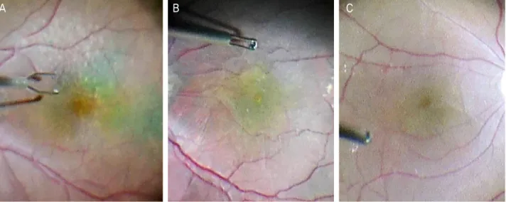
A B C
Figure 1. Surgeon’s view. Visual quality of the stained internal limiting membrane (ILM). (A) Good stain. (B) Fair stain. (C) Poor
stain.여한다.9-14 특히, 망막전막의 수술에서 내경계막을 함께 제 거해 줌으로써 망막전막의 재발을 줄이고 황반주름을 호전 시키며 시력향상을 가져온다는 여러 보고가 있다.15-20 그러 나 얇고 투명한 조직인 내경계막은 육안적 관찰이 용이하 지 않아 실제 수술적 제거의 어려움이 있었으며 이를 극복 하기 위하여 2000년도에 내경계막만 선택적으로 착색시키 는 인도시아닌그린(ICG, Indocyanine green)의 사용이 소개 되었고14 이후 많은 연구에서 ICG 용액을 사용함으로써 내 경계막의 시인성과 수술의 용이성을 향상시키고 망막손상 의 위험성을 줄일 수 있음을 보여주었다.14,18,21-23
최근 내경계막 제거 시 ICG는 가장 흔히 사용되고 있는 염료이나, 현재까지도 ICG 용액을 희석하는 방법은 표준화 되지 않아 다양한 농도로 사용되고 있다.22-26 Kwok et al27 은 내경계막을 효율적으로 염색할 수 있는 최소농도를 구 하기 위해 ICG의 농도를 달리하여 수술의 용이성과 임상적 결과 및 ICG 독성을 비교하였는데 ICG 농도가 높을수록 시인성은 향상되나 안전성에 대한 연구가 더 필요하다고 보고하였으며 다른 여러 연구에서도 높은 ICG 농도가 망막 독성을 증가시킬 수 있다고 하였다.26,28-30 이에 저자들은 ICG 용액의 농도는 일정하게 한 상태에서, 평형염액(BSS) 보다 비중이 큰 5% 포도당액을 용매로 사용하여 내경계막 제거를 시행하고, 이를 BSS를 용매로 사용한 경우와 수술 의 용이성 및 수술 결과를 비교해 보고자 한다.
대상과 방법
본원에서 특발성 망막전막으로 진단받고 유리체절제술 시행 시 ICG 용액을 이용하여 내경계막 염색을 시행하였던 환자 중 6개월 이상 경과관찰 가능하였던 연속된 환자 27
명 27안을 대상으로 의무기록을 통하여 후향적으로 조사하 였다. 수술 전 다른 황반부 질환이 함께 동반되어 있었던 경우와 속발성 망막전막을 일으킬 수 있는 안내 병증의 기 왕력이 있는 경우, 망막전막 외에 시력을 저하시킬 만한 안 질환이 있는 경우는 대상에서 제외하였다. 내경계막 제거 시 사용한 ICG 용액의 용매의 종류에 따라 BSS를 이용한 군 15안(I군)과 5% 포도당액을 사용한 12안(II군)으로 나누 어 분석하였다. 내경계막 제거 시 사용한 염색 용액의 혼합 은 ICG (Diagnogreen® Injection: Daiichi Pharmaceutical, Tokyo, Japan) 25 mg을 증류수 1 mL로 완전히 용해시킨 후 이 중 0.1 mL를 I군에서는 0.9 mL BSS와 혼합하고, II군 에서는 0.9 mL 5% 포도당액과 혼합하여 최종적으로 2.5 mg/mL (0.25%) 농도가 되도록 하였다.
수술 전 증상기간을 조사하고 최대교정시력 측정, 전안 부 및 수정체 검사, 안저검사를 시행하였으며 스펙트럼영 역 빛간섭단층촬영(SD-OCT, Spectral domain optical coher- ence tomography, Spectralis, Heidelberg Engineering GmbH, Heidelberg, Germany) 분석을 통한 망막전막의 심한 정도 를 비교하였다.
수술은 숙련된 단일 술자에 의해 23게이지 경결막 무봉 합 유리체절제술(Accurus®, Alcon, USA)을 시행하였다. 먼 저 후유리체박리를 일으킨 후, 후유리체 및 망막전막을 제 거하였다. 내경계막을 염색하기 위하여 관류액의 주입을 멈추고 I군에서는 BSS와 혼합된 0.25% ICG 용액을, II군에 서는 5% 포도당용액과 혼합된 ICG 용액을 황반부에 0.1 mL 주입하고 10초 후 재관류하여 ICG 용액을 제거하였다.
염색된 내경계막은 안내겸자를 이용하여 중심와로부터 약 3-4배 유두직경 크기의 원형으로 제거하였으며 그 후 주변 망막의 유리체절제술을 추가로 시행하였다. 백내장 수술이
Table 1. Comparison of patients’ demographics between two groups
Group I* Group II† Total p-value
No. of eyes 15 12 27
Age (years) 63.7 ± 6.0 66.0 ± 6.8 64.7 ± 6.3 0.464‡
Sex (M:F) 9:6 4:8 13:14 0.168§
Periods of Sx (months) 16.4 ± 23.7 11.2 ± 14.0 13.9 ± 19.4 0.496‡
Baseline BCVA (log MAR) 0.55 ± 0.26 0.40 ± 0.22 0.49 ± 0.25 0.172‡
Lens state (phakic:pseudophakic) 15:0 10:2 25:2 0.188§
Phacovitrectomy:Vitrectomy 14:1 10:2 24:3 0.569§
ERM stage (%) 1.000§
Stage 1 7 (46.7) 5 (41.7) 12 (44.4)
Stage 2 7 (46.7) 6 (50.0) 13 (48.1)
Stage 3 1 (6.7) 1 (8.3) 2 (7.4)
Values are presented as mean ± SD.
BCVA = best corrected visual acuity; ERM = epiretinal membrane; Sx: symptom.
*Using balanced salt solution (BSS) as solvent of indocyanine green; †Using 5% glucose solution as solvent of indocyanine green; ‡Mann- Whitney U-test; §Fisher’s exact test.
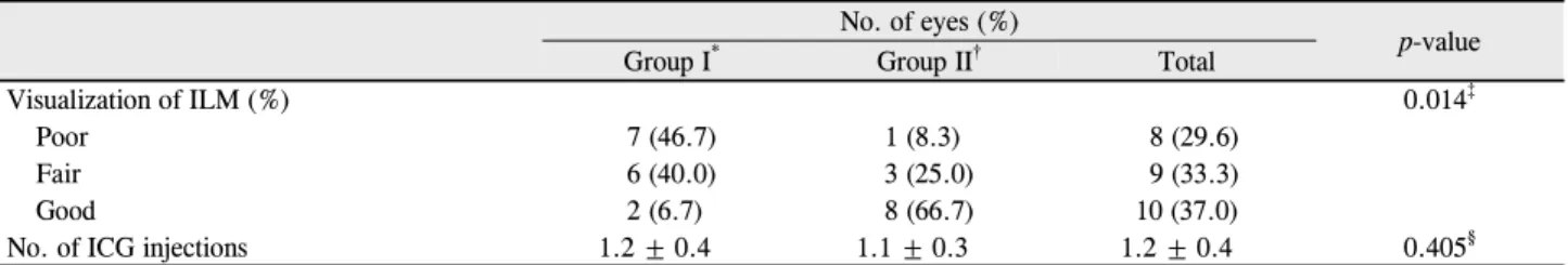
Table 2. Visualization of internal limiting membrane and number of indocyanine green (ICG) injection with different solvents
No. of eyes (%)
p-value
Group I* Group II† Total
Visualization of ILM (%) 0.014‡
Poor 7 (46.7) 1 (8.3) 8 (29.6)
Fair 6 (40.0) 3 (25.0) 9 (33.3)
Good 2 (6.7) 8 (66.7) 10 (37.0)
No. of ICG injections 1.2 ± 0.4 1.1 ± 0.3 1.2 ± 0.4 0.405§
Values are presented as mean ± SD.
ILM = internal limiting membrane.
*Using balanced salt solution (BSS) as solvent of indocyanine green; †Using 5% glucose solution as solvent of indocyanine green; ‡Fisher’s exact test; §Mann-Whitney U-test.
필요한 경우에는 유리체절제술 전 12시 방향의 투명각막절개 를 통하여 초음파유화술을 먼저 시행하고 유리체 절제술이 모두 끝난 다음 마지막에 접형 인공수정체를 삽입하였다.
망막전막의 심한 정도는 OCT를 통하여 분석하였으며 1 단계는 내측 망막의 불규칙한 주름을 일으킬 수 있으나 망 막전막의 가장자리는 보이지 않는 미세한 전막의 경우, 2단 계는 망막전층의 변형을 일으키며 망막전막의 가장자리가 보이고 망막전막의 절반 미만의 부분에서 아래의 망막과 혈관구조가 불분명하게 관찰될 정도의 상당한 전막의 경우, 그리고 3단계는 망막전막의 절반 이상의 부분에서 아래의 망막과 혈관구조가 불분명하게 관찰될 정도의 두껍고 뚜렷 한 혼탁이 있는 전막의 경우로 나누었다.27
내경계막의 시인성은 녹화된 영상 파일을 분석하여 Good, Fair, Poor 3단계로 나누었으며, Good은 밝은 녹색으로 염 색된 경우, Fair는 내경계막이 연한 녹색으로 염색된 경우, Poor은 내경계막이 뚜렷이 구분이 되지 않는 염색의 경우 로 나누었다(Fig. 1).27
수술 후 경과는 술 후 6개월째 빛간섭단층촬영을 통한 망막전막의 성공적인 제거여부와 황반주름의 호전여부 및
중심와 윤곽의 회복여부를 파악하여 해부학적 성공여부를 비교하였고, 기능적 성공여부는 수술 전과 비교하여 술 후 6개월째 한천석 시력표상 두 줄 이상의 시력호전을 보이는 경우로 정의하였으며, 술 후 6개월과 술 전의 시력을 비교 하기 위해 Log 스케일(LogMAR)로 환산하였다. 그 외 망막 전막의 재발여부, 술 후 합병증으로 망막색소상피의 위축, 망막박리 등을 확인하였다.
각 용액의 삼투압은 Advanced® 2020 multi-sample osmometer 를 이용하여 측정하였으며, pH는 Blood gas and electrolytes analyzer ABL800 BASIC®을 이용하여 측정하였다. 비중은 OHAUS PRECISION Advanced® 410을 이용하여 각 용액 의 단위부피당 질량을 측정한 후 밀도를 구하고, 표준물질 인 물의 밀도를 1 g/mL로 보고 각 용액의 밀도와 물의 밀 도의 비를 구하였다.
통계학적 분석은 PASW statistics v. 18.0을 사용하여 두 군 간의 비교는 Mann-Whitney U-test, Fisher’s exact test, Chi-squre test를, 수술 전후 시력호전은 Wilcoxon signed rank test를 이용하였으며 수술의 해부학적 결과와 시력과 의 상관관계는 Pearson 상관계수를 이용하였다. p값이 0.05
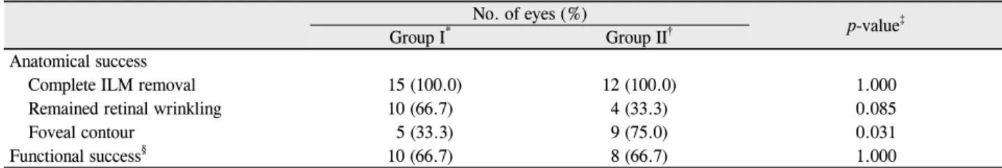
Table 3. Comparison of the anatomical and functional success after surgery between two groups
No. of eyes (%) p-value‡
Group I* Group II†
Anatomical success
Complete ILM removal 15 (100.0) 12 (100.0) 1.000
Remained retinal wrinkling 10 (66.7) 4 (33.3) 0.085
Foveal contour 5 (33.3) 9 (75.0) 0.031
Functional success§ 10 (66.7) 8 (66.7) 1.000
ILM = internal limiting membrane.
*Using balanced salt solution (BSS) as solvent of indocyanine green; †Using 5% glucose solution as solvent of indocyanine green; ‡Chi-square test; §Improvement visual acuity more than 2 lines.
Table 4. Comparison of the BCVA (log MAR) changes after surgery between two groups
BCVA (log MAR) Group I* Group II† p-value‡
Baseline 0.55 ± 0.26 0.40 ± 0.22 0.172
Postoperative 6 months 0.26 ± 0.26 0.20 ± 0.17 0.675
Changes 0.29 ± 0.28 0.20 ± 0.25 0.478
p-value§ 0.004 0.029
Values are presented as mean ± SD.
BCVA = best corrected visual acuity.
*Using balanced salt solution (BSS) as solvent of indocyanine green; †Using 5% glucose solution as solvent of indocyanine green; ‡Mann- Whitney U-test; §Wilcoxon signed rank test.
미만인 경우에 통계적으로 유의하다고 판단하였다.
결 과
전체 27명의 대상안 중 I군이 15안, II군이 12안이였으며, 남자와 여자의 비율은 I군이 9:6, II군이 4:8로 통계적으로 유의한 차이는 없었다. 평균연령은 I군이 63.7세, 2군이 66.0세로 유의한 차이는 없었으며 증상기간은 I군이 평균 16.4개월, II군이 11.2개월로 I군이 더 길었으나 통계적으로 유의하지 않았다. 초진 시 시력은 I군이 0.55 logMAR, II군 이 0.40 logMAR로 II군이 더 좋았으나 통계적 의의는 없었 다. 3단계로 분류한 망막전막의 심한 정도에서도 두 군 간 의 유의한 차이는 없었다(Table 1).
ICG 용액 염색 후 내경계막의 시인성은 BSS를 용매로 이 용한 I군에서는 Poor가 가장 많았고(46.7%), 5% 포도당액을 용매로 이용한 II군에서는 Good이 가장 많았으며(66.7%), II 군이 통계적으로 유의하게 시인성이 좋았다(Fisher’s exact test, p=0.014, Table 2). ICG 용액을 주입한 횟수는 I군이 평균 1.2회로 II군의 평균 1.1회보다 많았으나 통계적으로 의의는 없었다.
두 군 모두에서 해부학적으로 망막전막 및 내경계막은 완 전히 제거되었으며, 술 후 6개월째 OCT에서 망막주름이 남 아있는 경우는 I군이 10안(66.7%), II군이 4안(33.3%)으로 통계적으로 유의하지는 않았으나 I군이 더 많았고, 중심와 윤곽이 회복된 경우는 I군이 5안(33.3%), II군이 9안(75.0%)
으로 통계적으로 유의하게 II군이 더 많았다(Chi-square test, p=0.031, Table 3).
기능적 성공은 술 후 한천석 시력표상 두 줄 이상의 시력 호전을 보이는 경우로 하였는데 I군에서는 10안(66.7%), II 군에서는 8안(66.7%)에서 두 줄 이상의 시력호전을 보였으 며 두 군 간의 유의한 차이는 없었다. 술 전과 수술 6개월 후 시력의 변화는 I군에서는 0.55에서 0.26 logMAR로, II군 에서는 0.40에서 0.20 logMAR로 두 군 모두 통계적으로 유 의하게 호전되었으며(Wilcoxon signed rank test, p=0.004, 0.029), 술 후 6개월째 최대교정시력은 I군이 0.26 logMAR, II군이 0.20 logMAR로 두 군 간에 통계적으로 유의한 차이 는 없었다(Table 4, Fig. 2). 술 후 I군에서는 1안(6.7%), II군 에서는 2안(16.7%)이 술 전에 비해 시력감소가 있었다.
망막전막의 재발은 두 군 모두에서 관찰되지 않았으며, 망막색소상피의 위축, 망막박리 등의 술 후 합병증도 두 군 모두에서 관찰되지 않았다.
두 군에서 이용한 ICG 용액의 성질을 비교해 보면, BSS-ICG 용액의 삼투압은 272 mOsm/kg, 5% 포도당액-ICG용액의 경 우는 256 mOsm/kg로 5% 포도당액을 용매로 이용한 경우 더 낮은 삼투압이 측정되었으며, pH는 BBS-ICG 용액의 경우 7.643으로 약한 알칼리성을, 5% 포도당액-ICG는 6.999로 거의 중성을 나타냈다. 두 용액의 비중을 비교해 보면, BSS-ICG 용액은 1.094, 5% 포도당액-ICG 용액은 1.140으로 5% 포도 당액을 용매로 이용한 경우 좀 더 잘 가라앉는 성질을 보였 다(Table 5).
Table 5. Measurement in osmolality, pH and specific gravity of two solutions
ICG solution mOsm/kg pH Specific gravity
BSS-ICG 272 7.643 1.094
5% glucose-ICG 256 6.999 1.140
ICG = indocyanine green; BSS = balanced salt solution.
0.55
0.26
0.40
0.20 0.1
0.2 0.3 0.4 0.5
0.6
Baseline Postop 6 mo
Group I Group II
BCVA (log MAR)
Baseline Postop 6 months
Figure 2. Changes in best corrected visual acuity (BCVA) be-
tween preoperation (Preop) and postoperation (Postop).고 찰
망막전막 수술에서 내경계막을 함께 제거하는 것은 망막 전막의 재발률을 줄일 수 있어 여러 술자에 의해 시행되고 있으며, 내경계막의 시인성을 높이기 위해 ICG 용액이 흔 히 사용되고 있다. 그러나 몇몇 연구에서는 ICG 용액이 망 막에 대한 독성을 가지고 있어 수술 후 기능적 결과를 저하 시킨다는 문제점을 제기해 왔는데20,29,31-35 Gandorfer et al29 과 Hillenkamp et al30은 망막손상을 일으키는 기전으로 ICG 용액의 농도와 삼투압, 조직과의 접촉시간, 제거 시 가해진 힘에 의한 견인 등을 제시하였고, Stalmans et al36은 ICG 용액의 용매 종류에 따라 다른 삼투압이 망막색소상피세포 의 손상과 관계된다고 보고하였으며 Konstantinidis et al18 은 용매의 종류와 삼투압, 용매 내 나트륨의 여부도 망막독 성과 연관된다고 보았다. 따라서 내경계막의 시인성은 극 대화하면서 ICG의 독성을 최소화시키기 위해 ICG 용액의 적절한 농도와 적절한 용매의 사용은 매우 중요함에도 불 구하고 표준화되지 않아 매우 다양한 방법으로 사용되고 있다. ICG 염색의 가장 효율적인 최소농도를 구하기 위해 Kwok et al27은 다른 농도의 ICG 용액을 사용하여 수술의 용이성과 결과를 비교하였다. 그러나 ICG 희석에 사용하는 용매에 따른 수술의 용이성과 결과를 임상적으로 연구한 논문은 없어, 이에 저자들은 주로 사용하는 두 가지 용매인 BSS와 5% 포도당액을 사용한 망막전막 수술을 임상적으 로 비교 분석하였다.
수술 중 내경계막의 시인성은 5% 포도당액을 사용한 II 군에서 통계적으로 유의하게 좋았으며(Fisher’s exact test, p=0.014) 따라서 수술의 편의성도 향상되었는데 이는 두 용 매의 비중의 차이로 인한 것으로 생각한다. 5% 포도당액 -ICG 용액의 비중이 1.140으로 BSS-ICG 용액의 비중 1.094보다 높았는데 유리체절제술 중 BSS로 채워진 안구 내로 ICG 용액을 주입하는 경우 BSS보다 비중이 높은 ICG 가 좀 더 잘 가라앉아 착색이 잘 되는 것으로 보인다.
술 후 해부학적 결과를 비교해 보면, 두 군에서 모두 망 막전막 및 내경계막은 완전히 제거되었으며 잔존망막주름 은 유의하진 않으나 BSS를 사용한 I군에서 더 많았고, 중심 와 윤곽의 호전은 통계적으로 유의하게 5% 포도당액을 사 용한 II군에서 더 좋았다(Chi-square test, p=0.031). 두 줄 이상 시력호전을 나타내는 기능적 성공률은 66.7%로 두 군 에서 동일하며 두 군 모두에서 술 후 통계적으로 유의하게 시력호전을 보였다. 더 잘 가라앉는 5% 포도당액 군을 용 매로 이용한 경우 내경계막의 시인성을 높여 수술을 용이 하게 하고 따라서 망막주름 및 중심와 윤곽의 회복을 향상 시켜 해부학적 성공률을 높일 수 있었으나 기능적 결과에 는 뚜렷한 차이가 없었다. Kim and Kim20의 연구에서 제시 하였듯이 특발성 망막전막이 시력감소를 일으키는 기전은 황반부를 왜곡시키는 것 외에도 망막전막이 황반부위를 덮 거나, 견인성 망막박리, 망막 내 부종을 동반한 혈관 유출, 그리고 축삭이동의 차단 등 여러 인자가 작용하므로 망막 전막이 제거된 후 잔존하는 망막주름만으로 시력저하를 야 기한다고 보기는 어려우며 망막주름의 호전 및 중심와 윤 곽의 회복과 기능적인 시력의 향상과의 직접적인 연관성을 판단하기는 어렵다. 본 연구에서도 잔존하는 망막주름 및 윤곽선 회복 여부와 시력변화 사이에는 통계학적으로 상관 관계가 없었다(Pearson상관계수 r=0.012, 0.169).
BSS-ICG 용액의 삼투압은 272 mOsm/kg, 5% 포도당액 -ICG 용액의 삼투압은 256 mOsm/kg로 5% 포도당액을 용 매를 사용한 경우가 더 낮았다. Stalmans et al36과 Sippy et al31은 ICG의 망막색소상피에 대한 독성은 용매의 저삼투 압과 관련된다고 하였다. 또한 Stalmans and Himpens37의 연구에서 저삼투압 용액에 노출되면 망막색소상피세포 내 로 Ca2+의 유입이 증가하여 세포독성을 일으킨다고 보고하 였다. 본 연구에서 5% 포도당액을 용매로 사용한 군에서 시력감소를 보인 경우가 더 많이 관찰되었는데(I군=6.7%,
II군=16.7%), 이는 낮은 삼투압으로 인한 독성과 관계된 것 이 아닌가 생각한다. 그러나 Haritoglou et al38은 다양한 농 도로 서로 다른 종류의 용매를 사용한 ICG용액의 흡수스펙 트럼을 측정하여 ICG용액의 농도가 높을수록 광민감성이 증가함으로써 망막색소상피의 광독성이 증가한다고 하였 으며 BSS보다 5% 포도당액을 사용하는 것이 광독성을 줄 일 수 있다고 하였고 Ho et al39은 Sodium이 없는 용매를 사 용하면 ICG로 인한 광독성을 줄일 수 있다고 보고하였다.
이 두 연구의 결과로 볼 때 5% 포도당액을 용매로 사용하 는 것은 광독성으로 인한 망막 손상을 줄이는 데는 도움이 된다. 본 연구에서 두 용매를 사용하였을 때 기능적 결과 측면에서 차이가 없었던 것은 망막 독성을 일으키는 여러 기전에 따른 영향이 상반되어 결론적으로 두 용매가 큰 차 이가 없었던 것으로 보이며 따라서 ICG의 독성과 용매와의 관계에 대하여 여러 측면에서 좀 더 체계적인 연구가 필요 할 것으로 생각한다.
결론적으로 내경계막 제거 시 ICG 용액의 용매로 5% 포 도당액을 사용하는 것은 BSS를 사용하는 것과 비교하여 볼 때 기능적 측면에서 비슷한 정도의 시력회복을 기대할 수 있을 뿐만 아니라 내경계막의 시인성을 향상시켜 수술의 용이성을 높이고 해부학적 성공률을 높일 수 있었다. 따라 서 내경계막 제거 시 5% 포도당을 ICG 용매로 사용하면 낮은 ICG 농도로도 내경계막의 시인성을 향상시킬 수 있으 므로 이를 긍적적으로 고려해 볼 필요가 있다고 생각한다.
그러나 이 연구는 적은 환자를 대상으로 하여 정확한 통계 적 비교가 어려우며 추적관찰 기간이 6개월로 비교적 짧아 장기적인 예후를 알기 힘들어 더 많은 환자를 대상으로 더 장기적인 추적관찰을 할 필요가 있다. 또한 ICG 용액의 독 성 기전을 좀 더 체계적으로 분석하여 ICG의 독성을 최소 화하면서 수술의 용이성을 높일 수 있도록 ICG 용액을 만 드는 방법을 표준화할 필요가 있다.
REFERENCES
1) Machemer R. The surgical removal of epiretinal macular mem- branes (macular puckers). Klin Monbl Augenheilkd 1978;173:
36-42.
2) Michels RG. Vitrectomy for macular pucker. Ophthalmology 1984;91:1384-8.
3) Margherio RR, Cox MS Jr, Trese MT, et al. Removal of epimacular membranes. Ophthalmology 1985;92:1075-83.
4) McDonald HR, Verre WP, Aaberg TM. Surgical management of idiopathic epiretinal membranes. Ophthalmology 1986;93:978-83.
5) Pesin SR, Olk RJ, Grand MG, et al. Vitrectomy for premacular fibroplasia. Prognostic factors, long-term follow-up, and time course of visual improvement. Ophthalmology 1991;98:1109-14.
6) Donati G, Kapetanios AD, Pournaras CJ. Complications of surgery for epiretinal membranes. Graefes Arch Clin Exp Ophthalmol
1998;236:739-46.
7) Benhamou N, Massin P, Spolaore R, et al. Surgical management of epiretinal membrane in young patients. Am J Ophthalmol 2002;
133:358-64.
8) Massin P, Paques M, Masri H, et al. Visual outcome of surgery for epiretinal membranes with macular pseudoholes. Ophthalmology 1999;106:580-5.
9) Zarbin MA, Michels RG, Green WR. Epiretinal membrane con- tracture associated with macular prolapse. Am J Ophthalmol 1990;
110:610-8.
10) Clarkson JG, Green WR, Massof D. A histopathologic review of 168 cases of preretinal membrane. Am J Ophthalmol 1977;84:1-17.
11) Michels RG. A clinical and histopathologic study of epiretinal membranes affecting the macula and removed by vitreous surgery.
Trans Am Ophthalmol Soc 1982;80:580-656.
12) Smiddy WE, Green WR, Michels RG, de la Cruz Z. Ultrastructural studies of vitreomacular traction syndrome. Am J Ophthalmol 1989;107:177-85.
13) Fine BS. Limiting membranes of the sensory retina and pigment epithelium. An electron microscopic study. Arch Ophthalmol 1961;66:847-60.
14) Burk SE, Da Mata AP, Snyder ME, et al. Indocyanine green-assisted peeling of the retinal internal limiting membrane. Ophthalmology 2000;107:2010-4.
15) Park DW, Dugel PU, Garda J, et al. Macular pucker removal with and without internal limiting membrane peeling: pilot study.
Ophthalmology 2003;110:62-4.
16) Kwok AK, Lai TY, Yuen KS. Epiretinal membrane surgery with or without internal limiting membrane peeling. Clinical and Experimental Ophthalmology 2005;33:379-85.
17) Bovey EH, Uffer S, Achache F. Surgery for epimacular membrane:
impact of retinal internal limiting membrane removal on functional outcome. Retina 2004;24:728-35.
18) Konstantinidis L, Uffer S, Bovey EH. Ultrastructural changes of the internal limiting membrane removed during indocyanine green assisted peeling versus conventional surgery for idiopathic mac- ular epiretinal membrane. Retina 2009;29:380-6.
19) Kim TW, Song SJ, Chung H, Yu HG. Internal limiting membrane peeling in surgical treatment of macular epiretinal membrane. J Korean Ophthalmol Soc 2005;46:989-94.
20) Kim YC, Kim KS. The effect of internal limiting membrane peel- ing in treatment of idiopathic epiretinal membrane. J Korean Ophthalmol Soc 2007;48:1067-72.
21) Gandorfer A, Messmer EM, Ulbig MW, Kampik A. Indocyanine green selectively stains the internal limiting membrane. Am J Ophthalmol 2001;131:387-8.
22) Da Mata AP, Burk SE, Riemann CD, et al. Indocyanine green-as- sisted peeling of the retinal internal limiting membrane during vi- trectomy surgery for macular hole repair. Ophthalmology 2001;
108:1187-92.
23) Kwok AK, Lai TY, Li WW, et al. Indocyanine green-assisted in- ternal limiting membrane removal in epiretinal membrane surgery:
a clinical and histologic study. Am J Ophthalmol 2004;138:194-9.
24) von Jagow B, Hoing A, Gandorfer A, et al. Functional outcome of indocyanine green-assisted macular surgery: 7-year follow-up.
Retina 2009;29:1249-56.
25) Lanzetta P, Polito A, Del Borrello M, et al. Idiopathic macular hole surgery with low-concentration infracyanine green-assisted peel-
= 국문초록 =
특발성 망막전막에서 내경계막 제거 시 인도시아닌그린 염색의 용매에 따른 수술결과 비교
목적: 특발성 망막전막 수술의 내경계막 제거 시 인도시아닌그린(ICG) 염색에 있어 용매에 따른 수술결과를 비교해 보고자 하였다.
대상과 방법: 특발성 망막전막으로 유리체절제술 시 내경계막 염색을 시행한 환자 27명 27안을 대상으로 후향적 비교 연구를 시행하였 다. 내경계막 제거 시 사용한 0.25% ICG 용액의 용매의 종류에 따라 평형염액(BSS)을 이용한 군(I군 15안)과 5% 포도당액을 사용한 군(II군 12안)으로 나누어 망막전막의 심한 정도, 증상기간, 수술 전후의 최대교정시력과 수술 중 내경계막의 시인성(Good, Fair, Poor) 및 술 후 합병증 유무를 비교하였다.
결과: 술 전 두 군 간의 망막전막의 심한 정도, 증상기간, 최대교정시력의 통계적으로 유의한 차이는 없었다. 술 전과 비교하여 술 후 6개월째 시력(logMAR)은 I군에서 0.55에서 0.26으로, II군은 0.40에서 0.20으로 두 군 모두 통계적으로 유의하게 증가하였으나 두 군 사이에 유의한 차이는 없었다. 술 중 인도시아닌그린에 의한 내경계막의 시인성은 Poor로 염색된 경우가 I군과 비교하여 II군에 서 통계적으로 유의하게 적었다(p=0.014). 두 군 모두 최종관찰까지 망막전막의 재발, 망막색소상피 위축 및 망막박리 등의 합병증은 없었다.
결론: 내경계막 제거를 위한 ICG 염색 시 BSS보다 비중이 큰 5% 포도당액을 사용할 경우 술 후 시력 회복에는 차이가 없이 수술 중 내경계막의 시인성을 향상시켜 수술의 용이성을 높일 수 있다.
<대한안과학회지 2014;55(6):847-853>
ing of the internal limiting membrane. Am J Ophthalmol 2006;142:
771-6.
26) Lai MM, Williams GA. Anatomical and visual outcomes of idio- pathic macular hole surgery with internal limiting membrane re- moval using low-concentration indocyanine green. Retina 2007;
27:477-82.
27) Kwok AK, Lai TY, Yew DT, Li WW. Internal limiting membrane staining with various concentrations of indocyanine green dye un- der air in macular surgeries. Am J Ophthalmol 2003;136:223-30.
28) Kanda S, Uemura A, Yamashita T, et al. Visual field defects after intravitreous administration of indocyanine green in macular hole surgery. Arch Ophthalmol 2004;122:1447-51.
29) Gandorfer A, Haritoglou C, Gass CA, et al. Indocyanine green-as- sisted peeling of the internal limiting membrane may cause retinal damage. Am J Ophthalmol 2001;132:431-3.
30) Hillenkamp J, Saikia P, Gora F, et al. Macular function and mor- phology after peeling of idiopathic epiretinal membrane with and without the assistance of indocyanine green. Br J Ophthalmol 2005;89:437-43.
31) Sippy BD, Engelbrecht NE, Hubbard GB, et al. Indocyanine green effect on cultured human retinal pigment epithelial cells: implication for macular hole surgery. Am J Ophthalmol 2001;132:433-5.
32) Enaida H, Sakamoto T, Hisatomi T, et al. Morphological and func- tional damage of the retina caused by intravitreous indocyanine green in rat eyes. Graefes Arch Clin Exp Ophthalmol 2002;240:
209-13.
33) Haritoglou C, Gandorfer A, Gass CA, et al. The effect of in- docyanine-green on functional outcome of macular pucker surgery.
Am J Ophthalmol 2003;135:328-37.
34) Gandorfer A, Haritoglou C, Gandorfer A, Kampik A. Retinal dam- age from indocyanine green in experimental macular surgery.
Invest Ophthalmol Vis Sci 2003;44:316-23.
35) Maia M, Haller JA, Pieramici DJ, et al. Retinal pigment epithelial abnormalities after internal limiting membrane peeling guided by indocyanine green staining. Retina 2004;24:157-60.
36) Stalmans P, Van Aken EH, Veckeneer M, et al. Toxic effect of in- docyanine green on retinal pigment epithelium related to osmotic effects of the solvent. Am J Ophthalmol 2002;134:282-5.
37) Stalmans P, Himpens B. Confocal imaging of Ca2+ signaling in cultured rat retinal pigment epithelial cells during mechanical and pharmacologic stimulation. Invest Ophthalmol Vis Sci 1997;38:
176-87.
38) Haritoglou C, Gandorfer A, Schaumberger M, et al. Light-absorb- ing properties and osmolarity of indocyanine-green depending on concentration and solvent medium. Invest Ophthalmol Vis Sci 2003;44:2722-9.
39) Ho JD, Chen HC, Chen SN, Tsai RJ. Reduction of indocyanine green-associated photosensitizing toxicity in retinal pigment epi- thelium by sodium elimination. Arch Ophthalmol 2004;122:871-8.