INTRODUCTION
It has long been recognized that breast cancers can invade and grow as metastases in bone (1). Some breast cancer cell lines induce osteolytic lesions in animal models that mimic the metastatic process in clinical breast cancer (2, 3), but the factors favoring the growth of breast cancer in bone remain to be resolved. Bone destruction is primarily mediated by osteo- clastic bone resorption (4), and cancer cells that have metasta- sized to bone stimulate the formation and activation of osteo- clasts next to metastatic foci (5). Recent research has identified several factors inducing osteoclast formation (4). If different types of metastatic cancer follow the same mechanism to cause osteolytic lesions, we may focus on this target to control and prevent the damage to bone.
Accumulating evidences indicate that receptor activator of NF-kB ligand (RANKL), also known as osteoclast differenti- ation factor, is the ultimate extracellular mediator that acts on osteoclast precursors to differentiate them into mature osteo- clasts in the presence of macrophage-colony stimulating fac- tor (M-CSF) (6, 7), and that many bone resorbing factors up-regulate RANKL expression in bone marrow stromal and osteoblastic cells (6, 8). Osteoprotegerin (OPG), also known
as osteoclastogenesis inhibitory factor, inhibits osteoclast devel- opment (9, 10) because it is a decoy receptor for RANKL to block its actions (6, 7). Thus, divergent mechanisms for bone resorption may converge to the regulation of RANKL activi- ties. Cancer cells induce formation of osteoclasts by secreting osteotropic cytokines, such as PTH-related protein (PTHrP) (11), interleukin-11 (IL-11) (12) and leukemia inhibitory fac- tor (LIF) (13), or by direct contact with bone marrow cells (1). The molecular mechanisms for the latter are yet unclear, although osteotropic factors secreted by cancer cells could induce RANKL. In order to reveal a mechanism for cancer- induced osteoclastogenesis, we examined RANKL and OPG expression in co-cultures of bone marrow stromal or osteoblas- tic cells with breast cancer cells inducing osteoclast-like cell formation in vitro.
MATERIALS AND METHODS Tissue specimens and animals
Thirty tissue specimens from breast cancer were obtained from thirty patients by surgical resection in KangDong Sacred
Hye-Rim Park, Soo-Kee Min, Hyun-Deuk Cho, Duck-Hwan Kim, Hyung Sik Shin, Young Euy Park
Department of Pathology, College of Medicine, Hallym University, Chuncheon, Korea
Address for correspondence Hye-Rim Park, M.D.
Department of Pathology, Hallym University Sacred Heart Hospital, 896 Pyungchon-dong, Dongan-gu, Anyang 431-070, Korea
Tel : +82.31-380-3935, Fax : +82.31-381-9646 E-mail : [email protected]
*This work was supported by the Hallym Academy of Sciences at Hallym University in Korea (2000-4-1).
541
Expression of Osteoprotegerin and RANK Ligand in Breast Cancer Bone Metastasis
Bone destruction is primarily mediated by osteoclastic bone resorption, and cancer cells stimulate the formation and activation of osteoclasts next to metastatic foci. Accu- mulating evidences indicate that receptor activator of NF-kB ligand (RANKL) is the ultimate extracellular mediator that stimulates osteoclast differentiation into mature osteoclasts. In contrast, osteoprotegerin (OPG) inhibits osteoclast development. In order to elucidate a mechanism for cancer-induced osteoclastogenesis, cells from a human breast cancer line, MDA-MB-231, were directly co-cultured with ST2, MC3T3- E1, or with primary mouse calvarial cells. Osteoclast-like cells and tartarate resistant acid phosphatase (TRAP) activities were then quantitated. We examined these cell lines and samples from breast cancer by RT-PCR for the expressions of OPG and RANKL mRNA. Compared to controls, co-culture of MDA-MB-231 cells with stromal or osteoblastic cells induced an increase in number of osteoclasts and TRAP activi- ties. MDA-MB-231 cells alone or breast cancer samples did not express RANKL mRNA. However, co-culture of these cancer cells with stromal or osteoblastic cells induced RANKL mRNA expression and decreased OPG mRNA expression. These experiments demonstrate that direct interactions between breast cancer and stromal or osteoblastic cells induce osteoclastogenesis in vitro through modulating RANKL expression.
Key Words : Breast Neoplasms; Bone and Bones; Neoplasm Metastasis; Osteoclasts
Received : 9 April 2003 Accepted : 9 May 2003
Heart Hospital, Hallym University Medical Center (Seoul, Korea). Tissue specimens were snap-frozen in liquid N2and stored at -70℃ until used. Male C57 black mice (6 weeks old) were obtained from Asan Institute for Life Science (Seoul, Korea). All procedures involving animals were approved by the institutional animal care committee.
Isolation and culture of neonatal mouse calvarial cells
Calvariae from 3 to 5-day-old neonatal C57 black mice were dissected and cut into small pieces. Following serial treatments with 0.1% collagenase (Sigma, St. Louis, MO, U.S.A.) and 0.2% protease (Sigma, St. Louis, MO, U.S.A.), cells were col- lected from the supernatant (fractions 2 to 5). Cells were cul- tured in -MEM (minimum essential media, GIBCO, Gai- thersburg, MD, U.S.A.) supplemented with 10% FCS (fetal calf serum, GIBCO, Gaithersburg, MD, U.S.A.), 100 IU/ L penicillin and 100 g/ L streptomycin (GIBCO, Gaithers- burg, MD, U.S.A.), at 37℃ in a humidified atmosphere with 5% CO2. In addition, mouse osteoblastic cell line MC3T3-E1 and bone marrow-derived stromal cell line ST2 were used as osteoblastic and stromal cells.
MDA-MB-231: a human breast cancer cell line
The MDA-MB-231 cell line was provided by the Korean Cell Line Bank (Seoul, Korea). Cells were cultured in RPMI (GIBCO, Gaithersburg, MD, U.S.A.) supplemented with 10%
FCS, 100 IU/ L penicillin, and 100 g/ L streptomycin (Life Technologies, Inc.) at 37℃ in a humidified atmosphere with 5% CO2. MDA-MB-231 cells were studied in co-culture sys- tems with mouse bone marrow stromal or osteoblastic cells.
As described previously (12), 2×10⁵cancer cells were inoc- ulated onto a 60 mm culture dish at 2 hr before seeding with bone marrow stromal or osteoblastic cells, prepared as described below. Serum-free conditioned media collected from the cul- tures after a 5-day incubation were kept at -20°C until re- quired. Aliquots of conditioned media were diluted with - MEM supplemented with 10% FCS and used at dilutions of 1:2 and 1:5.
Mouse bone marrow culture for osteoclast formation
Bone marrow cells of 6-week-old male C57 black mice were obtained from femora and tibiae, and were cultured as previ- ously described (14). Non-adherent marrow mononuclear cells were prepared and then re-suspended in -MEM supplement- ed with 10% FCS at 106cells/ L. Cells were plated in 96 well plates (1×10⁶cells/well) to test the effects of conditioned me- dia on the formation of osteoclasts. Cultures were maintained in a humidified atmosphere of 5% CO2at 37℃ for 8 days.
Cultures were fed once in every other day by removing half of the medium and replacing it with an equal volume of fresh medium.
Tartarate-resistant acid phosphatase (TRAP) staining
On the eighth day of bone marrow culture, cells were fixed in 10% formalin neutral buffer solution for 10 min and stained with an Acid Phosphatase Leukocyte Kit (Sigma Diagnostics, St. Louis, MO, U.S.A.) for 1 hr at 37℃. TRAP-positive multi- nucleated cells with more than three nuclei per well were counted.
TRAP activity
For TRAP activities, 100 L of culture media were mixed with 100 L of 0.2 M sodium acetate buffer, 16 L of 0.1 M pNPP, and 4 L of 2.0 M L(+) tartarate for 1 hr at 37℃. We added 16 L of 0.5 M NaOH and measured the optical density at 405 nm, using an ELISA plate reader.
RT-PCR for OPG and RANKL mRNA expression Adherent bone marrow cells (2×10⁵/60 mm dish) were co- cultured with or without cancer cells (2×10⁵/60 mm dish) as described above. After 5 days, total RNAs were extracted by using RNeasy Kits (Qiagen, Santa Clarita, CA, U.S.A.) and then reversely transcribed according to the instructions of the manufacturer. Total RNAs from thirty tissue specimens of breast cancer were also extracted by using the same kits. Primer sequences (Bioneer, Korea): OPG, 5′-AACCCCAGAGCGAA- ACAC-3′(sense), 5′-AAGAAGGCCTCTTCACAC-3′(anti- sense), RANKL, 5′-GGTCGGGCAATTCTGAATT-3′ (sense), and 5′-GGGGAATTACAAAGTGC-3′(antisense).
PCR was performed on a 9600 thermal cycler (Perkin Elmer, Norwalk, CT, U.S.A.), at the following conditions: at 94℃
for 1 min (1 cycle), at 55℃ for 40 sec, and at 72℃ for 1 min (32 cycles) for OPG; at 94℃ for 1 min (1 cycle), at 62℃ for 1 min, and at 72℃ for 1 min (32 cycles) for RANKL. For semi-quantitative RT-PCR, one g of total RNA was treated with DNase I, reversely transcribed, and subjected to PCR for OPG and RANKL. Mouse -actin mRNA expression was also examined by RT-PCR as an internal control.
Statistical analysis
All the experiments were repeated at least three times. Data were expressed as mean±SE and analyzed by one-way analy- sis of variance. An unadjusted p value of less than 0.05 was considered to be significant.
RESULTS
Cancer cells adhered to bone marrow cells induce formation of TRAP-positive multinucleated cells
Direct co-cultures of mouse bone marrow cells with MDA-
MB-231 cells, without osteotropic agents, generated some TRAP-positive multinucleated cells.
MDA-MB-231 conditioned medium increases the for- mation of osteoclasts in mouse bone marrow cultures
To test the effects of the factor (s) released from the tumor cells on the formation of osteoclasts, bone marrow cells were cul- tured either in the presence or the absence of MDA-MB-231 conditioned medium from co-culture with osteoblastic (MC- 3T3-E1) or stromal (ST-2) cells. The use of conditioned medi- um, even at the highest dilution (1:5), significantly increased the number of TRAP (+) multinucleated cells. The mean number of TRAP (+) multinucleated cells/field was 16.4 in treated cultures, compared with 8.6 in controls (Fig. 1). TRAP activities were also significantly increased with the addition of the conditioned media from co-culture (Fig. 2).
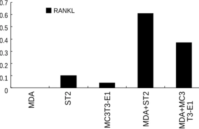
Co-culture of cancer cells with bone marrow cells induces RANKL expression
In order to clarify the mechanism whereby cancer cells induce osteoclast-like, TRAP-positive multinucleated cell to form in vitro through their direct contact with bone marrow cells, we examined the expression of RANKL mRNA by RT-PCR.
Cancer cell lines or tissue specimens from breast cancer did not express RANKL mRNA. Co-cultures of MDA-MB-231 with osteoblastic (MC3T3-E1) or stromal (ST-2) cells induced the RANKL mRNA expression (Fig. 3).
Co-culture of cancer cells with bone marrow cells reduces OPG expression
Osteoblastic (MC3T3-E1) or stromal (ST-2) cells alone exp- ressed OPG. Co-culture of MDA-MB-231 cells with osteo- blastic (MC3T3-E1) or stromal (ST-2) cells reduced the expres- sion of OPG. Cancer cells alone barely expressed OPG (Fig. 4).
1% FBS MDA
Osteoclast No.
ST2 MC3T3-E1 NDA+ST2 MDA+MC3 T3-E1
16 14 12 10 8 6 4 2 0
Fig. 1.Effects of conditioned media on osteoclast generation in bone marrow cultures. Conditioned media from co-culture of MDA- 231 with ST2 or MC3T3-E1 cells increase TRAP-positive multinu- cleated cell formation, compared to controls.
1% FBS MDA ST2 MC3T3-E1 NDA+ST2
0.36 0.35 0.34 0.33 0.32 0.31 0.3 0.29
Fig. 2 .Effects of conditioned media on TRAP activity in bone mar- row cultures. Conditioned media from co-culture of MDA-231 with ST2 or MC3T3-E1 cells increase TRAP activity, compared to con- trols.
TRAP
MDA+MC3 T3-E1
MDA
RANKL
ST2 MC3T3-E1 MDA+ST2 MDA+MC3 T3-E1
0.7 0.6 0.5 0.4 0.3 0.2 0.1 0
Fig. 3. Expression of RANKL mRNA assessed by RT-PCR. The re- sults were shown as a ratio of RANKL to actin mRNA. Co-culture of MDA-231 with ST2 or MC3T3-E1 cells induces expression of RAN- KL mRNA.
MDA
OPG
ST2 MC3T3-E1 MDA+ST2 MDA+MC3 T3-E1
1.4 1.2 1 0.8 0.6 0.4 0.2 0
Fig. 4.Expression of OPG mRNA assessed by RT-PCR. Co-culture of MDA-231 with ST2 or MC3T3-E1 cells inhibit expression of OPG mRNA.
DISCUSSION
Breast cancers commonly cause osteolytic metastases in bone, a process that depends upon osteoclast-mediated bone resorp- tion, but the mechanism of osteoclast activation is not yet clear.
In the present study we assessed whether a well-known human breast cancer cell line (MDA-MB-231) could cause osteoclasts to form in mouse bone marrow cultures. This cell line increases osteoclast formation in vitro and forms osteolytic bone metas- tases in mice (6). We demonstrated that in bone marrow cul- tures, the conditioned medium harvested from MDA-MB-231 increased the formation of multinucleated TRAP-positive cells.
These data suggest that MDA-MB-231 produces factors that increase the formation of osteoclasts in bone marrow culture.
Teti et al. (4) observed that melanomas as well as breast car- cinomas could induce osteoclast formation in vitro. Experi- ments where bone marrow cells contacted tumor cells direct- ly showed similar results to those where bone marrow cells were incubated in serum-free media conditioned by tumor cells. This effect was dose-dependent and indicated that cell- cell contact was not required. Furthermore, similar effects were observed whether the conditioned media were added to the bone marrow cultures during the first 1-6 days or the last 6-10 days of osteoclast formation. Ono et al. (12) also exam- ined mechanisms by which the breast cancer cell line, BALB/
c-MC, induces osteoclast formation. BALB/c-MC stimulated osteoclast formation through direct contact with bone marrow cells, and involved PGE2released from bone marrow cells. Co- culture of either mouse melanoma B16 cells or breast cancer BALB/c-MC cells with mouse bone marrow cells induced osteoclast-like cells, which were not observed when the cancer cells were segregated from the bone marrow cells (10). Thus, some tumor types seem to require direct contact with marrow cells to induce osteoclast formation, whereas other tumor types do not require direct contact.
When bone-seeking cancer cells stimulate osteoclast forma- tion, the process involves several humoral factors, such as PTH- rP (2), IL-11 (15), LIF (16), and PGE2(12). However, since the list was compiled from studies of several types of cancer cells, these factors may not be universally used. Central to the process is a group of TNF receptor and TNF ligand family members. RANKL, also known as ODF, OPGL or TRANCE, promotes osteoclast formation by interacting with the receptor RANK, and the process is inhibited by the decoy receptor, OPG (7, 17-22). It is now considered as a direct mediator of many osteotropic factors (5). Our experiment revealed that neither bone marrow stromal cells nor breast cancer cells alone expressed RANKL mRNA. However, co-culture of these can- cer cells with bone marrow stromal cells, or with osteoblastic cells, induced RANKL expression. Moreover, when bone mar- row stromal cells were co-cultured with cancer cells, the bone marrow cells inhibited OPG. Thus, enhanced osteoclast for- mation in the presence of cancer cells might be due to the increase in RANKL activities. These results suggest that in
metastases where bone is damaged, interactions between can- cer cells and bone marrow cells could induce RANKL and sup- press OPG.
Overproduction of PTHrP by breast cancer cells may be im- portant in determining their ability to establish and grow in bone. Although Martin (7) found that breast cancers do not produce RANKL themselves, they do produce PTHrP, and this can influence bone cells to produce RANKL and decrease OPG, thereby favoring osteoclast formation and metastasis growth. Thomas et al. (11) determined that the breast cancer cell lines MDA-MB-231, MCF-7, and T47D as well as prima- ry breast cancers do not express RANKL but express OPG and RANK. Because M-CSF and RANKL are essential for osteoclast formation, Mancino et al. (6) hypothesized that MDA-231 cells could stimulate osteoclastogenesis by produc- ing one or both of these cytokines themselves or by stimulat- ing bone marrow stromal/osteoblastic cells to produce them.
Soluble RANKL alone, or MDA-231 cells without added RANKL did not support osteoclast formation from hematopoi- etic cells. However, co-culture of MDA-231 cells with hema- topoietic cells and added soluble RANKL stimulated signif- icant osteoclast formation, indicating that MDA-231 cells produce M-CSF. In contrast to their ability to produce M- CSF, MDA-231 do not produce RANKL. However, when co-cultured with the murine bone marrow stromal cell line UAMS-33, MDA-231 cells increased their RANKL expres- sion significantly. These results indicate that MDA-231 cells could cause osteoclasts to form, partly by secreting M-CSF, and partly by stimulating RANKL expression in host stro- mal/ osteoblastic cells. IL-6 type cytokines may be involved in stimulating RANKL expression.
Accumulating evidences indicate that RANKL produced by bone marrow stromal cells is the final extracellular regulator of osteoclast development. OPG is a soluble decoy receptor for RANKL, and antagonizes its activities (20, 21). Thus, the ratio of RANKL to OPG could regulate osteoclastogenesis.
When adherent bone marrow cells were studied, the presence of MDA-MB-231 breast cancer cells significantly decreased the concentration of OPG in culture media. This might incre- ase the levels of free RANKL, enhancing osteoclast formation.
Because RANKL acts directly on hematopoietic osteoclast precursors (20), observations shown here and by others suggest that when cancer cells induce osteoclast development, it is primarily by causing bone marrow cells to express RANKL.
Therefore, RANKL and OPG may be useful targets for treat- ing bone metastases.
To elucidate the mechanism of osteoclastogenesis and bone destruction in a mouse model of breast cancer, Kitazawa et al.
(5) used in situ hybridization to study the expression of RAN- KL mRNA. In early stages of bone invasion, spindle-shaped mesenchymal cells and osteoblasts on the bone surface expre- ssed RANKL. At this stage, TRAP-positive osteoclasts were already seen next to cancer cells. Cancer cells did not express RANKL but did express PTHrP. At a later stage, when cancer
cells had invaded and started to damage the bone, RANKL mRNA was detected mainly on the osteoblastic cells around the eroded bone surface. TRAP-positive osteoclasts at the same surfaces also expressed RANKL. Prior to direct invasion by the cancer cells, RANKL expression was confined to the osteo- blastic cell lineage, suggesting that cancer-derived factors like PTHrP plays a central role in inducing RANKL, eventually leading to bone destruction. The activities of OPG were stud- ied in a syngeneic tumor model of humoral hypercalcemia of malignancy. OPG blocked tumor-induced increases in bone resorption and hypercalcemia and rapidly normalized blood calcium. In tumor-bearing mice, OPG treatments reduced osteoclast activities. The potent effects of OPG in this model suggest it could be used therapeutically (13).
In conclusion, cell-cell interaction between cancer and bone marrow cells possibly stimulates osteoclast-like cell formation by inducing RANKL expression and suppressing OPG secre- tion. This mechanism may be ubiquitous in bone destruction in malignancy because in different tumor types, the actions converge of RANKL. In addition, characterization of this sim- ple model of tumor-induced osteoclast generation identified possible molecular targets for treating osteolytic metastases.
ACKNOWLEDGEMENTS
We thank Jung Wha Kim (Asan Institute for Life Science) for her excellent technical support.
REFERENCES
1. Mundy GR. Mechanisms of bone metastasis. Cancer 1997; 80: 1546- 56.
2. Guise TA. Parathyroid hormone-related protein and bone metastases.
Cancer 1997; 80 (8 Suppl): 1572-80.
3. Guise TA, Mundy GR. Cancer and bone. Endocr Rev 1998; 19: 18-54.
4. Teti A, Festuccia C, Perez M, Migliaccio S, Micaroni M, Bologna M, Faraggiana T. Characterization of tumor-induced osteoclastoge- nesis in vitro. J Bone Miner Res 1999; 14(Suppl 1): S321.
5. Kitazawa S, Kitazawa R. RANK ligand is a prerequisite for cancer- associated osteolytic lesions. J Pathol 2002; 198: 228-36.
6. Mancino AT, Klimberg VS, Yamamoto M, Manolagas SC, Abe E.
Breast cancer increases osteoclastogenesis by secreting M-CSF and upregulating RANKL in stromal cells. J Surg Res 2001; 100: 18-24.
7. Martin TJ. Osteoclast formation in bone diseases of cancer and inflam- mation. (Abstract in Bone&Tooth Society Meeting) J Bone Miner Res 2000; 15: 1212.
8. Yi B, Williams PJ, Niewolna M, Story B, Mundy GR, Yoneda T. Re- duction of osteoblastic bone metastases by the bisphosphonate Iban- dronate. J Bone Miner Res 1999; 14(Suppl 1): S322.
9. Grano M, Mori G, Minielli V, Cantatore FP, Colucci S, Zallone AZ.
Breast cancer cell line MDA-231 stimulates osteoclastogenesis and bone resorption in human osteoclasts. Biochem Biophys Res Commun
2000; 270: 1097-100.
10. Chikatsu N, Takeuchi Y, Tamura Y, Fukumoto S, Yano K, Tsuda E, Ogata E, Fujita T. Interactions between cancer and bone marrow cells induce osteoclast differentiation factor expression and osteoclast-like cell formation in vitro. Biochem Biophys Res Commun 2000; 267:
632-7.
11. Thomas RJ, Guise TA, Yin JJ, Elliott J, Horwood NJ, Martin TJ, Gille- spie MT. Breast cancer cells interact with osteoblasts to support osteo- clast formation. Endocrinology 1999; 140: 4451-8.
12. Ono K, Akatsu T, Murakami T, Wada S, Nishikawa M, Kugai N, Yamamoto M, Matsuura N, Nagata N. Mouse mammary carcinoma cell line (BALB/c-MC) stimulates osteoclast formation from mouse bone marrow cells through cell-to-cell contact. Bone 1998; 23: 27-32.
13. Capparelli C, Kostenuik PJ, Morony S, Starnes C, Weimann B, Van G, Scully S, Qi M, Lacey DL, Dunstan CR. Osteoprotegerin prevents and reverses hypercalcemia in a murine model of humoral hypercal- cemia of malignancy. Cancer Res 2000; 60: 783-7.
14. Kodama Y, Takeuchi Y, Suzawa M, Fukumoto S, Murayama H, Yam- ato H, Fujita T, Kurokawa T, Matsumoto T. Reduced expression of Interleukin-11 in bone marrow stromal cells of senescence-acceler- ated mice (SAMP6): Relationship to osteopenia with enhanced adi- pogenesis. J Bone Miner Res 1998; 13: 1370-7.
15. Morinaga Y, Fujita N, Ohishi K, Zhang Y, Tsuruo T. Suppression of interleukin-11-mediated bone resorption by cyclooxygenases inhibitors.
J Cell Physiol 1998; 175: 247-54.
16. Akatsu T, Ono K, Katayama Y, Tamura T, Nishikawa M, Kugai N, Yamamoto M, Nagata N. The mouse mammary tumor cell line, MMT- 060562, produces prostaglandin E2 and leukemia inhibitory factor and supports osteoclast formation in vitro via a stromal cell-dependent pathway. J Bone Miner Res 1998; 13: 400-8.
17. Nagai M, Sato N. Reciprocal gene expression of osteoclastogenesis inhibitory factor and osteoclast differentiation factor regulates osteo- clast formation. Biochem Biophys Res Commun 1999; 257: 719-23.
18. Hofbauer LC, Khosla S, Dunstan CR, Lacey DL, Boyle WJ, Riggs BL.
The roles of osteoprotegerin and osteoprotegerin ligand in the para- crine regulation of bone resorption. J Bone Miner Res 2000; 15: 2-12.
19. Suda T, Takahashi N, Udagawa N, Jimi E, Gillespie MT, Martin TJ.
Modulation of osteoclast differentiation and function by the new mem- bers of the tumor necrosis factor receptor and ligand families. Endocr Rev 1999; 20: 345-57.
20. Yasuda H, Shima N, Nakagawa N, Yamaguchi K, Kinosaki M, Mochizuki S, Tomoyasu A, Yano K, Goto M, Murakami A, Tsuda E, Morinaga T, Higashio K, Udagawa N, Takahashi N, Suda T. Osteo- clast differentiation factor is a ligand for osteoprotegerin/osteoclasto- genesis-inhibitory factor and is identical to TRANCE/RANKL. Proc Natl Acad Sci USA 1998; 95: 3597-602.
21. Lacey DL, Timms E, Tan HL, Kelley MJ, Dunstan CR, Burgess T, Elliott R, Colombero A, Elliott G, Scully S, Hsu H, Sullivan J, Hawkins N, Davy E, Capparelli C, Eli A, Qian YX, Kaufman S, Sarosi I, Shal- houb V, Senaldi G, Guo J, Delaney J, Boyle WJ. Osteoprotegerin lig- and is a cytokine that regulates osteoclast differentiation and activation.
Cell 1998; 93: 165-76.
22. Simonet WS, Lacey DL, Dunstan CR, Kelley M, Chang MS, Luthy R, Nguyen HQ, Wooden S, Bennett L, Boone T, Shimamoto G, DeRose
M, Elliott R, Colombero A, Tan HL, Trail G, Sullivan J, Davy E, Bu- cay N, Renshaw-Gegg L, Hughes TM, Hill D, Pattison W, Campbell P, Sander S, Van G, Tarpley J, Derby P, Lee R, Amgen EST Program,
Boyle WJ. Osteoprotegerin: A novel secreted protein involved in the regulation of bone density. Cell 1997; 89: 309-19.