INTRODUCTION
The Lambert-Eaton myasthenic syndrome (LEMS) is an uncommon presynaptic neuromuscular junction disorder. It is associated with cancer in 50% to 60% of cases, of which the overwhelming majority are small cell lung carcinomas (SCLC). LEMS associated with atypical pulmonary carcinoid tumor is extremely rare (1-3). Recently, Burns et al. reported two cases of LEMS with atypical pulmonary carcinoids which remitted after treatment (1). Atypical carcinoid tumors make up about 11% of bronchopulmonary carcinoids which rep- resent 1 to 5% of all lung tumors (6). The 5-yr survival rate is about 60%, which is intermediate between that of typical carcinoids (95%) and small cell carcinomas (2%) (6). We describe here a rare case of LEMS with atypical pulmonary carcinoid tumor, which have shown transient clinical and electrophysiological remission after surgical resection and chemotherapy.
CASE REPORT
A 65-yr-old man presented with progressive weakness of legs in February 2001. He has noticed worsening difficulty in climbing stairs before five months, which seemed to be worse in the early mornings and then improved later in the daytime. He has been smoking two packs of cigarettes a day
for 40 yr. The family history was unremarkable. Neurologi- cal examination showed mild proximal arm and leg weakness (MRC grade 4+) and hypoactive tendon reflexes. Cranial nerve and sensory examinations were normal. Nerve conduction study revealed low amplitude compound muscle action poten- tials (CMAPs) in peroneal and posterior tibial nerves with normal conduction velocities. In suspicion of LEMS, repeti- tive nerve stimulation test (RNST) was performed on abduc- tor digiti minimi (ADM) and tibialis anterior (TA) muscles using Oh’s methods (4). Despite some potentiation of CMAP after 30 sec of maximum voluntary contraction and incremen- tal response at high rate (50 Hz) of stimulation (HRS) for 1 sec in both muscles, the results did not fully satisfied com- monly used diagnostic criteria for LEMS (Table 1). On axon- ally stimulated single fiber electromyography (S-SFEMG), the jitter on the extensor digitorum communis muscle (EDC) was markedly increased (mean of mean consecutive difference:
158 sec, upper normal limit; 25 sec) with frequent impulse blockings (67%). Tensilon (Edrophonium) test and anti-ace- tylcholine receptor antibody assay were negative. The amount of salivary secretion measured by chewing gauze for 5 min was within normal limit (7.88 mg for 5 min, low normal limit; 7.5 mg for 5 min) (4). The second RNST 3 weeks later showed classical LEMS pattern; 133% increment in CMAP after short exercise, and 109% of incremental response at 50 Hz stimulation for 2 sec in ADM (Fig. 1). Chest radiography on admission revealed a nodule in right upper lung and chest
Jae-Hyeok Lee, Jin-Hong Shin, Dae-Seong Kim, Dae Soo Jung, Kyu-Hyun Park, Min-Ki Lee*, Jee-Yeon Kim�
Department of Neurology, Pulmonology*, and Pathology�, College of Medicine, Pusan National University, Busan, Korea
Address for correspondence Dae-Seong Kim, M.D.
Department of Neurology, Pusan National University Hospital, 10 1-ga, Ami-dong, Seo-gu, Busan 602-739, Korea
Tel : +82.51-240-7672, Fax : +82.51-245-2783 E-mail : [email protected]
753 J Korean Med Sci 2004; 19: 753-5
ISSN 1011-8934
Copyright � The Korean Academy of Medical Sciences
A Case of Lambert-Eaton Myasthenic Syndrome Associated with Atypical Bronchopulmonary Carcinoid Tumor
The Lambert-Eaton myasthenic syndrome (LEMS) is typically recognized as a para- neoplastic syndrome associated with a small cell lung carcinoma (SCLC), whereas LEMS with other neuroendocrine lung tumors, including carcinoids or large cell lung carcinoma, are highly unusual. Here, we report a rare case of LEMS with atypical bronchopulmonary carcinoid tumor: A 65-yr-old man presented with progressive leg weakness and a diagnosis of LEMS was made by serial repetitive nerve stimulation test. Chest CT revealed a lung nodule with enlargement of paratracheal lymph nodes, and surgically resected lesion showed pathological features of atypical carcinoid tumor. We concluded that LEMS could be associated with rare pulmonary neuroen- docrine tumor other than SCLC, which necessitates pathologic confirmation followed by aggressive treatment for optimal management in these rare cases.
Key Words : Lambert-Eaton Myasthenic Syndrome; Carcinoid Tumor; Electrodiagnosis
Received : 3 July 2003 Accepted : 6 October 2003
754 J.-H. Lee, J.-H. Shin, D.-S. Kim, et al.
CT disclosed a speculated, 1.4 cm-sized enhancing nodule and enlarged right upper paratracheal lymph node (Fig. 2A).
The material from CT-guided needle biopsy showed malig- nant tumor cells, which was initially read out as adenocarci-
noma. After two serial courses of chemotherapy with pacli- taxol and carboplatin, the nodule in right upper lobe and mediastinal lymph node were resected in May 2001. Detailed microscopic examination showed mosaic pattern with cellu-
CMAP Date
Muscle Rest (mV) PEF (%) 2 Hz 3 Hz 5 Hz 1 sec 2 sec Jitter % blocking
LRS (%) HRS (%) S-SFEMG*
2001. 2. 23 ADM 8.2 +70 -21 -22 -17 +30 158� 67
EDB 1.6 +88 -33 -33 -25 +74
3.16 ADM 5.8 +133 -31 -34 -31 +78 +109 144� 100
7.20 ADM 10.3 +23 -2 -6 -4 +10 33� 0
2002. 5.18 ADM 10.3 +54 -25 -31 -26 +22 +43
8.29 ADM 6.7 +75 -31 -35 -33 +145
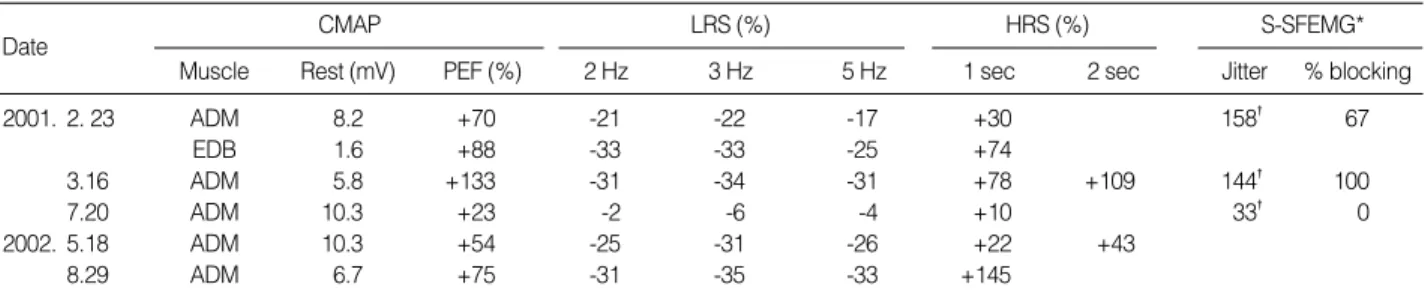
Table 1.Serial data of repetitive nerve stimulation test and stimulated single fiber electromyography
CMAP, compound muscle action potential; LRS, low rate of stimulation; HRS, high rate stimulation; S-SFEMG, stimulated single fiber EMG; PEF, pos- texercise fascilitation; ADM, abductor digiti minimi; EDB, extensor digitorum brevis; +, incremental response; -, decremental response.
*S-SFEMG was performed on the extensor digitorum communis muscle (EDC) at stimulation rate of 10 Hz. �Increased mean of mean consecutive dif- ference (upper normal limits; 25 sec).
Fig. 1.Postexercise fascilitation and incremental response at high rate of stimuration in the abductor digiti minimi muscle. Note definite facilitation at high rate of stimulation is achieved by prolonged stimulation for 2 sec in second test. (A) Compound muscle action potential (CMAP) before exercise. (B) CMAP after 30 sec of exercise. (C) Response at 50/sec stimulation for 1 sec. (D) Response at 50/sec stimu- lation for 2 sec.
2001.2.23 (First test) 2001.3.16 (Second test)
5 mV 5 msec
5 mV 5 msec
5 mV 5 msec
2.5 mV 0.2 sec
2.5 mV 0.2 sec
2.5 mV 0.4 sec 5 mV
5 msec
A
C
B A B
C D
Fig. 2.(A) CT scan of the chest shows a 1.4 cm nodule (arrow) in the posterior segment of right upper lobe. (B) Atypical carcinoid tumor with mosaic patterns separated by thin fibrovascular stroma. The tumor cells have central round nuclei with abundant cytoplasm (H&E ×100).
A B
Lambert-Eaton Myasthenic Syndrome and Atypical Bronchopulmonary Carcinoid Tumor 755
lar pleomorphism and increased mitotic activity, which is considered intermediate between that of typical carcinoid and small cell carcinoma. Immunohistochemical stains for neu- roendocrine markers, such as chromogranin A, synaptophysin, and neuron-specific enolase were all positive and was finally interpreted as atypical carcinoid (Fig. 2B). The antibody titer against the P/Q type voltage-gated calcium channel, sent after surgical resection was negative (less than 1.0 pmol/L; upper limit of normal; 20 pmol/L). After surgery, his strength has improved to almost premorbid level. The typical RNS fea- tures of LEMS normalized 2 months later (Table 1) and S- SFEMG findings also improved. However, his improvement was shortlived. In May 2002, he was reevaluated due to clin- ical worsening, showing a waddling of gait and fatigable weakness of proximal muscles in all limbs. Tumor recurrence was detected on chest CT. RNST abnormalities recurred as the same patterns as the initial test; normal CMAP, decremen- tal response at LRS, and mild incremental response at HRS.
It was 3 months after tumor recurrence that 145% of incre- mental response at HRS was achieved (Table 1). Despite sev- eral additional cycles of chemotherapy, he remained mildly disabled.
DISCUSSION
We describe a rare case of LEMS associated with atypical bronchopulmonary carcinoid tumor. No more than 5 cases could be found in English literatures (1-3), all of which, how- ever, lacked description of detailed clinical features and elec- trophysiological patterns.
LEMS results from an autoimmune attack directed against the voltage-gated calcium channels (VGCCs) on the presy- naptic motor nerve terminal. It was first described as a para- neoplastic syndrome in patients with lung cancer but we now know about half of the patients with LEMS do not have can- cer. When tumor occurs in LEMS, it is usually SCLC (5, 9).
Recently, Burns et al. (1) reported LEMS associated with other pulmonary neuroendocrine carcinomas with prolonged remission; two patients with atypical carcinoids and one pa- tient with large cell neuroendocine carcinoma. In the spec- trum of neuroendocrine tumors of the lung, which can be divided into the typical carcinoid, atypical carcinoid, SCLC and large cell neuroendocrine carcinoma, atypical carcinoids occupy the middle ground in terms of pathological features as well as natural history and prognosis (6). LEMS in the setting of underlying atypical carcinoid lung tumor, unlike those associated with SCLC, may be better responsive to treat- ment with variable long-term remission (1). In our case, clin- ical and electrophysiological remission was achieved by sur- gical resection and chemotherapy, but failed to remain in pro- longed remission after tumor recurrence.
It is noteworthy that our case initially did not satisfy the electrophysiological criteria of LEMS, and the follow-up study 3 weeks later revealed marked facilitation of CMAP compat- ible with LEMS. It is known that electrophysiologic findings of LEMS may occasionally overlap with those seen in myas- thenia gravis; a decremental response at low stimulations rate, normal CMAP amplitudes and absent facilitation at high stimulation rate. Furthermore, facilitation up to 50% can also be seen in myasthenia gravis (7), complicating electrophysi- ologic diagnosis not to be straightforward between the two myasthenic syndromes. Otherwise, it is believed that these patterns represent a mild form of LEMS, based on the obser- vation that many LEMS patients show these patterns as they improve with treatment (8). Our case demonstrates how re- peated serial RNST can help diagnosis when initial findings are equivocal. In addition, prolonged stimulation up to 2 sec or more may be needed for full extent of facilitation (8).
We concluded that LEMS could be associated with pulmo- nary neuroendocrine tumor other than SCLC, which necessi- tates pathologic confirmation followed by aggressive treatment for optimal management in these rare cases.
REFERENCES
1. Burns TM, Juel VC, Sanders DB, Phillips LH II. Neuroendocrine lung tumors and disorders of the neuromusclular junction. Neurology 1999; 52: 1490-1.
2. Elrington G, Newsom-Davis J. Clinical presentation and current immunology of the Lambert-Eaton myasthenic syndrome. In Lisak RP. eds. Handbook of Myasthenia Gravis and Myasthenic Syndromes.
New York: Marcel Dekker, 1994: 81-102.
3. Gutmann L, Phillips LH 2nd, Gutmann L. Trends in the association of Lambert-Eaton myasthenic syndrome with carcinoma. Neurology 1992; 42: 848-50.
4. Oh SJ. Principles of Clinical Electromyography, Case studies. Bal- timore, USA: Williams & Wilkins, 1998.
5. Tim RW, Massey JM, Sanders DB. Lambert-Eaton myasthenic syn- drome: Electrodiagnostic findings and response to treatment. Neu- rology 2000; 54: 2176-8.
6. Corrin B. Pathology of the Lungs, London: Churchil-Livingstone, 2000.
7. Sanders DB. Lambert-Eaton myasthenic syndrome: clinical diagno- sis, immune-mediated mechanisms, and update on therapies. Ann Neurol 1995; 37 (Suppl 1): S63-73.
8. Oh SJ. Diverse electrophysiological spectrum of the Lambert-Eaton myasthenic syndrome. Muscle & Nerve 1989; 12: 464-9.
9. Moon JS, Sunwoo IN, Kim SM, Lee SA, Cho KH, Park KD, Kim WK, Choi BO, Chun HY. Clinical analysis of 12 Korean Lambert- Eaton myasthenic syndrome (LEMS) patients. Yonsei Med J 1999;
40: 454-9.