Abstract
Purpose: The aim of study was to evaluate the effect of chemically surface modification with hydrophilicity on various physiochemical parameters which are involved in in vitro osteogenesis and in vivo new bone formation.
Materials and Methods: Commercially pure grade IV titanium disks with 12 mm diameter and 1mm height were manufactured by computerized numerical control machining. The sandblasted with large-grit alumina and acid etched (SA) surface was prepared by sandblasting with 250~500 μm Al2O3 grit and acid etching in hydrochloric and sulfuric acid according to the proprietary process. The chemically activated calcium-modified SA (CA) surface was prepared by a controlled process for the protection of carbon adsorption after SA surface treatment. After surface treatment, we verified the surface characteristics between two surfaces by scanning electron microscope (SEM), contact angle measurement. The biological efficiency and capability of chemically activated surface was evaluated by various in vitro tests and an in vivo push-in test.
Results: SEM investigations for macro and micro topography between SA and CA surface indicated no optical differences, but the carbon contents and wettability were different between the two surfaces. The CA surface showed lower carbon contents and higher wettability compared to the SA surface. Albumin adsorption, platelet adsorption and activation on chemically activated CA surface were dramatically enhanced compared with hydrophobic SA surface. Also, these hydrophilic CA surface showed a higher osteoblastic response such as cell adhesion, proliferation, alkarine phosphatase activity and mineralization.
Conclusion: In this study, we verified that chemistry and wettability of titanium surface were important variables in determining protein and osteoblast response. A chemically activated and super hydrophilic CA surface may play roles in stimulating the bone formation and ultimately enhanced initial implant stability compared with a hydrophobic SA surface.
Key Words: calcium-modified sandblasted with large-grit alumina and acid etched surface, hydrophilic, osseointegration, sandblasted with large-grit alumina and acid etched surface, titanium surface, wettability
In-vitro and In-vivo Evaluation of Early Bone Response to Hydrophilic Calcium- modified Sandblasted with Large-grit Alumina and Acid Etched Surface and Hydrophobic Sandblasted with Large-grit Alumina and Acid Etched Surface
Hong-Young Choi, Su-Kyung Kim, Jae-June Park, Tae-Gwan Eom
Department of Implant Research, Implant R&D Center, Osstem Implant Co., Ltd., Busan, Korea
Reprint requests: Hong-Young Choi
Surface Research Team, Implant R&D Center, Osstem Implant Co., Ltd., 33, Geoje-daero 214beon-gil, Yeonje-gu, Busan 611-801, Korea Tel: 82-70-7016-4433, Fax: 82-51-851-4341
E-mail: [email protected]
Received for publication: June 24, 2014 Accepted for publication: June 27, 2014
Copyright © 2014. The Korean Academy of Oral & Maxillofacial Implantology
This is an Open Access article distributed under the terms of the Creative Commons Attribution Non-Commercial License (http://creativecommons.org/licenses/by-nc/3.0/) which permits unrestricted non-commercial use, distribution, and reproduction in any medium, provided the original work is properly cited.
implant protocol proposed by Branemark has been altered, with quite a few cases being currently treated with immediate loading protocols. Nowadays there are several factors contributing to the success of the early osseointegration of titanium dental implants. The implant loading to bone is influenced by various fac- tors, including surface chemistry, surface energy and surface topography36. These surface parameters are decisive during the bone healing period in the peri
implant region. Immediately upon implantation dental implants get exposed to the patient’s blood and surface interactions between blood components such as blood cells and fibrinogen will influence the extent of blood coagulation, fibrin fiber formation and acute inflam- mation7,8. Complement activation and the activation of platelets and leukocytes on an implant surface has been described extensively9,10. The chemistry and topography of the medical implant surfaces were dem- onstrated to be able to influence interactions with all blood components11,12. After implantation, implant sur- faces are also in contact with body fluids and interact with a number of proteins and different cell types. Cell adhesion is one of the initial stages for subsequent proliferation and differentiation of osteoblastic cells producing bony tissue and extracellular matrix, which will ensure a high implant contact. It has been shown that osteoblastic cell adhesion, growth and differenti- ation are related to surface chemistry, energetic and roughness6,13,14. Among them, the surface free energy and hydrophilicity of an implant surface play decisive roles during the initial interaction with proteins and cells in bone15,16. A hydrophilic implant surface is assumed to be advantageous during the cascade of
I Introduction
Dental implants were initially used just as alter- native to removable prostheses or as a means to provide additional stability to removable resto- rations. Nowadays, the clinical indications of dental implants have increased substantially, whereas patients demand a lot more than improvement of mas- tication. They have high aesthetic expectations, they prefer as shorter treatment time as possible and they want reliable solutions to their dental problem with as few complications and failure rates as low as possible.
On the other hand, the popularity of implants and their increased demand has made dentists look for implant system with more straightforward surgical procedures, shorter osseointegration time, clinical flexibility and a variety of prosthetic components to meet varying needs. As a result, most of dental implants companies have been researched continuously in the search for the next, more improved, more reli- able and successful successor that will replace the current state of products, by trying to improve on, its limitations and widen its clinical usage indications.
Different implant surface topography have been stud- ied and introduced to solve this limitations, but resto- ration achieved by early or immediate loading still remains controversial compared with the reliability of enoseeous implants1,2.
All this has led to significant achievements the last few decades that are meant to primarily benefit the patient. Most importantly, the initial twostage
events that occurs during osseointegration. It has been reported that osteoblasts cultured on chemically pure and hydrophilic surfaces express higher levels of dif- ferentiation markers, such as alkaline phosphatase and osteocalcine17,18.
We have been verified that rough surfaces were also superior to smooth surfaces in promoting osteogenic induction of the human osteosarcoma cell line MG63, determined by increased osteocalcin and osteoprote- gerin19. Increased osteogenic differentiation measured by elevated alkaline phosphatase activity and nodule formation is associated with decreased proliferation on rough versus smooth surfaces in vitro. Moreover, the osteoblast cell number on rough sandblasted with largegrit alumina and acid etched (SA) surfaces has been verified to be much lower than on smooth surfac- es or plastic 24 hours after reaching confluence, yet concomitant elevation in osteocalcin and alkaline phosphatase production resulted from accelerated osteogensis, indicating that cell number and prolifera- tion is not a determinant of osteogenic responses to rough Ti surfaces. It is assumed that the reason of lower cell number on SA surface compared to smooth surface is attributed to the hydrophobicity of SA sur- face by carbon adsorption in air.
The various parameters of an implant surface have been shown to be a major influence on the evolution and properties of the implanttissue interface and the longterm success or failure of integration with the body. Recent studies on implant surface have been shown that surface chemistry is another key factor for affecting early osteogenic activity. The surface chemi- cal composition of titanium implants affects the
hydrophilicity of the surface. Highly hydrophilic sur- faces seem more desirable than hydrophobic ones in view of their interactions with biological fluids, cells and tissues.
The aim of the study was to examine the perfor- mance between hydrophobic SA surface and hydrophilic calciummodified SA surface in related to invitro osteogenesis and invivo new bone fromation. More specifically, we wanted to find out if the clinical advantage of hydrophilic CA implants can be attributed to any changes in the surfaces. In addition, chemically activated surface chemistry that leads to greatly increased wettability and hydrophilicity of the Ti sur- face was evaluated for potential benefit to osteogenic responses and bone apposition.
II Materials and Methods
1. Preparing of titanium samples
Titanium disks (12 mm in diameter and 1 mm in thickness) of commercially pure titanium were pre- pared by machining. SA and CA disks samples are manufactured in the same way; their only difference lies at the last stage of the production process. The SA disks are drystored after sandblasting and acid etching, whereas CA disks are rinsed under protective environment for preventing carbon adsorption on Ti surface and then stored in CaCl2 solution. The process results in a more active hydrophilic surface (CA sur- face), with higher surface energy and less hydrocarbon contamination from atmospheric environment. This
surface remains active until the implant actually sur- gically inserted into the bone by the surgeon, where the primary interaction with the aqueous biosystem is therefore accelerated.
Cylindrical titanium rods (1 mm in diameter and 2 mm in length) were also prepared for an in vivo bio- mechanical testing.
2. Surface characteristics
The prepared titanium disks were sterilized with gamma irradiation in a sealed container. The gamma
sterilized disks were stored in a accelerating chamber (at 55oC) during 6 weeks for reliability under normal use condition and unknown variable use environment.
The surface topography of SA and CA surface was examined using a scanning electron microscopy (SEM, JSM6480LV; Jeol, Tokyo, Japan) employing a solid state backscattered detector operated in 20 kV accel- erating voltage. The chemical composition of two sur- faces was examined using auger electron spectroscopy (AES, PHI 700; ULVACPHI Inc., Kanagawa, Japan) employing 10 kV/10 nA electron beam energy.
The surface wettability was tested using Phoenix contact angle meter (Surface Electro Optics, Suwon, Korea) several times for each sample. The sessile drop method was used to measure contact angle in this experiment on a plane surface with sheep blood. These are the most common measurements made with video instruments. An approximately 5 μl droplet of sheep blood was suspended from the tip of the microliter syringe. Once the drop settled on the surface, an image of the droplet was captured using a charge cou- pled device camera attached to the equipment. The
camera looks at the drop from its side and measures the liquidvapor profile and the liquidsolid baseline and the software solves for the contact angle. Both SA and CA disks were measured individually.
3. Protein adsorption, platelet adhe- sion and activation
The amount of albumin adsorbed on SA and CA disks was determined using a commercially available bicin- choninic acid assay (BCA) total protein kit via a spec- trophotometer. Purity of proteins was confirmed by the manufacturer. Three hundred microliters of each protein solutions of each protein solutions (1 mg/μl protein/saline solution) was pipette onto Ti disks in a sixwell plate. The study was then conducted in a sterile humidified incubator at 37oC over time. The nonadherent proteins were removed and washed twice using saline. The removed solution was saved and recorded as total volume. A 100 μl aliquot of the initial and removed solution were mixed with 150 μl of micro BCA working reagent in a 96well plate and incubated at 37℃ for 2 hours. Protein concentrations were analyzed using the micro BCA protein assay and measured using micro plate reader at 595 nm. Each protein concentration was calibrated using a standard curves. The degree of adsorption was determined by subtracting the residual protein from the initial added protein. Measurements were performed in triplicate for each time point.
Platelet adherence was quantified by a photospectro- metric measurement based on kinetic determination of lactate dehydronase (LDH) activity. After thorough washing, adherent platelets were lysed by the addition
by the addition of 200 μl of 0.2% Triton (Sigma
Aldrich, St. Louis, MO, USA) to the well. After 1 hour incubation at room temperature, 150 μl of each lysate was collected and mixed with 2.5 ml of 0.24 mM NADA, and 0.5 ml of 9.76 mM pyruvate. The LDH activity in the lysate was then determined by recording the change in absorbance at 340 nm. A calibration curve was used to relate platelet number after lysing washed bulk platelets with LDH activity.
For qualitative SEM examination of adherent plate- lets, 500 μl of 2.5% glutaraldehyde in Dulbecco’s phosphate buffered saline (DPBS) was added to each well, and incubated overnight at 4oC. The fixed sam- ples were subsequently dehydrated through graded ethanol solutions, critical points dried, and sputter gold coated, for SEM.
4. Cell attachment and proliferation
The MG63 cells (ATCC, Manassas, VA, USA) is a non
transformed cell line established from human osteosar- coma. These cells were routinely grown in Dulbecco's modified Eagle's medium supplemented with 10% fetal bovine serum (FBS; HyClone, Salt Lake City, UT, USA) and 0.1 mg/ml penicillin and streptomycin (P/S) (HyClone). Cell were subcultured twice a week using trypsin/ethylenediaminetetraacetic acid and maintained at 37oC in a humidified atmosphere of 5% CO2 in air.
Medium was completely renewed every two days.
The cell adhesion assay of the MG63 cells was per- formed. MG63 cells were grown to approximately 80%
confluence prior to cell adhesion assay. Mg63 cells were cultured either onto the various titanium disc or in the culture plastic in 24 multiwell plate at a densi-
ty of 1×10⁵ cells. After 1 hour, culture media were removed and rinsed three times by DPBS. And the cells fixed using 4% formaldehyde solution for 1 hour at 4oC and were rinsed three times by DPBS. Then the viable cells were stained using cresyl violet which stained ribonucleic acid and were eluted using citric acid.
Finally colorimetric measurement of cresyl violet was performed on a DTX 880 (Beckman Coulter, CA, USA) with optical density reading at 590 nm. Also, A scan- ning electron microscope was employed for morpholog- ical characterization of MG63 cells adhered to SA and CA surfaces.
The cell proliferation assay of the MG63 cells was performed using CellTiter 96 Aqueous Non
Radioactive Cell Proliferation Assay kit (Promega, Madison, WI, USA) according to the manufacturer’s protocol. MG63 cells were cultured either onto the various titanium disc or in the culture plastic in 24 multiwell plate at a density of 1×10⁵ cells. After indicated times, culture media were removed and 0.5 kl MTS/phenazine methosulfate/media mixture was added in each well for 1 hour. Finally colorimetric measurement of formazan dye was performed on a DTX 880 with optical density reading at 490 nm.
Results were expressed as optical density and relative MTS activity as compared to control.
5. Immuno-fluorescent analysis of the actin cytoskeleton and vinculin
Cells were seeded at the initial seeding density of 1×10⁵ cells and after 2 hours of incubation they were fixed with 3.7% neutral buffered formation. The cells were then permeabilized with 0.25% Triton X100
(SigmaAldrich) solution, blocked with 1% BSA solu- tion and incubated for 1 hour at room temperature with Alexa (488)conjugated phalloidin (5 U/ml, Invitrogen, Carlsbad, CA, USA). After washing with PBS the nucleus was counterstained with Hoechst 33528 (Hoechst AG, Frankfurt, Germany) and the samples were mounted in aqueous mounting medium (Dako Faramounts; Dako, Carpinteria, CA, USA) and evalu- ated under a fluorescence microscope.
6. Cell differentiation - ALP activity and mineralization
Alkaline phosphatase (ALP) activity was examined as previously descri bed2023, in MG63 cells cultured either onto the various titanium disc or culture plastic in 24 multiwell plates (1×10⁵ cells/well). Osteogenic medium (0.5 M dexamethasone, 50 g/ml Lascorbic acid, 10 mM bglycerol phosphate) was completely renewed every two days and after indicated times, cells were washed twice with DPBS and collected in 0.1%
ALP lysis buffer. In order to extract the dissolved pro- teins from the crude cell lysates, the supernatants were centrifuged at 13,000 rpm for 10 minuts. The protein concentrations were determined using a BCA reagent (Pierce, Rockford, IL, USA). For ALP activity measurement with isolated cell supernatants, 100 g total protein were incubated with 2 mg/ml pNPP (as a chromogenic substrate, SigmaAldrich) using ALP assay buffer (pH 9.75 in 0.1 M glycine containing 1 mM MgCl2) at 37oC for 2 hours. The degree of colorimetic reaction was measured by DTX 880 for an optical den- sity measurements at 405 nm.
The alizarin reds staining with experimental sam-
ples were performed as reported elsewhere2426 with a minor modifications. Briefly, MG63 cells were cul- tured either onto the various titanium disc or culture plastic in 24 multiwell plates (1×10⁵ cells/well).
Osteogenic medium was completely renewed every two days and after indicated times. Cells were washed twice with DPBS and fixed in 10% (v/v) formaldehyde (Junsei, Tokyo, Japan) for 20 minuts at room temper- ature and rinsed with triple distilled H2O. Cells were stained with 2% alizarin reds (Sigma), pH 4.2, for 20 minuts with gentle agitation. After aspiration of the unstained dye, the wells were washed five times with distilled H2O while shaking for 5 minuts. For quantifi- cation of staining, 0.5 kl 10% (v/v) acetic acid was added to each well, and the plate was heated to 70oC~80oC for 1 hour. The cells was then scraped from the plate and centrifuged at 13,000 rpm for 10 minuts.
The supernatant was transferred to a new etube and the 0.4 volume of 10% (v/v) ammonium hydroxide added to neutralize the acid. Absorbance of extracted alizarin reds in acetic acid solution (0.2 kl) was mea- sured at 405 nm. Amount of alizarin reds was deter- mined according to an alizarin reds standard curve and normalized to the total protein of the cell lysates using BCA method (Pierce, Rockford, IL, USA).
7. Implant biomechanical push-in-test
Eightweekold male SpragueDawley rats were anesthetized by inhalation with 1%~2% isoflurane.
After their legs were shaved and scrubbed with 10%
providoneiodine solution, the distal aspects of the femurs were carefully exposed via skin incision and muscle dissection. The flat surfaces of the distal
femurs were selected for implant placement. The implant site was prepared 11 mm from the distal edge of the femur by drilling with a 0.8 mm round burr and enlarged using reamers. Profuse irrigation with sterile isotonic saline solution was used for cooling and cleaning. One cylindrical titanium implant was placed into a femur. Surgical sites were then closed in layers.
The established implant biomechanical pushintest was used to assess the biomechanical strength of boneimplant integration. At weeks 2 of healing, femurs containing a cylindrical implant were harvested and embedded into resin with the top surface of the implant level. The testing machine (Instron 8841;
Instron Corp., Canton, MA, USA) equipped with a 2 kN load cell and a pushing rod was used to load the
implant vertically downward at a crosshead speed of 1 mm/min. The pushin value was determined by mea- suring the peak of the loaddisplacement curve.
After the pushin test at week 2, the bone/implant complex was carefully retrieved and soaked in agitated water for 1 hour and dried under heat and vacuum.
After being gold sputtercoated, the specimens were examined by SEM.
III Results
1. Surface characteristics
The morphological features of SA and CA disk were
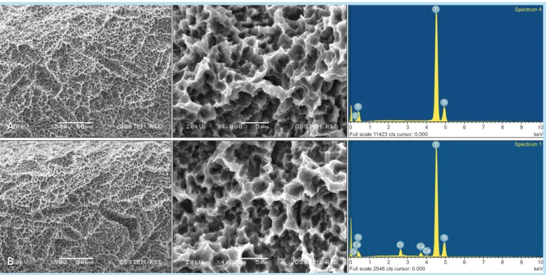
Fig. 1. The surface topography (×500) and energy-dispersive X-ray spectroscopy (×4,000) of the SA (A) and CA (B) surface. SA: sandblasted with large-grit alumina and acid etched, CA: calcium-modified SA.
Hong-Young Choi et al. : In-vitro and In-vivo Evaluation of Early Bone Response to Hydrophilic Calcium-modified Sandblasted with Large-grit Alumina and Acid Etched Surface and Hydrophobic Sandblasted with Large-grit Alumina and Acid Etched Surface. Implantology 2014
demonstrated in Fig. 1. The topography looks similar, with a hierarchical structure comprised of irregularly rounded shape domains, incorporating more rounded grooves with sharpedged and overhanging crater
like micropores. The smooth and amorphous structure of the submicron topography observed at high magni- fications, possibly resembles the oxide film formed during the acidic treatment on the previously sand- blasted irregular surfaces.
Representative AES spectra obtained from SA and CA surface are also presented in Fig. 2. On all samples Ti, O, C, and N signals were always detected by AES.
The dominance of the Ti and O signals shows that the surface consists mainly of a titanium oxide layer. The relatively strong C signal can be assigned mainly to surface contamination by absorbed carboncontaining molecules, which is a normal observation for air
exposed surfaces. The C signal is not as intense on CA spectra, due to the fact it is transferred from the
etching process to aqueous storage under controlled environments and, hence, less exposure to surface contaminants takes place.
The contact angle of SA surface displayed 128 degree for water and 90 degree for sheep blood (Fig. 3). The difference on contact angle between water and sheep blood was assumed the differences of polarity and density between 2 liquids. On the other hand, with regards to the CA surface, it was difficult to form a satisfactory profile image of a sessile drop. They exhibited complete wetting forming a layer of water across the surface, with a contact angle approaching zero (less than 5 degrees), thus the CA surfaces are defined as having super hydrophilic properties. The same results were found in case of blood.
2. Adsorption of protein, platelet and platelet activation
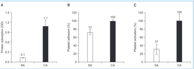
First, albumin adsorption on SA and CA surfaces Fig. 2. Auger spectra of two surfaces hydrophobic SA surface (A), hydrophilic CA surface (B). SA: sandblasted with large- grit alumina and acid etched, CA: calcium-modified SA.
Hong-Young Choi et al. : In-vitro and In-vivo Evaluation of Early Bone Response to Hydrophilic Calcium-modified Sandblasted with Large-grit Alumina and Acid Etched Surface and Hydrophobic Sandblasted with Large-grit Alumina and Acid Etched Surface. Implantology 2014
was examined at pH 7.0. The amount of albumin adsorbed on hydrophilic CA surfaces during the 1h incubation was 11fold greater than that adsorbed on hydrophobic SA surfaces (Fig. 4A).
Higher platelet adhesion was observed on hydrophilic CA surfaces, which was slightly higher than hydropho- bic SA surfaces (Fig. 4B). SEM showed increased num- ber of red blood cells present on CA surfaces, in com- parison of SA surfaces. The agglomeration of red blood cells appeared more extensive on hydrophilic CA sur-
faces compared to hydrophobic SA surfaces. Also, platelet activation on hydrophilic CA surface was 3fold greater than that of hydrophobic SA surfaces (Fig. 4C).
3. Cell attachment and proliferation
The attachment and proliferation of MG63 cells on the SA and CA surfaces were evaluated, with the results shown in Fig. 5. The cell attachment and proliferation results were expressed as the number of cells normal- ized to that of the control surface after expected time Fig. 3. The wettability results of SA and CA surface for water and blood. (A) The contact angle for deionized water on SA and CA surface. (B) The contact angle for sheep blood on SA and CA surface. SA: sandblasted with large-grit alumina and acid etched, CA: calcium-modified SA.
Hong-Young Choi et al. : In-vitro and In-vivo Evaluation of Early Bone Response to Hydrophilic Calcium-modified Sandblasted with Large-grit Alumina and Acid Etched Surface and Hydrophobic Sandblasted with Large-grit Alumina and Acid Etched Surface. Implantology 2014
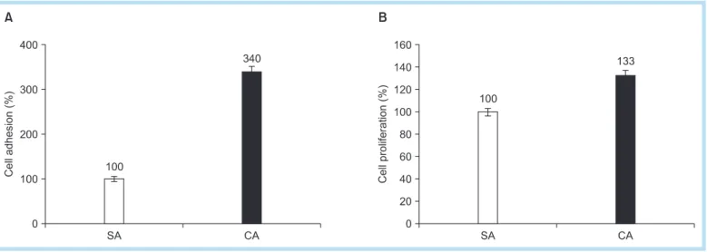
cultured on sample surfaces. The cell adhesion data of the MG63 cells on the different surfaces are presented in Fig. 5. Statistical testing of the data, using a two
way analysis of variance and a multiple comparison test (Turkey), revealed that after 1hr of incubation in the CA surfaces, the adherent cells were significantly higher than the SA surface and the culture plate.
Fig. 6 shows SEM images of spread cells on SA and CA surfaces. At 10 min after beginning the incubation, most of the cells on SA surface were in the early stage of cell spreading (stage 1 and 2). SA surface which have a rough morphology and hydrophobic surface property were connected to the surface by a few filo- podia, whereas CA surface which have a same rough morphology but hydrophilic surface property had a greater number of cells at more advanced stage of spreading (stage 4). On CA surface, fully spread cells were more frequently observed and very close contact
with the underlying surface.
To examine the proliferation of osteoblastic cells in CA surface, we carried out a measurement of MTS activity on MG63 cells after 1 day in culture. Results indicated that cell proliferation on CA surface was increased after 1 day of culture than the SA surface.
To confirm the above findings, an additional experi- ment based on the morphology of the cell adhesion as well as a quantitative evaluation of the cell was con- ducted using microscopic observations via immunoflu- orescent staining (Fig. 7). Generally, the figures of merged image show that the cells on the CA surface were stretched filopodia compare to the SA surface, indicating an improved attachment ability of the cells.
The extremely of the cell attachment can also be determined based on the actin filaments (red color), and much longer and brighter actin filaments found in CA surface than SA surface, which enhance cell adhe- Fig. 4. Attachment of blood plasma protein to hydrophobic SA and hydrophilic CA surface. (A) Protein adsorption. (B) Platelet adhesion. (C) Platelet activation. OD: optical density, SA: sandblasted with large-grit alumina and acid etched, CA:
calcium-modified SA.
Hong-Young Choi et al. : In-vitro and In-vivo Evaluation of Early Bone Response to Hydrophilic Calcium-modified Sandblasted with Large-grit Alumina and Acid Etched Surface and Hydrophobic Sandblasted with Large-grit Alumina and Acid Etched Surface. Implantology 2014
sion property. Additionally, elements of focal adhe- sions, vinculins (green color), were visualized using a confocal laser microscope.
4. Expression of focal adhesion pro- tein, vinculin
Confocal microscopic images of osteoblasts after
antivinculin and antiactin staining are shown in Fig. 8. After 3 hours incubation, size and shape of the cells were remarkably different between cultures on SA and CA surfaces. The expression of vinculin was observed in cells both on the SA and CA surfaces.
However, the localization of vinculin at the tip of the stretching cytoplasmic projections was only seen in Fig. 5. Representative scanning electron microscope micrographs (×3,000) showed the morphology and quantity adherent blood-clot and platelets on SA surface (A), CA surface (B). SA: sandblasted with large-grit alumina and acid etched, CA:
calcium-modified SA.
Hong-Young Choi et al. : In-vitro and In-vivo Evaluation of Early Bone Response to Hydrophilic Calcium-modified Sandblasted with Large-grit Alumina and Acid Etched Surface and Hydrophobic Sandblasted with Large-grit Alumina and Acid Etched Surface. Implantology 2014
osteoblasts on the CA surface.
5. Cell differentiation - ALP activity and mineralization
Generally, ALP activity was affected by the surface wettability. In addition osteoblastic cell differentiation was assessed by measuring the ALP activity normalized to total protein content. On hydrophilic CA surfaces, we observed a little enhancement of ALP activity as com- pared with hydrophobic SA surface after 7 days. These results indicate that osteoblastic cells cultured in direct contact with hydrophilic CA surfaces increased their capability to express ALP. We use the alizarin reds staining for quantification of mineralization induced by osteoblastic cells. As shown Fig. 9, similar pattern in ALP activity data, MG63 cells cultured on CA surfaces had increased a little in mineralization.
6. Push-in test
The strength of osseointegration evaluated by the pushin test was increased about 50% by CA surface at week 2 of healing as shown in Fig. 10. The new bone formation after pushin test was observed by SEM and EDX. The significant part of the SA surfaces lacked bone tissue as shown in SEM images and low Ca/Ti ratio of EDX analysis. In contrast, CA surfaces were covered with more dense bone tissue. The Ca/Ti ratio was consistently higher for SA surfaces regardless of the location of the specimen.
IV Discussion
Osteoblastic cells play a critical role in the early stages of ossteointegration. It is widely accepted that surface topography and roughness influence the early
Fig. 6. MG-63 cellular activity on SA and CA surface for cell attachment and proliferation. (A) Cell adhesion. (B) Cell proliferation. SA: sandblasted with large-grit alumina and acid etched, CA: calcium-modified SA.
Hong-Young Choi et al. : In-vitro and In-vivo Evaluation of Early Bone Response to Hydrophilic Calcium-modified Sandblasted with Large-grit Alumina and Acid Etched Surface and Hydrophobic Sandblasted with Large-grit Alumina and Acid Etched Surface. Implantology 2014
healing stages of bone integration2729. Also, surface properties such as wettability, surface topography and charge are known to affect endothelial cells attach- ment and growth likely by altering the rate of the amount of adsorbed proteins and their conformational change. The effect of surface materials on erythrocyte aggregation and platelet adhesion/activation becomes a
chief parameter in haemocompatibility studies.
Additionally, it has been shown that chemical modifi- cation resulting in increased hydrophilicity leads to improved osseointegration in vivo30,31 and osteogenic properties in vitro32,33.
However, little is known about the regulatory mech- anism between the osteoblastic cell response and sur- Fig. 7. Scanning electron microscope images showing the morphology of spread cells on SA (A) and CA (B) surfaces after 10 minuts of incubation (A: ×500, B: ×2,000). SA: sandblasted with large-grit alumina and acid etched, CA: calcium- modified SA.
Hong-Young Choi et al. : In-vitro and In-vivo Evaluation of Early Bone Response to Hydrophilic Calcium-modified Sandblasted with Large-grit Alumina and Acid Etched Surface and Hydrophobic Sandblasted with Large-grit Alumina and Acid Etched Surface. Implantology 2014
face wettability on the titanium discs. In this study, we focused on the effect of surface wettability during the early stage of cell behavior and bone apposition. This study was provided the evidence that the surface wet- tability is an important regulating factor of the blood coagulation, early cell response and bone apposition.
To improve surface wettability, a CA surface has been produced by the same procedure as SA surface treat- ment but being protected under controlled atmosphere
and submerged in the CaCl2 solution.
We examined osteogenic properties and bone apposi- tion between two surfaces in various ways, using the cresyl violet staining, MTS assay, ALP activity assay, alizarin redS staining, pushin measurement and scanning electron microscopy. In all cases, our results show that hydrophilic CA surfaces are much higher performance than hydrophobic SA surfaces as follows:
increased adsorption of protein, increased platelet Fig. 8. Initial behavior of osteoblast on hydrophilic CA surface and hydrophobic SA surface. SA:
sandblasted with large-grit alumina and acid etched, CA: calcium- modified SA.
Hong-Young Choi et al. : In-vitro and In-vivo Evaluation of Early Bone Response to Hydrophilic Calcium-modified Sandblasted with Large-grit Alumina and Acid Etched Surface and Hydrophobic Sandblasted with Large-grit Alumina and Acid Etched Surface. Implantology 2014
adhesion/activation, increased attachment of osteo- blasts, facilitated osteoblast spread, increased prolif- eration of osteoblasts, promoted osteoblastic differ- entiation and increased initial mechanical stability.
These results provided unequivocal evidence that sur- face wettability is an important factor that enhances the protein adsorption, blood coagulation and osteo- blastic cell response. Also, these properties should not be considered as independent from each other. The strong correlation between impaired blood coagulation followed by impaired wound healing underlines the importance of blood coagulation for wound healing, later followed by implant integration34,35. The first step of dental implant contact with blood is adhesion of plasma proteins to the surface. Then the same hap- pens with platelets, but the intensity of this process is dependent on the type of proteins adhered to the sur- face. Some proteins, e.g., fibrinogen, stimulate the platelet adhesion, while others inhibit this process.
Platelets adhered to the surface of implant become activated: their shape and size change, the content of granules is released and finally platelets aggregate.
This results in a hemostatic system activation and a blood clot formation. Additionally, increased protein adsorption may have promoted osteoblastic attachment via enhanced interaction between proteins and cellular integrins. Increased osteoblastic proliferation may have caused the promoted differentiation due to the increased celltocell interaction. Future studies need to address the initial behavior of other protein and cell type.
Being a more direct and sensitive predictor of new bone formation than invitro osteoblastic cell response, biomechanical pushin testing is utilized to assess implant anchorage in the surrounding bone in implant. The hydrophilic CA surface showed a higher value of bonetitanium interfacial strength than hydrophobic SA surface after 2 weeks healing periods Fig. 9. Effect of surface wettability on the osteoblastic differentiation of MG-63 cells. (A) ALP activity after 7 days. (B) Mineralization after 2 weeks. SA: sandblasted with large-grit alumina and acid etched, CA: calcium-modified SA.
Hong-Young Choi et al. : In-vitro and In-vivo Evaluation of Early Bone Response to Hydrophilic Calcium-modified Sandblasted with Large-grit Alumina and Acid Etched Surface and Hydrophobic Sandblasted with Large-grit Alumina and Acid Etched Surface. Implantology 2014
which indicates that the hydrophilic surface stimulated bone growth and accelerated formation of the bone
titanium interface. In this study, the hydrophilic CA surface is proven to have beneficial effects on the osteoblastic cell response and bone growth.
V Conclusion
In summary, this study examined the effect of chemically modified hydrophilic CA surface on protein adsorption, platelet adhesion/activation, osteoblastic Fig. 10. Bone-titanium integration evaluated by biomechanical push-in test and new bone formation. (A) Push-in test value at 2 weeks. (B) Ca/Ti ratio after push-in test at 2 weeks. (C) New bone formation on SA and CA specimen after 2 weeks healing period (×500). SA: sandblasted with large-grit alumina and acid etched, CA: calcium-modified SA.
Hong-Young Choi et al. : In-vitro and In-vivo Evaluation of Early Bone Response to Hydrophilic Calcium-modified Sandblasted with Large-grit Alumina and Acid Etched Surface and Hydrophobic Sandblasted with Large-grit Alumina and Acid Etched Surface. Implantology 2014
cell adhesion, proliferation, differentiation. Hydrophilic CA surface also great increased the new bone forma- tion and bonetitanium interfacial strength in the early healing period after implantation. We believe that the enhancement of weattability on implant surface is feasible in clinical practice. In conclusion, CA surface enhanced the bioactivity of the traditional SA surface by integrating the advantages of surface wettability.
References
1. Chiapasco M. Early and immediate restoration and loading of implants in completely edentulous patients. Int J Oral Maxillofac Implants. 2004;
19(Suppl): 76-91.
2. Ganeles J, Wismeijer D. Early and immediately restored and loaded dental implants for single-tooth and partial-arch applications. Int J Oral Maxillofac Implants. 2004; 19(Suppl): 92-102.
3. Albrektsson T, Wennerberg A. Oral implant surfaces: part 2--review focusing on clinical knowledge of different surfaces. Int J Prosthodont.
2004; 17: 544-564.
4. Esposito M, Coulthard P, Thomsen P, et al. The role of implant surface modifications, shape and material on the success of osseointegrated dental implants. A Cochrane systematic review. Eur J Prosthodont Restor Dent. 2005; 13: 15-31.
5. Puleo DA, Thomas MV. Implant surfaces. Dent Clin North Am. 2006;
50: 323-338.
6. Zhao G, Schwartz Z, Wieland M, et al. High surface energy enhances cell response to titanium substrate microstructure. J Biomed Mater Res A. 2005; 74: 49-58.
7. Anderson JM. Biological response to materials. Annu Rev Mat Res.
2001; 31: 81-110.
8. Arvidsson S, Askendal A, Tengvall P. Blood plasma contact activation on silicon, titanium and aluminium. Biomaterials. 2007; 28: 1346-1354.
9. Smith BR, Rinder HM, Rinder CS. Interaction of blood and artificial surfaces. In: Loscalzo J, Schafer AI, eds. Thrombosis and Hemorrhage.
3rd ed. Philadelphia: Lippincott; 2003. p. 865-900.
10. Jackson SP. The growing complexity of platelet aggregation. Blood.
2007; 109: 5087-5095.
11. Park JY, Gemmell CH, Davies JE. Platelet interactions with titanium:
modulation of platelet activity by surface topography. Biomaterials.
2001; 22: 2671-2682.
12. Stanford CM. Surface modification of biomedical and dental implants and the processes of inflammation, wound healing and bone formation.
Int J Mol Sci. 2010; 11: 354-369.
13. Cooper LF, Masuda T, Yliheikkilä PK, et al. Generalizations regarding the process and phenomenon of osseointegration. Part II. In vitro studies.
Int J Oral Maxillofac Implants. 1998; 13: 163-174.
14. Anselme K. Osteoblast adhesion on biomaterials. Biomaterials. 2000;
21: 667-681.
15. Kieswetter K, Schwartz Z, Dean DD, et al. The role of implant surface characteristics in the healing of bone. Crit Rev Oral Biol Med. 1996; 7:
329-345.
16. MacDonald DE, Deo N, Markovic B, et al. Adsorption and dissolution behavior of human plasma fibronectin on thermally and chemically modified titanium dioxide particles. Biomaterials. 2002; 23: 1269-1279.
17. Yoshinari M, Matsuzaka K, Inoue T, et al. Superhydrophilicity promotes cell proliferation. J Jpn Assoc Regen Dent. 2004; 2: 20-28.
18. Buser D, Broggini N, Wieland M, et al. Enhanced bone apposition to a chemically modified SLA titanium surface. J Dent Res. 2004; 83: 529- 533.
19. Park SJ, Bae SB, Kim SK, et al. Effect of implant surface microtopography by hydroxyapatite grit-blasting on adhesion, proliferation, and differentiation of osteoblast-like cell line, MG-63. J Korean Assoc Oral Maxillofac Surg. 2011; 37: 214-224.
20. Engvall E. Enzyme immunoassay ELISA and EMIT. Methods Enzymol. 1980; 70: 419-439.
21. Jones JV, Mansour M, James H, Sadi D, Carr RI. A substrate amplifi- cation system for enzyme-linked immunoassays. II. Demonstration of its applicability for measuring anti-DNA antibodies. J Immunol Methods. 1989; 118: 79-84.
22. Harada M, Hiraoka BY, Fukasawa K, Fukasawa KM. Purification and properties of bovine dental-pulp alkaline-phospatase. Arch Oral Biol.
1982; 27: 69-74.
23. Jung K, Pergande M. Influence of inorganic phosphate on the activity determination of isoenzymes of alkaline phosphatase in various buffer systems. Clin Chim Acta. 1980; 102: 215-219.
24. Gregory CA, Gunn WG, Peister A, Prockop DJ. An alizarine red-based assay of mineralization by adherent cells in culture: comparison with
cetylpyridinium chlroride extraction. Anal Biochem. 2004; 329: 77-84.
25. Malladi P, Xu Y, Chiou M, Giaccia AJ, Longaker MT. Effect of reduced oxygen tension on chondrogenesis and osteogenesis in adipose-derived mesenchymal cells. Am J Physiol Cell Physiol. 2006; 290: C1139- C1146.
26. Sudo H, Kodama HA, Amagai Y, Yamamoto S, Kasai S. In vitro differentiation and calcification in a new clonal osteogenic cell line derived from newborn mouse calvaria. J Cell Biol. 1983; 96: 191-198.
27. Wennerberg A, Albrektsson T. Effects of titanium surface topography on bone integration: a systematic review. Clin Oral Implants Res. 2009;
20(Suppl 4): 172-184.
28. Buser D, Schenk RK, Steinemann S, et al. Influence of surface characteristics on bone integration of titanium implants. A histomorphometric study in miniature pigs. J Biomed Mater Res. 1991;
25: 889-902.
29. Abrahamsson I, Berglundh T, Linder E, et al. Early bone formation adjacent to rough and turned endosseous implant surfaces. An experimental study in the dog. Clin Oral Implants Res. 2004; 15: 381- 392.
30. Lang NP, Salvi GE, Huynh-Ba G, et al. Early osseointegration to hydrophilic and hydrophobic implant surfaces in humans. Clin Oral Implants Res. 2011; 22: 349-356.
31. Qu Z, Rausch-Fan X, Wieland M, et al. The initial attachment and subsequent behavior regulation of osteoblasts by dental implant surface modification. J Biomed Mater Res A. 2007; 82: 658-668.
32. Rausch-fan X, Qu Z, Wieland M, et al. Differentiation and cytokine synthesis of human alveolar osteoblasts compared to osteoblast-like cells (MG63) in response to titanium surfaces. Dent Mater. 2008; 24:
102-110.
33. Zhao G, Raines AL, Wieland M, et al. Requirement for both micron- and submicron scale structure for synergistic responses of osteoblasts to substrate surface energy and topography. Biomaterials. 2007; 28: 2821- 2829.
34. Masaki C, Schneider GB, Zaharias R, et al. Effects of implant surface microtopography on osteoblast gene expression. Clin Oral Implants Res.
2005; 16: 650-656.
35. Xu Z, Xu H, Ploplis VA, et al. Factor VII deficiency impairs cutaneous wound healing in mice. Mol Med. 2010; 16: 167-176.