Primary Hepatic Angiosarcoma with Portal Vein Thrombosis and Satellite Hepatic Nodules: A Case Report
Mi Jeong Kim, M.D., Jung Hyeok Kwon, M.D., Koo Jeong Kang, M.D. , Yong Hoon Kim, M.D. , Yu Na Kang, M.D.
Department of Diagnostic Radiology, Department of Surgery , Department of Pathology ,
Keimyung University School of Medicine, Daegu, Korea
Abstract
Key Words :
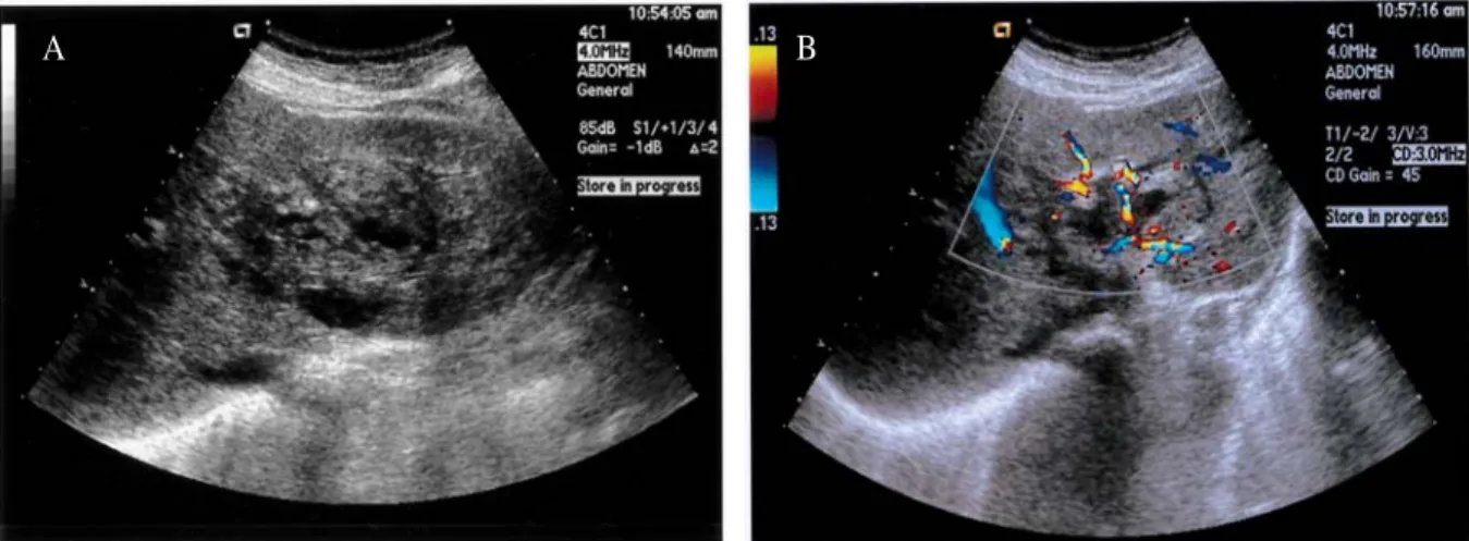
Fig. 1. Gray scale and color Doppler ultrasonogram of hepatic angiosarcoma. (A) Transverse US scan of the liver shows a large heterogeneous mass with multiple hypoechoic lesions in the left lobe. (B) Color Doppler US scan reveals the presence of intratumoral vessels within this mass.
B A
196
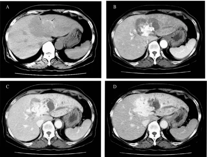
Fig. 2. Computed tomographic findings of hepatic angiosarcoma. (A) Precontrast CT scan shows a large hypoattenuating mass with marginal hyperattenuating lesions (arrows) suggesting focal areas of hemorrhage.
(B) Arterial phase contrast-enhanced CT scan shows strong enhancement at the central portion of the tumor, thrombosis of P2 branch, and decreased parenchymal enhancement in the lateral segment. (C) Portal phase contrast-enhanced CT scan shows progressive centrifugal enhancement of the tumor, portal vein thrombosis of P2 branch, and multiple small hypoattenuating nodules in the lateral segment. (D) Equilibrium phase contrast- enhanced CT scan shows non-filling of contrast materials into a quarter of this mass corresponding to hyperattenuating lesions on precontrast scan.
A B
C D
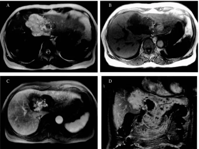
Fig. 3. Magnetic resonance findings of hepatic angiosarcoma. (A) T2-weighted MR image shows very hyperintense components (arrows) in three quarters and heterogeneous hypointense components (arrowheads) in one quarter of this mass. There reveals slightly increased signal intensity in the lateral segment. A nodule is noted in the right lobe of the liver. (B) T1-weighted GRE MR image shows hypointense components (arrows) in three quarters and heterogeneously hyperintense components (arrowheads) in one quarter of this mass. There reveals slightly decreased signal intensity in the lateral segment. A nodule is noted in the right lobe of the liver. (C) Gadolinum-enhanced T1-weighted fat-suppressed GRE MR image obtained 30 sec after initiation of contrast agent administration shows irregular-shaped strong central enhancement, (D) Delayed-phase gadolinium T1 -weighted coronal image reveals progression of contrast enhancement with persistent nonenhancing portion of the hepatic mass and numerous hypointense satellite nodules.
A B
C D
198
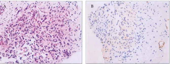
Fig. 4. The histopathologic and immunohistochemical findings of hepatic angiosarcoma. (A) Microscopic finding shows characteristic histologic pattern, which is a scaffold-like or tectorial growth of atypical cells between liver cell plates (H&E, 400). (B) Immunohistochemical staining for endothelial markers (Factor VIII-related antigen) is positive in neoplastic endothelial cells.
B A
1. Koyama T, Fletcher JG, Johnson CD, Kuo MS, Notohar K, Burgart LJ. Primary hepatic angiosarcoma: findings at CT and MR imaging.
Radiology 2002;222:66-73.
2. , , , .
: 1 .
1986;22:1061-5.
3. , , , , , .
: 1 . 1997
;36:1033-6.
4. Buetow PC, Buck JL, Ros PR, Goodman ZD.
Malignant vascular tumors of the liver: radiologic -pathologic corrleation. Radiographics 1994;14:153 -66.
5. Ludwig J, Hoffman HN. Hemangiosarcoma of the liver. Spectrum of morphologic changes and clinical findings. Mayo Clin Proc 1975;50:255-63.
6. Itai Y, Teraoka T. Angiosarcoma of the liver mimicking cavernous hemangioma on dynamic CT. J Comput Assist Tomogr 1989;13:910-2.
7. Peterson MS, Baron RL, Rankin SC. Hepatic angiosarcoma: findings on multiphasic contrast -enhanced helical CT do not mimic hepatic hemangioma. Am J Roentgenol 2000;175:165-70.
8. Cohen J, Edelman RR, Chopra S. Portal vein thrombosis: a review. Am J Med 1992;92:173-82.
9. Primentel Cauduro SK, Petrovic LM, Sodeman TC, Ishitani MB, Menon KV, et al. Unsuspected primary hepatic angiosarcoma associated with portal vein thrombosis complicating cirrhosis. Liver Transplantation 2002;8:1080-1.
10. Hertzanu Y, Oeiser J, Zirkin H. Massive bleeding after fine needle aspiration of liver angiosarcoma.
Gastrointest Radiol 1990;15:43-6.