Improving amber suppression activity of an orthogonal pair of
Saccharomyces cerevisiae tyrosyl-tRNA synthetase and a variant of E.
coli initiator tRNA, fMam tRNA CUA , for the efficient incorporation of unnatural amino acids §
Eyob Tekalign
†, Ju-Eon Oh
†, and Jungchan Park*
Department of Bioscience and Biotechnology, Hankuk University of Foreign Studies, Yongin 17035, Republic of Korea
효율적인 비천연 아민노산 도입을 위한 효모균 타이로신-tRNA 합성효소와 대장균 시작 tRNA 변이체의 엠버써프레션 활성증가 §
이욥테칼린
†・ 오주연
†・ 박중찬*
한국외국어대학교 생명공학과
(Received December 4, 2018; Revised December 17, 2018; Accepted December 18, 2018)
†These authors contributed equally to this work.
§Supplemental material for this article may be found at http://www.kjom.
org.
*For correspondence.
E-mail: [email protected];Tel.: +82-31-330-4355; Fax: +82-31-330-4566
The orthogonal pair of Saccharomyces cerevisiae tyrosyl-tRNA synthetase (Sc YRS) and a variant of E. coli initiator tRNA, fMam tRNA
CUAwhich recognizes the amber stop codon is an effective tool for site-specific incorporation of unnatural amino acids into the protein in E. coli. To evolve the amber suppression activity of the orthogonal pair, we generated a mutant library of Sc YRS by randomizing two amino acids at 320 and 321 which involve recognition of the first base of anticodon in fMam tRNA
CUA. Two positive clones are selected from the library screening with chloramphenicol resistance mediated by amber suppression. They showed growth resistance against high concentration of chloramphenicol and their IC
50values were approximately 1.7~2.3 fold higher than the wild type YRS. In vivo amber suppression assay reveals that mutant YRS-3 (mYRS-3) clone containing amino acid substitutions of P320A and D321A showed 6.5-fold higher activity of amber suppression compared with the wild type. In addition, in vitro aminoacylation kinetics of mYRS-3 also showed approximately 7-fold higher activity
than the wild type, and the enhancement was mainly due to the increase of tRNA binding affinity. These results demonstrate that optimization of anticodon recognition by engineered aminoacyl tRNA synthetase improves the efficiency of un- natural amino acid incorporation in response to nonsense codon.
Keywords: amber suppression, aminoacyl-tRNA synthetase, anticodon recognition, genetic code expansion, unnatural amino acid
The development of a method that allows the cell to geneti-
cally encode additional amino acids other than 20 canonical
amino acids might enable the evolution of proteins, or even
entire organisms, with new or enhanced properties. Expanding
genetic code of organisms via a pair of orthogonal aminoacyl-
tRNA synthetase (aaRS) and its cognate tRNA is a widely used
method, and so far, more than 167 unnatural amino acids (UAA)
are being incorporated in diverse organisms such as bacteria,
yeast, plant, and mammalian cells (Liu and Schultz, 2010; Chin,
2017; Mukai et al., 2017). The key requirement to genetically
encode UAAs is the design of novel aaRS/suppressor tRNA
pair that can specifically incorporate UAA into protein in response to the nonsense triple codon or quadruplet codon (Liu et al., 1997; Wang et al., 2001; Anderson et al., 2004). Moreover, the aaRS/tRNA pair must be ‘orthogonal’ to the host, meaning that the aaRS should not aminoacylate the endogenous tRNAs of the host and the tRNA should not be aminoacylated by the host endogenous aaRSs (Liu et al., 1997; Liu and Schultz, 2010). Several orthogonal aaRS/tRNA pairs have been used for this purpose in E. coli, which include pairs derived from Saccharomyces cerevisiae (Sc) such as DRS/tRNA
Asp, QRS/
tRNA
Gln, and YRS/tRNA
Tyr(Liu et al., 1997; Pastrnak et al., 2000; Ohno et al., 2001), a pair of Methanocaldococcus jannaschii (Mj) YRS/tRNA
Tyr(Wang et al., 2001), and a hybrid pair of Sc YRS/fMam tRNA
CUA,a variant of E. coli initiator tRNA
2fMet(Kowal et al., 2001).
For the site-specific incorporation of UAAs, orthogonal amber suppressor tRNAs are commonly used because amber codon is not only the least used stop codon (about 7% of total genes) but also rarely terminates essential genes in E. coli (Nakamura et al., 2000). However, changes in base sequences of the anticodon of amber suppressor tRNAs deteriorate tRNA recognition activity of the orthogonal aaRS, which result in low yields of protein products containing desired UAAs. Thus, efforts towards optimization of suppressor tRNA recognition by its orthogonal aaRS pair are highly desirable to increase efficiency and fidelity of UAA incorporation. In this regard, studies for the crystal structure of a ternary complex of YRS/
tRNA
Tyr/tyrosine from M. jannaschii were very informative to evolve Mj YRS, which enables to enhance aminoacylation activity to the amber suppressor tRNA (Kobayashi et al., 2003;
Wang et al., 2015).
Sc YRS and a variant of E. coli initiator tRNA
2fMet(fMam tRNA
CUA) function as an effective orthogonal tool for site- specific incorporation of UAAs into protein in E. coli (Kowal et al., 2001). However, Sc YRS which originally recognizes tyrosine anticodon GUA poorly aminoacylated fMam tRNA
CUAbecause its anticodon was changed to CUA complementary to amber stop codon, and showed weak amber suppression activity (Edan et al., 2014). Thus, to improve amber suppression efficiency of the orthogonal pair of Sc YRS and fMam tRNA
CUA, here we applied the directed evolution approach to Sc YRS based on the x-ray crystal structure of Sc YRS-tRNA
Tyr-tyrosine-AMP complex.
According to the crystal structure, the first anticodon G34 is flipped out from the back-bone of tRNA and specifically interacts with Pro320 and D321 residues of Sc YRS (Tsunoda et al., 2007). Since the second and third anticodon bases, U35 and A36, are same to those of the cognate tRNA
Tyr, and have fewer base-specific interactions with the synthetase, we excluded residues consisting of the other part of anticodon-binding pocket from the mutation targets. After screening a library generated by randomly mutating the two residues, P320 and D321, two mutant clones were selected and their aminoacylation activity was measured both in vivo and in vitro.
Materials and Methods
E. coli strains, culture media, and plasmids
E. coli strain DH10B [F
-mcrA Δ(mrr-hsdRMS-mcrBC) Φ 80lacZΔM15 Δ lacX74 deoR recA1 araD139 Δ(ara-leu)7697 galU galK rpsL nupG tonA] (Invitrogen) and a genetically modified strain DH10B(Tn:lacZam), which contains an amber- mutated lacZ gene in the DH10B chromosome (Kim et al., 2009), were used in this study. LB and GMML media supple- mented with antibiotics (100 μg/ml ampicillin, 25 μg/ml tetra- cyclin, and 0~600 μg/ml chloramphenicol), if necessary, were used for bacterial culture. Concentrated M9 salt solution (5X) was prepared by dissolving 30 g Na
2HPO
4, 15 g KH
2PO
4, 2.5 g NaCl, 5 g NH
4Cl in 1 L deionized water. GMML medium was prepared by adding 2 mM MgSO
4, 0.1 mM CaCl
2,0.4% glucose, 1% glycerol, 0.3 mM Leu to 1X M9 salt solution.
Chloramphenicol acetyltransferase (CAT) gene in the vector frame of pQE30 (Qiagen) was deleted by restriction digestion of PvuII and NcoI and subsequent self-ligation. Sc YRS gene from pKK-YRS*9 (Kowal et al., 2001) was subcloned into CAT-deleted pQE30 by using restriction sites of BamH1 and PstI, thereby constructing Sc YRS expression vector pQE-YRS.
The pACamG encodes a CAT gene containing an amber mutation at the 27
thresidue and a suppressor tRNA derived from mutant E. coli initiator tRNA gene trnfMT2:A71/T35A36/G72 (fMam tRNA
CUA) (Chow and RajBhandary, 1993).
Mutant library construction
For the random mutation of P320 and D321 residues of Sc
(A) (B)
(C) (D)
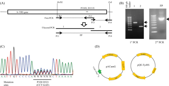
Fig. 1. Generation of a random mutation library of Sc YRS at P320 and D321 residues. (A) Strategy for library construction by PCR. P12 primer contains randomly degenerated sequences encoding P320 and D321 residues. (B) Agarose gel electrophoresis of PCR products. Both the first (left) and the second (right) PCR products migrate at the expected position. (C) Electropherogram of nucleotide sequence encompassing mutation sites from the final PCR product indicates randomized nucleotide sequence encoding P320 and D321 residues. (D) Diagram of plasmids used in this experiments.
YRS, we performed PCRs composed of 2 steps. As shown in Fig. 1A, we carried out the first round PCR with degenerative primers encoding P320X and D321X amino acid residues.
After combining purified PCR products of the first round PCR at equimolar ratio, the second round PCR was performed with P11 and P14 primers that contain BstX1 and PstI restriction sites, respectively. The primer sequences used in PCR were as follows; P11; 5'-AGATGTTGATTGCCAATTTGGTGG-3', P12; 5'-GATAGCGTCAGCAACAATTTTTAGNNNNNN TGGGGACAATTTTTC-3', P13; 5'-CTAAAAATTGGTGT TGCTGACGCTATC-3', P14; 5'-AAAAACTGCAGTTACA ATTTGGTTTCCTCTAG-3'. After 30 cycles of PCR, each product was analyzed by electrophoresis on 1% agarose gel.
The final PCR product was purified and cut with BstX1 & Pst1 restriction enzymes and subcloned into pQE-YRS plasmid at the same restriction sites. The purified final PCR product was also directly sequenced to verify the randomized mutation.
To measure the library titer, 1 μl of ligation reaction was transformed to DH10B competent cells (Invitrogen) via electro- poration (2.0 kv, 200 Ω, 25 μF), rescued with SOC media for 40 min, and spread (100 μl) on LB plates containing 100 μg/ml ampicillin. After overnight culture, library titer was measured
by counting the growing colonies (CFU), and the library size was estimated at 5.5 × 10
3CFU/μl.
Library screening
For library screening, 2 μl of library construct was trans- formed into DH10B competent cells containing pACamG by electroporation. The transformants were allowed to grow on LB plates containing 100 μg/ml ampicillin, 25 μg/ml tetracycline, and different concentration of chloramphenicol such as 100, 200, and 300 μg/ml for 16 h at 37°C.
Eight clones were isolated from the first screen on LB plates
containing 300 μg/ml of chloramphenicol and further analyzed
for their chloramphenicol resistance. Same amount of seed
culture of each clone was inoculated into LB broth medium and
GMML medium containing 100 μg/ml ampicillin, 25 μg/ml
tetracycline, and increasing concentrations of chloramphenicol
(0~1,000 μg/ml). Growth rate was determined by measuring
optical density of the bacterial cultures at 600 nm every 6 h. The
clone containing pQE-YRS which expresses wild-type YRS
(wt-YRS) was also tested as a control. Finally, two positive
clones (mYRS-3 and mYRS-5) out of eight clones have been
selected and their YRS DNA sequences were read in both
directions to reveal amino acid substitution.
β-galactosidase assay for in vivo amber suppression activity
E. coli DH10B (Tn:lacZam), which contains amber-mutated lacZ gene in the genomic DNA (Kim et al., 2009), was cotrans- formed with pACamG and plasmid expressing wt-YRS, mYRS-3, or mYRS-5. The transformant of each mutant was cultured overnight in 1 ml LB broth media containing 100 μg/ml ampi- cillin and 25 μg/ml tetracycline. Next day, same volume of each seed culture (1:50) was inoculated into 2 ml of fresh LB broth media containing 100 μg/ml ampicillin and 25 μg/ml tetracycline.
After 4 h culture, 0.5 mM IPTG was added to the culture to induce YRS expression for 4 h. Bacterial culture (1.5 ml) was harvested and washed once with Z buffer (60 mM Na
2HPO
4, 40 mM NaH
2PO
4, 10 mM KCl, 1 mM MgSO
4, 50 mM 2- mercaptoethanol, pH7.0). Cell pellet was resuspended in 50 μl of lysis buffer (1 mg/ml lysozyme, 30 mM Tris-Cl pH8.0, 1 mM EDTA, 20% Sucrose) and incubated for 4 min at room temperature. After adding 100 μl of Z buffer, 15 μl of 0.1%
SDS, and 30 μl of chloroform to the cell resuspension, cells were broken by vortexing for 10 sec. After centrifugation at 12,000 rpm for 10 min, the aqueous layer was taken for β- galactosidase assay. Enzyme reaction mixture consisting of 100 μl of cell extract, 200 μl of Z buffer, and 30 μl of 5 mM 4- methylumbelliferyl-β-D-galactopyranoside (MUG) was incubated for 30 min at 37°C. Reaction was stopped by adding 500 μl of 1 M Na
2CO
3and fluorescence was measured at 360 nm excitation and 440 nm emission. In addition, protein quantitation was carried out with the same cell extracts by the Bradford method (Bio-Rad protein assay) and the protein concentration of each cell extract was used for normalization of β-galactosidase activity.
Recombinant YRS purification
To test in vitro aminoacylation activity of mutant YRS, pQE- YRS expressing either wt-YRS or mYRS-3 was transformed into E. coli M15 strain. Each transformant was cultured in 200 ml LB containing 100 μg/ml ampicillin and protein expression was induced by the treatment of 1 mM IPTG for 4 h. Since the recombinant YRS proteins were produced with an N-terminal 6xHis tag, they were purified by the immobilized metal ion affinity chromatography of HiPur cobalt resin (ThermoFisher
Scientific) at 4°C according to manufacturer’s protocol. Briefly, instead of phosphate buffer, Tris-based buffer was used for protein purification to prevent contamination of inorganic phosphate in the in vitro aminoacylation reaction. The compositions of buffers were as follows; lysis buffer (50 mM Tris-HCl, 300 mM NaCl, and 10 mM 2-mercaptoethanol, pH 7.4), washing buffer (50 mM Tris-HCl, 300 mM NaCl, and 10 mM imidazole, pH 7.4), and elution buffer (50 mM Tris-HCl, 300 mM NaCl, and 150 mM imidazole, pH 7.4). Eluted protein was dialyzed in dialysis buffer (50 mM Tris-HCl and 150 mM NaCl, pH 7.4) with cut-off molecular size of 8 kDa. After dialysis, protein concentration was measured by the Bradford method (Bio-Rad protein assay).
In vitro aminoacylation assay
In vitro aminoacylation assay was performed in aminoacylation buffer (30 mM HEPES, 140 mM NaCl, 30 mM KCl, and 40 mM MgCl
2), 1 mM tyrosine, 1 mM DTT, 200 μM ATP, and 200 U/ml inorganic pyrophosphataes (PPiase), 10 μg/ml of wt-YRS or mYRS-3, and 0~25.8 μM of fMam tRNA
CUA. The aminoacylation reactions (30 μl total reaction volume) were performed in clear, flat-bottom 96-well plates and incubated for 30 min at 37°C. Reactions were stopped by adding 20 μl of 0.3 M EDTA and 100 μl of malachite green reagent (Echelon Biosciences). After incubating for 30 min at room temperature for color development, absorbance was measured at 620 nm by using Infinite M200 Pro Spectrophotometer (Nano-Quent).
Inorganic phosphate production was calculated from the standard curve produced according to manufacturer’s protocol.
Statistical analysis
The experiments were repeated at least more than 3 time to obtain reproducibility, the mean, and standard deviation. For statistical analysis, Student’s unpaired t-test was performed.
Results and Discussion
Generation of mutant library of Sc YRS randomizing the amino acid residues at 320 and 321
For directed evolution of amber suppression activity of the
orthogonal pair of Sc YRS and fMam tRNA
CUA, we generated
(A)
(B)
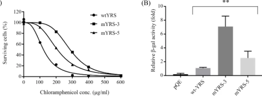
Fig. 2. Amber suppression activity of mutant YRS clones. (A) Chloramphenicol-resistant growth derived by amber suppression of CATam gene. Bacterial clones containing pACamG plus either wild type YRS or mutant YRSs selected from library screening were cultured in GMML medium containing increasing amounts of chloramphenicol up to 600 μg/ml. After 36 h culture, optical density of the bacterial culture was measured at 600 nm. Relative growth rate was presented as percentile compared to the bacterial growth without chloramphenicol. (B) Comparison of in vivo β-galactosidase activity produced by YRS-mediated amber suppression in DH10B (Tn:lacZam) strain. The plasmids expressing the indicated YRS were co-transformed with pACamG into E. coli DH10B (Tn:lacZam) and their amber suppression activities were analyzed by β-galactosidase assay. **P < 0.01.
Table 1. Amino acid sequences of mutant YRS and their IC50 values of chloramphenicol
Residue numbera
Cm IC50b (µg/ml)
320 321
wt YRS Pro Asp 122
mYRS-3 Ala Ala 286
mYRS-5 Ala His 207
aNumber of amino acid residues of Sc TyrRS
bChloramphenicol concentration showing 50% of growth inhibition in GMML medium
a mutant library of YRS by randomizing the nucleotide sequence encoding two amino acid residues, P320 and D321, which are known to be involved in recognition of the first anticodon base G34 in X-ray crystallography (Tsunoda et al., 2007). A degene- rated primer was used to randomize the mutation sites and two rounds of PCR were carried out to get the final PCR product encoding C-terminal half of YRS. The randomized mutations were confirmed by sequencing the final PCR products. The mutant library was generated by replacing the C-terminal half of Sc YRS gene with the final PCR products by using restriction sites BstXI and PstI (Fig. 1A). The experimental library titer was calculated as 5.5 × 10
3CFU/μl, which is sufficient enough considering the theoretical maximum diversity of randomizing 6 base pairs, i.e. 4
6≒ 4.1 × 10
3.
Library screening and characterization of selected clones Using chloramphenicol resistance derived by amber supp- ression of CAT amber gene in pACamG, the mutant library was screened on LB plates containing increasing amounts of chlo- ramphenicol. Eight clones were selected from the first screening on LB plates containing 300 μg/ml of chloramphenicol. After additional analysis for their chloramphenicol-resistant growth, two positive clones (mYRS-3 and mYRS-5) were finally chosen for further characterization of their amber suppression activity.
Assay for the amber suppression-mediated chloramphenicol resistance in the minimal medium of GMML showed two mutant clones had strong resistance at the higher concentrations of
chloramphenicol than wt-YRS (Fig. 2A), and their IC
50values of chloramphenicol were 122, 286, and 207 μg/ml for wt-YRS, mYRS-3, and mYRS-5, respectively (Table 1). For additional in vivo amber suppression assay, we have used a reporter strain E. coli DH10B (Tn:lacZam) which contains a single copy of amber-mutated lacZ gene in the genomic DNA (Kim et al., 2009). After co-transforming pACamG and YRS expression plasmids to the strain, β-galactosidase activity was measured from the cell extracts. Compared to wt-YRS, mYRS-3, and mYRS-5 showed approximately 6.5-fold and 2.5-fold higher activities, respectively (Fig. 2B).
DNA sequencing of the mutant YRSs revealed Pro320 residue
has been changed to alanine in both mutants and Asp321
residue to alanine and histidine in mYRS-3 and mYRS-5,
respectively (Table 1). P320 residue was known to involve
hydrophobic interaction with the base of G34 of tRNA
Tyrand
also participate in a hydrophobic patch composed of Phe254,
Pro257, Pro319, and Pro320 where accommodates the second and third anticodon bases U35 and A36 in the crystal structure of the ternary complex of Sc YRS associated with Tyr-AMP analog and tRNA
Tyr(Tsunoda et al., 2007). Ala substitution at Pro320 residue may maintain the hydrophobic interaction with the base of C34 of fMam tRNA
CUAand, in addition, confer more flexible and spacious structure for the placement of the other anticodon bases than Pro. On the other hand, Asp321 was known to interacts specifically with N1 and O6 atoms of G34 through bifurcated hydrogen bonds, thereby playing as a key residue for specific recognition of G34 (Tsunoda et al., 2007).
Similarly, crystal structure of Mj YRS complexed with its cognate tRNA and L-tyrosine has shown that Asp286, the residue homologous to Asp321 of Sc YRS, interacts with N1 and N2 atoms of G34 by two hydrogen bonds (Kobayashi et al., 2003). In addition, when Asp286 of Mj YRS was replaced with other larger amino acids such as glutamine, tyrosine, and arginine residues, the aminoacylation activity of Mj YRS mutants for the amber suppressor tRNA which has C34 in place of G34 significantly increased. D286R mutation, which showed the highest activity, exhibited an 8-fold higher activity than the wild type Mj YRS (Kobayashi et al., 2003). Those results were well-matched with the fact that cytosine base at position 34 of the amber suppressor tRNA is smaller than guanine. However, inconsistent with Mj YRS, our Sc YRS mutants showed sub- stitution of Asp321 with alanine or histidine which are not larger amino acids than aspartate. Moreover, D321A mutant which has smaller side chain than histidine exhibited higher in vivo amber suppression activity than D321H mutant (Fig. 2).
Although the overall anticodon recognition mode of archaeal Mj YRS and eukaryotic Sc YRS is quite similar, there is a difference in conformation surrounding G34. The hydrophobic interaction between G34 and Pro320 in Sc YRS is replaced by the stacking interaction between G34 and His283 in Mj YRS. In addition, directed evolution of several UAA-incorporating aaRSs derived from Mj YRS for improving their binding affinity to the amber suppressor tRNA revealed that substitutions of amino acids interacting with G34 are different among aaRSs, indicating that the conformation of anticodon recognition pocket may be affected by the structure of the other part of aaRS (Wang et al., 2015). Thus, D321A or D321H mutations in the Sc YRS impli- cate delicate structural differences in the binding pocket of
anticodon as well as the other part of the protein.
Previously, we had generated Sc YRS(D321R) mutant by site-directed mutagenesis and tested in vivo amber suppression activity of the mutant. In contrast to our expectation, cells transformed with plasmid expressing D321R mutant showed very low level of CAT amber suppression activity (Supplementary data Fig. S1). The low level of amber suppression activity appears to be involved in low copy numbers of D321R mutant expression plasmid because the activity of β-lactamase (the antibiotic resistant marker of D321R mutant expression plasmid) of the same cell extract was also very low. In addition, the expression level of Sc YRS(D321R) was also at least 10-fold less than that of wild-type YRS when judged from SDS-PAGE analysis in the purification process of recombinant wt-YRS and YRS(D321R) proteins from the same amount of cell culture (Supplementary data Fig. S2). Taken together, these results suggest that Sc YRS(D321R) protein might be toxic to the cells or very labile to be degraded inside cells. Moreover, when we analyzed tyrosylation kinetics of YRS and its D321R mutant to fMam tRNA
CUAwith the purified recombinant proteins, K
mvalue of D321R mutant was decreased by approximately 2-fold, indicating that D321R mutation increases binding affinity of YRS to fMam tRNA
CUA. D321R mutation increases k
cat/K
mby approximately 2-fold through lowering the K
mvalue (Supple- mentary data Fig. S3). The corresponding D286R mutation of Mj YRS increased tyrosylation activity to Mj tRNA
Tyr(G34C) by approximately 65-fold of k
cat/K
mby lowering its K
mvalue (Kobayashi et al., 2003). Thus, these results demonstrate D321R mutation of Sc YRS is not essential as much as D286R mutation of Mj YRS in recognition of C34 in the amber suppressor tRNA, and also support the idea that the delicate structure of anticodon binding pocket of Sc YRS may be different from that of Mj YRS.
Tyrosylation kinetics of wt-YRS and mYRS-3 to fMam tRNA
CUATo compare tyrosylation activity of wt-YRS and the mutant,
wt-YRS and mYRS-3 proteins having an N-terminal 6xHis tag
were expressed in E. coli M15 strain and purified by immo-
bilized metal affinity chromatography. SDS-PAGE analysis of
the purified recombinant proteins showed high quality of puri-
fication without any significant contaminant, and production
(A) (B)
Tyrosylation kinetics of Sc YRS and mYRS-3 to fMam tRNACUA
kcat (s-1) Km (µM) kcat/Km
YRS 0.197 1.041 0.19
mYRS-3 0.188 0.135 1.39
Fig. 3. Tyrosylation kinetics of wild type YRS and mYRS-3 to fMam tRNACUA. (A) SDS-PAGE analysis of purified recombinant wt-YRS and mYRS-3 proteins from the same culture volume. (B) Tyrosylation kinetics of wt-YRS and mYRS-3 to the amber suppressor tRNA, fMam tRNACUA, were determined from the Lineweaver-Burk reciprocal plot.