Differential Sialic Acid Content and Hemoglobin-binding Activity of Precursor Prohaptoglobin and Mature Haptoglobin
Joo-Hyun Lee, Mi-Kyung Oh and In-Sook Kim*
Department of Medical Lifescience, College of Medicine, The Catholic University of Korea, Seoul 06591, Korea Received April 7, 2017 /Revised May 10, 2017 /Accepted May 11, 2017
Mature haptoglobin (Hp) is a plasma glycoprotein and acts as an antioxidant by scavenging cell-free hemoglobin (Hb). Prohaptoglobin (proHp) is an unprocessed Hp precursor which is present a little in circulation. However, the biological function of proHp remains unknown. To investigate the struc- tural and functional differences between proHp and Hp, we prepared recombinant proHp isoforms and compared their sialic acid content and Hb-binding capacity with those of mature isoforms. When proHp samples were analyzed by Western blot under non-reducing conditions, proHp1 was detected as one band of approximately 130 kDa and proHp2 as multiple bands >200 kDa, in the manner of mature Hp1-1 and Hp2-2, respectively. On the native polyacrylamide gel under non-reducing and non-denaturing conditions, both proHp isoforms migrated more slowly than their mature Hp counterparts. In addition, the lectin-based ELISA assay demonstrated that the content of sialic acid in proHp1 and proHp2 was much less than in Hp1-1 and Hp2-2. The Hb-binding capacity of proHp was also lower than those of mature Hp. These findings indicate that proHp and Hp are similar in the size and polymerization pattern, but different in sialic acid content and Hb-binding activity. It sug- gests precursor proHp may exert different functions in circulation than does mature Hp.
Key words : Antioxidant, haptoglobin, hemoglobin-binding, prohaptoglobin, sialic acid
*Corresponding author
*Tel : +82-2-2258-7240, Fax : +82-2-2258-7240
*E-mail : [email protected]
This is an Open-Access article distributed under the terms of the Creative Commons Attribution Non-Commercial License (http://creativecommons.org/licenses/by-nc/3.0) which permits unrestricted non-commercial use, distribution, and reproduction in any medium, provided the original work is properly cited.
Journal of Life Science 2017 Vol. 27. No. 6. 632~639 DOI : https://doi.org/10.5352/JLS.2017.27.6.632
Introduction
Haptoglobin (Hp) is a plasma glycoprotein that is synthe- sized in the liver and secreted into the bloodstream. Hp lev- els in plasma increase in response to inflammatory reactions, immune activation, and malignant tumors and decrease in response to chronic liver disease and intravascular hemol- ysis [10, 13, 26]. Accordingly, plasma concentration of Hp has been considered a clinical indicator of inflammatory and hemolytic diseases.
In humans, two major Hp alleles, Hp1 and Hp2, have been identified and are expressed as three Hp phenotypes, name- ly, Hp1-1 (Hp1/Hp1), Hp2-2 (Hp2/Hp2), and Hp2-1 (Hp1/
Hp2) [5]. Hp proteins consist of two polypeptides, the a-chain and the b-chain, which form either a heterotetramer or polymer. The b-chain is identical in the three Hp pheno- types, but the a-chain is expressed as two isoforms, specifi-
cally, a1 for the Hp1 allele and a2 for the Hp2 allele. The a2 polypeptide has an additional cysteine residue, which fa- cilitates polymer formation through disulfide bridge con- nections. Therefore, Hp2-2 exists in a polymeric form [(α2β)n,
where n ≥ 3)], and Hp1-1 exists as a tetramer [(a1b)2]. The heterozygote Hp2-1 also forms polymers [(a1b)2+(α2β)n,
where n ≥ 0] [12].
Hp contains four N-glycosylation sites in the β-chain at which several complex glycan structures have been identi- fied [29]. Biantennary (~70%) and triantennary glycans (30%) are the major structures, and tetraantennary glycans are ob- served less frequently (1-2%). In addition, Hp N-glycans have mono-, di-, or tri-sialylated structures via terminal sialylation. The N-acetylglucosamine-linked fucose moiety has been also found in Hp glycans [20]. Recent glyco- mics-based research has indicated that the alterations of Hp glycosylation, particularly increased fucosylation and sialy- lation, are associated with several human cancers [2, 16, 20].
Therefore, the glycosylation patterns of Hp are of interest for clinical applications as cancer biomarkers [20, 29].
The characteristic function of Hp is high-affinity binding to cell-free hemoglobin (Hb). When Hb is released into the plasma from ruptured erythrocytes during trauma or hemo- lytic diseases, Hp immediately noncovalently combines with free Hb to form an irreversible Hp-Hb complex. The interact-
A
B
Fig. 1. ProHp1 and proHp2 expressed in human MDA-MB 231 breast cancer cells. (A) Schematic structures of proHp1, proHp2, Hp1-1, and Hp2-2. ProHp1 is a product of the Hp1 gene and processed to mature Hp1-1 by cleavage at the indicated site (▲). ProHp2 is a product of the Hp2 gene and processed to Hp2-2 in the same manner. The amino acid numbering begins at valine without the signal peptide of 18 amino acids. ★ = N-glycosylation site (B) MDA-MB 231 cells were transfected with human Hp1 or Hp2 cDNA and cultured in serum-free medium. After 48 hr of incubation, the CM was collected.
Hp1-transfected MDA-MB 231 CM (30 μl) and purified Hp1-1 (Sigma, 90 ng) were electrophoresed on a 12% SDS-poly- acrylamide gel under reducing conditions. After blotting, the Hp isoforms were detected using antibodies against Hp and Hp α-chain (left). Hp2-transfected cells CM and purified Hp2-2 (Sigma) were analyzed using the same procedure (right).
All experiments were performed independently three times, and representative results are shown.
ing regions of Hp are in the β-chain, and each Hp β-chain stoichiometrically binds with one Hb dimer (αβ) [1, 14].
Because the Hp-Hb complex is rapidly scavenged via the CD163 receptors of activated monocytes and macrophages [11], the primary function of Hp has been thought to be pro- tecting tissues from free-Hb mediated oxidative damage [12, 30].
In hepatocytes, the Hp gene is expressed in a precursor form, prohaptoglobin (proHp), in which the α- and β-sub- unit are linked within a single polypeptide [8, 15] (Fig. 1A).
In the endoplasmic reticulum, proHp is site-specifically cleaved by a C1r-like protein (C1r-LP), which contains the serine protease domain of complement C1r [27]. The cleaved products, the α- and the β-chain, are held together by disul- fide bonds and form mature Hp [(αβ)n, where n = 2 in Hp1-1 and n ≥ 3 in Hp2-2]. After being fully processed within the cell, the mature form of Hp is released into circulation
[8]. However, our previous study showed the presence of proHp in serum from patients with liver cancer [15]. The significance of the secretion of unprocessed proHp and its biological function remain unknown. To investigate the structural and functional differences between proHp and mature Hp, we examined the sialic acid content and Hb- binding capacity of the two forms.
Materials and Methods
Human serum samples
Serum samples from healthy individuals (Hp1-1 type, 4 males and 1 female aged 45~57 years; Hp2-2 type, 3 males and 2 females aged 40~58 years) were provided by the Seoul St. Mary’s Biobank (Seoul, Korea). All procedures were ap- proved by the Institutional Review Board of the Catholic University of Korea (Seoul, Korea).
Cell culture and conditioned medium collection MDA-MB 231 human breast cancer cells, A549 human lung carcinoma cells and MH7A synoviocyte cells were maintained in RPMI medium (WelGENE, Daegu, Korea) supplemented with 10% fetal bovine serum (FBS; Gibco Laboratories, Gaithersburg, MD, USA). COS-7 kidney cells were maintained in DMEM medium (WelGENE) supple- mented with 10% FBS. The cells were transfected with hu- man Hp1 or Hp2 open reading frame (ORF) cDNA, which were cloned into the pCR3.1 vector, using Lipofectamine™ 2000 (Invitrogen, Carlsbad, CA, USA) and cultured in se- rum-free medium. After 48 hr of incubation, the conditioned medium (CM) was harvested and used as the source of proHp1 or proHp2 [15].
Western blot analysis
Human serum and the CM samples were electrophoresed on 12% SDS-polyacrylamide gels under reducing conditions or, to detect the Hp-Hb complex, on 7.5% native poly- acrylamide gels. The proteins were then transferred to nitro- cellulose membranes and blocked with 5% skim milk. The immunoreactions were performed with rabbit polyclonal an- ti-human Hp antibody (Sigma-Aldrich, Saint Louis, MO, USA) and polyclonal anti-human Hp α-chain antibody [17].
The immunoreactive proteins were detected with an en- hanced chemiluminescence detection kit (Amersham Biosci- ences, Buckinghamshire, UK) using an LAS-3000 lumines- cent image analysis system (Fujifilm, Tokyo, Japan).
Determination of the concentrations of Hp isoforms The concentrations of proHp1/2 in CM and Hp1-1/2-2 in serum were measured using an enzyme-linked im- munosorbent assay (ELISA) with purified Hp2-2 as a stand- ard [15]. Hp1-1 and Hp2-2 samples, which were purified from normal human plasma and lyophilized, were pur- chased from Sigma-Aldrich and reconstituted with de- ionized water (1 mg/ml) according to the manufacturer’s directions.
Sialic acid assay using reverse lectin-based ELISA The sialic acids in proHp and mature Hp were detected using a Sambucus nigra agglutinin (SNA) lectin-based ELISA assay [28]. Briefly, each well of a 96-well ELISA plate was coated with 100 μl of SNA (10 μg/ml; Sigma-Aldrich) and incubated at 37℃ for 2 hr. After washing with PBST (0.1%
Tween 20 in PBS), the wells were blocked with 3% BSA in
PBST for 1 hr at room temperature. CM samples containing either proHp1 or proHp2 (0.02 μg/100 μl or 0.05 μg/100 μl, respectively) were added to each well. Purified Hp1-1 and Hp2-2 (Sigma-Aldrich) were diluted with control CM ob- tained from vehicle-transfected cells and applied to the wells in the same doses as the corresponding proHp samples.
After the plate was incubated for 1 hr at room temperature, the lectin-bound Hp isoforms were reacted with anti-human Hp antibody conjugated to horseradish peroxidase (Abcam, Cambridge, UK). The color reaction was developed with a tetramethylbenzidine (TMB) substrate solution (GenDEPOT, Barker, TX, USA) and stopped by adding 2 M H2SO4. The absorbance was measured at 450 nm using a Victor3 ELISA reader (PerkinElmer, Boston, MA, USA)
Analysis of the Hb-binding capacity of proHp and mature Hp
Each well of a 96-well ELISA plate was coated with hu- man HbA0 (5 μg in 100 μl PBS; Sigma-Aldrich), incubated at 4℃ overnight, washed with PBS containing 0.1% Tween 20, and blocked with 3% skim milk for 2 hr at room temperature. 100 μl samples of either proHp or Hp (0.25 μg/
ml or 0.5 μg/ml, respectively) were added to the wells and incubated for 2 hr at room temperature. The captured Hp isoforms were measured using the same procedure de- scribed for the lectin-based ELISA.
Statistical analysis
Statistical analysis was assessed differences between the results by the unpaired Student's t-test. Data were analysed using graphpad prism (version 5.04) software (GraphPad software, Inc., San Diego, CA). p-value<0.01 was considered significant.
Results
Preparation of human proHp1 and proHp2 Human proHp isoforms were expressed in various cell lines such as MDA-MB 231 human breast cancer cells, A549 lung cells, MH7A synoviocyte cells, and COS-7 kidney cells.
When Hp1 or Hp2 cDNA was transfected into MDA-MB 231 human breast cancer cells and the CM was analyzed by Western blot under reducing conditions, single polypeptides of approximately 57 or 68 kDa, respectively, reacted with Hp antibodies (Fig. 1B). The same sized proHp isoforms were also detected in the CM from A549 lung cells, MH7A
A
B C
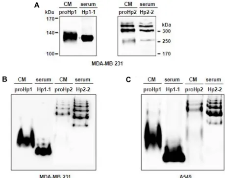
Fig. 2. ProHp and mature Hp have the same polymerization, but different pIs. (A) Normal human sera (Hp1-1 type or Hp2-2 type) and CM samples from MDA-MB 231 cells were electrophoresed on a 7.5% SDS-polyacrylamide gel under non-reducing conditions (i.e., without β-mercaptoethanol and heating). The Hp isoforms were then detected with Western blotting using anti-human Hp antibody. (B) Human sera (Hp1-1 type or Hp2-2 type) and CM samples from MDA-MB 231 cells were analyzed by Western blotting performed under non-denaturing as well as non-reducing conditions, in which the samples were electrophoresed on a 7.5% polyacrylamide gel without SDS and β-mercaptoethanol. (C) Human sera and CM samples from A549 cells were analyzed using the same procedure. Pooled human sera (5 sera with Hp1-1 type or 5 sera with Hp2-2 type) were used, and all CM samples were used after dialysis against PBS. The figures shown represent a typical result obtained from three independent experiments.
synoviocyte cells, and COS-7 kidney cells transfected with the Hp genes (data not shown). However, Hp1-1 purified from normal human serum had two separate chains, the α
1-chain (12 kDa) and the β-chain (45 kDa). Hp2-2 purified from 2-2 phenotype serum contained a 21-kDa α2-chain and the 45-kDa β-chain (Fig. 1B). These findings indicate that the cells secrete unprocessed proHp, but not mature Hp.
Therefore, in this study, the CM of Hp gene-transfected cells and normal human serum were used as the source of proHp and mature Hp, respectively.
Structural differences between proHp and mature Hp
Because Hp1-1 exists as a hetero tetramer of (α1β)2 and Hp2-2 as a polymer of (α2β)n [12], we tested whether proHp1 and proHp2 have the same polymerization as the mature Hp isoforms. After SDS-polyacrylamide gel electrophoresis under non-reducing conditions, the proHp samples were an- alyzed by Western blot. As shown in Fig. 2A, proHp1 was detected as one band of approximately 130 kDa and proHp2
as multiple bands >200 kDa, in the manner of Hp1-1 and Hp2-2, respectively. This indicates that proHp1 is dimeric in the form of (α1-β)2, and proHp2 is polymeric in the form of (α2-β)n. On the native polyacrylamide gel under non-re- ducing and non-denaturing conditions, however, both proHp isoforms migrated more slowly than their mature Hp counterparts (Fig. 2B, Fig. 2C). These findings suggest that proHp is similar in size and polymerization pattern to ma- ture Hp, but its isoelectric point (pI) is higher than that of mature Hp.
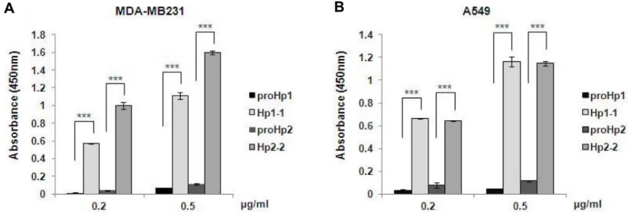
Lower sialic acid content in proHp
The pI differences of the proteins may be due to a differ- ing content of charged sugars. Since Hp has terminally sialy- lated N-glycans [20], we compared the content of sialic acid within proHp and mature Hp. In an ELISA assay using the sialic acid-binding SNA lectin, the content of sialic acid in proHp1 and proHp2 was much less than in purified Hp1-1 and Hp2-2 (Fig. 3A, Fig. 3B). Because the Hp1-1 and Hp2-2 was tested in the presence of control CM prepared from vec-
A B
Fig. 3. Low sialic acid content in proHp1 and proHp2. (A) MDA-MB 231 CM (100 μl) containing proHp1 or proHp2 (0.2 μg/ml or 0.5 μg/ml, respectively) was added to SNA-coated wells in an ELISA plate. Purified Hp1-1 or Hp2-2 (Sigma) was diluted with control CM obtained from vehicle-transfected cells and used in the same dose as the corresponding proHp samples.
The lectin-bound Hp isoforms were probed with anti-human Hp antibody conjugated to horseradish peroxidase. After reacting with TMB, the absorbance was measured at 450 nm. The results are expressed as the mean ± S.D. of triplicate experiments.
***p<0.001 vs. Hp1-1 or Hp 2-2. (B) Analysis of the CMs from A549 cell cultures was carried out using the same method.
All experiments were performed independently at least twice, and the representative data are shown.
tor-transfected cells, the low sialic acid levels observed for proHp are not due to inhibitory effects of other sialylated proteins in the CM.
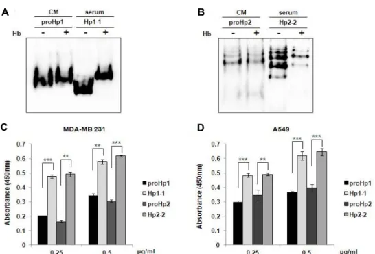
Hb-binding activity of proHp and mature Hp Hp-Hb complex formation is an important part of the function of mature Hp [13]. To investigate the functional dif- ferences between proHp and mature Hp, we examined whether proHp binds to free Hb. When proHp1 and proHp2 CM samples, either without or with an excess of human Hb, were analyzed by Western blot under native conditions, proHp-Hb complexes were not observed in Hb-supple- mented samples. However, for Hp1-1 and Hp2-2 in serum, we detected bands with higher molecular mass that corre- spond to the expected bands of the Hp-Hb complexes (Fig.
4A, Fig. 4B).
For further confirmation, modified ELISA was performed in an Hb-coated 96-well plate. As shown in Fig. 4C and Fig.
4D, the Hb-binding capacities of proHp1 and proHp2 were lower than those of mature Hp1-1 and Hp2-2, respectively.
Therefore, it is likely that the Hb-binding affinity of the pre- cursor proHp is weaker compared to that of mature Hp.
Discussion
Some proteins are synthesized and secreted as inactive precursors. These precursors are site-specifically cleaved to yield the active forms of the proteins, which have lower mo-
lecular weights than the precursors. Hp protein is also syn- thesized as the precursor proHp and processed to mature Hp by site-specific cleavage within cells [8]. Interestingly, proHp and mature Hp have the same amino acid composi- tion and sequences but with different subunit connections (Fig. 1A). Although mature Hp is the major form in serum, a small fraction of proHp has been detected as well [15].
However, it is unclear whether proHp is only an inactive precursor or a unique protein that has different functions from mature Hp. Our findings in this study indicate that proHp and Hp are similar in size and polymerization pat- tern, but different in sialic acid content and Hb-binding capacity. Due to the α-β junction site being uncleaved in proHp, we think that proHp may be less flexible and that this rigidity may affect its sialylation and Hb binding.
Although the biological function of proHp is still unclear, modulation of tight-junction permeability, as well as activa- tion of B-cell survival and differentiation, have been sug- gested as functions of proHp [9, 25]. In addition, we pre- viously demonstrated that recombinant proHp upregulates vascular endothelial growth factor (VEGF) expression in en- dothelial cells and promotes angiogenesis via VEGF re- ceptor-2 signaling [15]. The in vitro angiogenic effect was shown at a low concentration (0.2 μg/ml) compared to the active dose of mature Hp (> 50 μg/ml) [6]. Taken together with our present findings, it seems likely that non-processed proHp has unique functions independent of mature Hp, rather than exclusively existing in circulation as an inactive
A B
C D
Fig. 4. Comparison of the Hb-binding activity of proHp and mature Hp. (A) ProHp1 CM (30 μl) and Hp1-1 serum (0.1 μl) were mixed with or without an excess of human Hb (1 μg). After incubation for 30 min at room temperature, the samples were electrophoresed on a native 7.5% polyacrylamide gel. The Hp isoforms and their complexes with Hb were detected with Western blots using antibody against Hp. (B) ProHp2 CM and Hp2-2 serum were mixed with or without an excess of human Hb, and analyzed using the same procedure. (C) ProHp1/2 CM samples (MDA-MB 231) and purified Hp1-1/2-2 were added to the Hb-coated wells of an ELISA plate. The captured Hp isoforms were probed with horseradish peroxidase-conjugated anti-Hp antibody. After reacting with TMB, the absorbance was measured at 450 nm. The results are expressed as mean
± S.D. of triplicate experiments. **p<0.01, ***p<0.001 vs. Hp1-1 or Hp 2-2. (D) The same experiments were performed with A549 CM. All experiments were performed independently three times, and the representative data are shown.
Hp precursor.
Mature Hp rapidly forms a stable Hp-Hb complex, and this complex is cleared from circulation via the CD163 re- ceptor on macrophages. Endocytosis via the CD163 receptor induces expression of heme oxygenase-1 and interleukin 10, which participate in anti-inflammation responses [18, 21].
However, Hp has conflictingly been reported as both an an- ti-inflammatory regulator and a pro-inflammatory mediator.
Several studies have shown the inhibitory effects of Hp on the production of pro-inflammatory cytokines in activated lymphocytes and monocytes [3, 4], and Hp knockout mice are more sensitive to lipopolysaccharide-induced inflamma- tory effects [4]. Autoimmune inflammation was also en- hanced in Hp-deficient mice [7]. On the contrary, recent studies demonstrated that Hp mediates MyD88-dependent inflammation and activates innate immunity in a mouse model of skin or heart transplantation, which leads to acute transplant rejection [23, 24]. The underlying mechanisms of these contradictory Hp functions are unclear. Because there are diverse Hp isoforms or variants in vivo, such as mature
Hp, proHp, differently glycosylated Hp, and plasmin- cleaved Hp fragments [17], it is thought that Hp may exert different functions in various cellular environments. Our current findings suggest that, based on its Hb-binding prop- erties, proHp has insufficient activity to act as an antioxidant or anti-inflammatory agent. However, it remains unknown if proHp participates in pro-inflammatory reactions.
In hemolytic diseases, such as sickle cell disease, the con- centration of free Hb in plasma greatly increases, which gen- erates reactive oxygen species, depletes nitrogen oxide, and induces endothelial expression of adhesion molecules. These responses are associated with pulmonary hypertension, is- chemic stroke, and nephropathy. To protect tissues from the harmful effects of Hb, intravenous Hp infusion therapy has been proposed [19, 22], which is based on the ability of Hp to rapidly remove free Hb from circulation by forming an Hp-Hb complex [11]. For therapeutic applications, the prepa- ration of large amounts of recombinant Hp protein is required. However, our results show that even in human cell lines, recombinant Hp is synthesized as a non-processed
proHp isoform, which has low Hb-binding activity. There- fore, to use recombinant Hp as a therapy or for functional studies, it must be determined whether the prepared protein is proHp or mature Hp.
Acknowledgements
The human bio-specimen (normal serum) used in this study was provided by the Seoul St. Mary’s Biobank. This work was supported by Mid-career Researcher Program through NRF grant funded by the MEST (2016R1A2B 1009425).
References
1. Andersen, C. B., Torvund-Jensen, M., Nielsen, M. J., de Oliveira, C. L., Hersleth, H. P., Andersen, N. H., Pedersen, J. S., Andersen, G. R. and Moestrup, S. K. 2012. Structure of the haptoglobin-haemoglobin complex. Nature 489, 456- 459.
2. Ang, I. L., Poon, T. C., Lai, P. B., Chan, A. T., Ngai, S. M., Hui, A. Y., Johnson, P. J. and Sung, J. J. 2006. Study of serum haptoglobin and its glycoforms in the diagnosis of hep- atocellular carcinoma: a glycoproteomic approach. J.
Proteome Res. 5, 2691-2700.
3. Arredouani, M., Matthijs, P., Van Hoeyveld, E., Kasran, A., Baumann, H., Ceuppens, J. L. and Stevens, E. 2003. Hapto- globin directly affects T cells and suppresses T helper cell type 2 cytokine release. Immunology 108, 144-151.
4. Arredouani, M. S., Kasran, A., Vanoirbeek, J. A., Berger, F.
G., Baumann, H. and Ceuppens, J. L. 2005. Haptoglobin dampens endotoxin-induced inflammatory effects both in vitro and in vivo. Immunology 114, 263-271.
5. Bowman, B. H. and Kurosky, A. 1982. Haptoglobin: the evo- lutionary product of duplication, unequal crossing over, and point mutation. Adv. Hum. Genet. 12, 189-261, 453-184.
6. Cid, M. C., Grant, D. S., Hoffman, G. S., Auerbach, R., Fauci, A. S. and Kleinman, H. K. 1993. Identification of haptoglo- bin as an angiogenic factor in sera from patients with sys- temic vasculitis. J. Clin. Invest. 91, 977-985.
7. Galicia, G., Maes, W., Verbinnen, B., Kasran, A., Bullens, D., Arredouani, M. and Ceuppens, J. L. 2009. Haptoglobin deficiency facilitates the development of autoimmune inflammation. Eur. J. Immunol. 39, 3404-3412.
8. Hanley, J. M., Haugen, T. H. and Heath, E. C. 1983.
Biosynthesis and processing of rat haptoglobin. J. Biol. Chem.
258, 7858-7869.
9. Huntoon, K. M., Russell, L., Tracy, E., Barbour, K. W., Li, Q., Shrikant, P. A., Berger, F. G., Garrett-Sinha, L. A. and Baumann, H. 2013. A unique form of haptoglobin produced by murine hematopoietic cells supports B-cell survival, dif- ferentiation and immune response. Mol. Immunol. 55, 345- 354.
10. Kormoczi, G. F., Saemann, M. D., Buchta, C., Peck- Radosavljevic, M., Mayr, W. R., Schwartz, D. W., Dunkler, D., Spitzauer, S. and Panzer, S. 2006. Influence of clinical factors on the haemolysis marker haptoglobin. Eur. J. Clin.
Invest. 36, 202-209.
11. Kristiansen, M., Graversen, J. H., Jacobsen, C., Sonne, O., Hoffman, H. J., Law, S. K. and Moestrup, S. K. 2001.
Identification of the haemoglobin scavenger receptor. Nature 409, 198-201.
12. Langlois, M. R. and Delanghe, J. R. 1996. Biological and clin- ical significance of haptoglobin polymorphism in humans.
Clin. Chem. 42, 1589-1600.
13. Levy, A. P., Asleh, R., Blum, S., Levy, N. S., Miller-Lotan, R., Kalet-Litman, S., Anbinder, Y., Lache, O., Nakhoul, F.
M., Asaf, R., Farbstein, D., Pollak, M., Soloveichik, Y. Z., Strauss, M., Alshiek, J., Livshits, A., Schwartz, A., Awad, H., Jad, K. and Goldenstein, H. 2010. Haptoglobin: basic and clinical aspects. Antioxid. Redox Signal. 12, 293-304.
14. Nagel, R. L. and Gibson, Q. H. 1971. The binding of hemo- globin to haptoglobin and its relation to subunit dissociation of hemoglobin. J. Biol. Chem. 246, 69-73.
15. Oh, M. K., Park, H. J., Lee, J. H., Bae, H. M. and Kim, I.
S. 2015. Single chain precursor prohaptoglobin promotes an- giogenesis by upregulating expression of vascular endothe- lial growth factor (VEGF) and VEGF receptor2. FEBS Lett.
589, 1009-1017.
16. Okuyama, N., Ide, Y., Nakano, M., Nakagawa, T., Yamana- ka, K., Moriwaki, K., Murata, K., Ohigashi, H., Yokoyama, S., Eguchi, H., Ishikawa, O., Ito, T., Kato, M., Kasahara, A., Kawano, S., Gu, J., Taniguchi, N. and Miyoshi, E. 2006.
Fucosylated haptoglobin is a novel marker for pancreatic cancer: a detailed analysis of the oligosaccharide structure and a possible mechanism for fucosylation. Int. J. Cancer 118, 2803-2808.
17. Park, H. J., Oh, M. K., Kim, N. H., Cho, M. L. and Kim, I. S. 2013. Identification of a specific haptoglobin C-terminal fragment in arthritic synovial fluid and its effect on inter- leukin-6 expression. Immunology 140, 133-141.
18. Philippidis, P., Mason, J. C., Evans, B. J., Nadra, I., Taylor, K. M., Haskard, D. O. and Landis, R. C. 2004. Hemoglobin scavenger receptor CD163 mediates interleukin-10 release and heme oxygenase-1 synthesis: antiinflammatory mono- cyte-macrophage responses in vitro, in resolving skin blisters in vivo, and after cardiopulmonary bypass surgery. Circ. Res.
94, 119-126.
19. Quimby, K. R., Hambleton, I. R. and Landis, R. C. 2015.
Intravenous infusion of haptoglobin for the prevention of adverse clinical outcome in Sickle Cell Disease. Med.
Hypotheses 85, 424-432.
20. Ratanasopa, K., Chakane, S., Ilyas, M., Nantasenamat, C.
and Bulow, L. 2013. Trapping of human hemoglobin by hap- toglobin: molecular mechanisms and clinical applications.
Antioxid. Redox Signal. 18, 2364-2374.
21. Schaer, C. A., Schoedon, G., Imhof, A., Kurrer, M. O. and Schaer, D. J. 2006. Constitutive endocytosis of CD163 medi- ates hemoglobin-heme uptake and determines the non-
초록:전구체 프로합토글로빈과 성숙 합토글로빈의 시알산 함량 및 헤모글로빈-결합력 비교
이주현․오미경․김인숙*
(가톨릭대학교 의과대학 의생명과학교실)
성숙 합토글로빈(haptoglobin, Hp)은 혈장 당단백질로서 혈중의 유리 헤모글로빈(hemoglobin, Hb)을 제거하고 항산화 작용을 한다. 프로합토글로빈(prohaptoglobin, proHp)은 Hp 전구체이며 혈중에 낮은 농도로 존재하지만 그 생리적 기능은 아직 확실하지 않다. 본 연구에서는 proHp와 Hp의 구조적, 기능적 차이를 연구하기 위하여 재조합 proHp를 제조하여 시알산과 Hb-결합력을 조사하고 성숙 Hp와 비교하였다. 비환원 조건의 Western blot 분석에서 proHp1은 약 130 kD의 하나의 밴드를 보였고 proHp2는 200 kDa 이상의 다중 밴드를 보였는데 이것은 대응되는 성숙 Hp1-1과 Hp2-2와 각각 동일한 양상이었다. 하지만 비환원 및 비변성 조건 하에서 수행된 전기영동 에서는 proHp가 Hp보다 더 느리게 이동하였다. 렉틴을 이용한 ELISA 분석에서 proHp는 Hp보다 산성 당인 시 알산 함량이 더 낮음을 알 수 있었다. 또한 proHp는 Hb과의 결합력도 더 낮았다. 이러한 결과들로부터 proHp는 Hp와 같은 양상으로 중합체를 형성하지만 시알산 함량과 Hb-결합력이 Hp와 다름을 알 수 있었고 혈중에서 전구 체인 proHp는 Hp와 다른 기능을 수행할 것임을 시사한다.
inflammatory and protective transcriptional response of macrophages to hemoglobin. Circ. Res. 99, 943-950.
22. Schaer, D. J., Buehler, P. W., Alayash, A. I., Belcher, J. D.
and Vercellotti, G. M. 2013. Hemolysis and free hemoglobin revisited: exploring hemoglobin and hemin scavengers as a novel class of therapeutic proteins. Blood 121, 1276-1284.
23. Shen, H., Heuzey, E., Mori, D. N., Wong, C. K., Colangelo, C. M., Chung, L. M., Bruce, C., Slizovskiy, I. B., Booth, C.
J., Kreisel, D. and Goldstein, D. R. 2015. Haptoglobin enhan- ces cardiac transplant rejection. Circ. Res. 116, 1670-1679.
24. Shen, H., Song, Y., Colangelo, C. M., Wu, T., Bruce, C., Scabia, G., Galan, A., Maffei, M. and Goldstein, D. R. 2012.
Haptoglobin activates innate immunity to enhance acute transplant rejection in mice. J. Clin. Invest. 122, 383-387.
25. Tripathi, A., Lammers, K. M., Goldblum, S., Shea-Donohue, T., Netzel-Arnett, S., Buzza, M. S., Antalis, T. M., Vogel, S.
N., Zhao, A., Yang, S., Arrietta, M. C., Meddings, J. B. and Fasano, A. 2009. Identification of human zonulin, a physio- logical modulator of tight junctions, as prehaptoglobin-2.
Proc. Natl. Acad. Sci. USA 106, 16799-16804.
26. Wang, Y., Kinzie, E., Berger, F. G., Lim, S. K. and Baumann, H. 2001. Haptoglobin, an inflammation-inducible plasma protein. Redox Rep. 6, 379-385.
27. Wicher, K. B. and Fries, E. 2004. Prohaptoglobin is proteo- lytically cleaved in the endoplasmic reticulum by the com- plement C1r-like protein. Proc. Natl. Acad. Sci. USA 101, 14390-14395.
28. Wu, J., Zhu, J., Yin, H., Buckanovich, R. J. and Lubman, D. M. 2014. Analysis of glycan variation on glycoproteins from serum by the reverse lectin-based ELISA assay. J.
Proteome Res. 13, 2197-2204.
29. Zhang, S., Shang, S., Li, W., Qin, X. and Liu, Y. 2016. Insights on N-glycosylation of human haptoglobin and its associa- tion with cancers. Glycobiology 26, 684-692.
30. Zhao, X., Song, S., Sun, G., Strong, R., Zhang, J., Grotta, J. C. and Aronowski, J. 2009. Neuroprotective role of hapto- globin after intracerebral hemorrhage. J. Neurosci. 29, 15819- 15827.