https://doi.org/10.15204/jkobgy.2020.33.3.040
連翹와 金銀花 에탄올 추출물의 항염증 효능 연구
1 동신대학교 한의과대학 한방부인과교실, 2 평동한의원 류효경 1 , 정민재 2 , 최유진 1 , 양승정 1 , 조성희 1
ABSTRACT
Research of the Anti-inflammatory Effects of Forsythiae Fructus and Lonicerae Flos Ethanol Extracts
Hyo-Kyung Ryu 1 , Min-Jae Jung 2 , Yu-Jin Choi 1 , Seung-Jeong Yang 1 , Seong-Hee Cho 1
1 Dept. of Korean Gynecology and Obstetrics, College of Korean Medicine, Dong-Shin University
2 Pyeong-Dong Korean Medical Clinic
Objectives: The purpose of this study was to investigate the anti-inflammatory effects of ethanol extracts from Forsythia viridissima Lindley 's fructus and Lonicera japonica Thunberg 's flos in vitro , which has been frequently used in inflammatory diseases.
Methods: In this experiment, the anti-inflammatory effects of ethanol extracts from Forsythia viridissima Lindley 's fructus and Lonicera japonica Thunberg 's flos were evaluated by checking the following substances of LPS-activated Raw264.7 cell: Prostaglandin E 2 (PGE 2 ), Nitric oxide (NO), Cyclooxygenase-2 (COX-2), inducible Nitric oxide synthase (iNOS), Interlukine-1β (IL-1β), Interlukine-6 (IL-6), Tumor necrosis factor-α (TNF-α), mitogen-activated protein kinase (MAPK), Inhibitor of kappa B-α (IκBα), Nuclear factor kappa B (NF-κB). And additionally measured reactive oxygen species (ROS) and free radicals to check the antioxidant effect of ethanol extracts from Forsythia viridissima Lindley 's fructus and Lonicera japonica Thunberg 's flos which affect inflammatory responses.
Results: As a result of measuring anti-inflammatory efficacy, PGE 2 , NO, IL-1β, IL-6, TNF-α production amounts were reduced in the ethanol extracts from Forsythia viridissima Lindley 's fructus and Lonicera japonica Thunberg 's flos groups compared with the control group, and decreased the amount of COX-2 mRNA, iNOS mRNA gene expression. Expression of MAPK (ERK, JNK, p38) pathway was decreased.
Expression of IκBα was increased and NF-κB was decreased. It is demonstrated that ethanol extracts from Forsythia viridissima Lindley 's fructus and Lonicera japonica Thunberg 's flos, by reducing NF-κB, regulate the expression of the inflammatory genes and reduce the inflammatory mediators. Ethanol extracts from Forsythia viridissima Lindley 's fructus and Lonicera japonica Thunberg 's flos also decreased ROS production and free radicals, which shown to have antioxidant efficacy and influence anti-inflammatory effects.
Conclusions: These data suggest that ethanol extracts from Forsythia viridissima Lindley 's fructus and Lonicera japonica Thunberg 's flos can be used to treat various inflammatory diseases.
Key Words: Forsythiae Fructus, Lonicerae Flos, Ethanol Extract, Anti-inflammatory Effect
3)
Corresponding author(Seong-Hee Cho) : Korean Hospital of Dong-Shin University, 141 Wolsan-ro, Namgu, Gwangju
Tel : 062-350-7213 E-mail : [email protected]
Ⅰ. 서 론
손상된 부위의 조직은 다양한 화학물 질을 유리하여 염증반응을 유도한다. 국 소 증상으로는 열, 통증, 발적, 종창이 있 고 전신 증상으로는 발열, 피로, 식욕감 퇴, 쇠약, 수면 양상의 변화 등이 있어 일상생활 전반에 불편을 초래한다. 또한 과도한 염증 반응은 국소부위 조직의 변 화와 순환 장애를 초래하며 특히 혈관 내 혈장과 혈구의 비정상적인 삼출과 염 증 발생 부위의 세포증식을 일으켜 인체 의 기관과 세포의 손상을 악화시킨다 1-4) . 염증치료의 상용 약물인 비스테로이드 성 소염진통제의 경우 부작용으로 궤양, 출혈을 동반한 위장관 질환이 가장 흔하 게 나타나고 5) , 신경색 6) , 간질성 신염 7) 등 신장질환의 발생도 보고되고 있다. 이러 한 소염진통제의 부작용을 줄이기 위한 관련 연구가 활발하게 진행되고 있으며 한의학적으로도 항염증 효과를 가진 단 일약재 및 처방에 대한 실험이 다양하게 이루어지고 있다.
천연물에 대한 실험연구 중 단일약재 로 연교와 금은화 추출물의 연구는 많이 진행되어 있다. 연교 추출물은 항염증 작 용을 하며 8-10) 항노화 11) , 항산화 12) , 멜라닌 생성 억제 및 미백 등의 효과 13) , 아토피 피부염 14) 등의 효과가 보고되었으며 금 은화 추출물은 항염증 효능 15-7) 항바이러 스 18) , 항균 19) , 미백 20) , 아토피 피부염 21) 역 류성 식도염 22) , 지방세포 분화 억제 23) , 항
소양 24) , 항암 25,26) 등의 효과가 보고되어
연교와 금은화 추출물이 염증성 질환과 여성피부질환 및 비만에 응용될 수 있음 을 시사한다. 더불어 연교는 질염균에 대
한 항균효과가 보고되었고 27) 금은화를 사 용한 처방은 여성 질환 중 대하 28) , 만성 골반염 29) 의 치료에 응용되고 있으며 금 은화와 연교가 함께 들어간 처방은 질염 30) 과 유선염 31) 에 활용되는 등 임상에서 여 성 질환에 흔히 사용된다.
이에 본 연구에서는 부인과 염증성 질 환의 다빈도 천연물인 연교와 금은화의 효능을 관찰하여 염증성 질환에 미치는 영향을 파악하기 위해 연교와 금은화의 에탄올 추출 실험을 통해 항염증 효과를 알아보았다.
Ⅱ. 재료 및 방법
1. 재 료 1) 시 료
본 실험에 사용한 연교 에탄올 추출물 ( Forsythia viridissima Lindley (의성개나 리)의 과실, 이하 FVL으로 표기), 금은화 에탄올 추출물( Lonicera japonica Thunberg (인동덩굴)의 꽃봉오리, 이하 LJT로 표 기)의 연교와 금은화는 각각 충남 금산 군, 경남 산청군에서 생산된 것을 사용 하였다.
2) 시 약
시약은 Dulbecco's Modified Eagle's Medium
(Gibco BRL, U.S.A.), Fetal bovine serum
(Gibco BRL, U.S.A.), Penicillin-Streptomycin
(Gibco BRL, U.S.A.), Sample loading
buffer(BIO-RAD Co., Japan), 30% Acrylamide
/Bis Solution 29:1(3.3%C) (BIO-RAD Co.,
Japan), Skim Milk(BD Co., France), Pierce
BCA Proein Assay Kit(Thermo Co., U.S.A.),
TEMED(BIO-RAD Co., Japan), Protease
inhibitor cocktail (Sigma Co., U.S.A),
Phosphatase inhibitor cocktail(Sigma Co., U.S.A), Total RNA prep kit(Intronbio, Korea), ECL solution (Intronbio, Korea), EZ-Cytox(Daeil Lab, Korea), NE-PER ™ Nuclear and Cytoplasmic Extraction Reagents (Thermo Co., U.S.A.), AccuPower CycleScript RT PreMix(Bioneer, Korea), NO assay kit (Intronbio, Korea), Primer ACTIN, iNOS, COX-2(Bioneer, Korea), PGE 2 (Prostaglandin E2) ELISA Kit(R&D systems Co., U.S.A.), Luminex Kit(Merck Co., U.S.A.), SYBR Green(Qiagen, Germany), Primary Antibody ERK, JNK, p38, IκBα, NF-κB, Actin(Cell signaling, Danvers, MA), Peroxidase IgG (Immuno Research PA, USA) 등을 사용 하였다.
3) 기 기
사용된 기기는 High-performance liquid chromatography(shimadzu corp, Japan), Mini-Protean tetra Cell(BIO-RAD Co., Japan), Rotary vacuum evaporator(EYELA, Japan), Extraction heating mantles(Misung, Korea), Freeze dryer(IlShinbiobase, Korea), Clean bench(Vision scientific Co., Korea), Autoclave(Sanyo Co., Japan), Vortex mixer (Vision scientific Co., Korea), Micro centrifuge (Hanil scientific, Korea), Deep-freezer (Sanyo Co., Japan), Ice-maker(Vision scientific Co., Korea), Plate shaker(Lab-Line Co., U.S.A.), Micro plate reader(Molecular Devices Co., U.S.A.), Nanodrop(Thermofisher, U.S.A.), Alpha Cycler 1 PCRmax(PCRmax, U.K.), Real time PCR(Qiagen, Germany), Chemidoc(Vilber Lourmat, France), Optical microscope(Carl ZEISS, Germany) 등을 사용하였다.
2. 방 법 1) 시료 추출
연교 30 g, 금은화 30 g에 각각 70%
EtOH 500 ml을 넣어 100℃에서 3시간 동 안 추출을 한 뒤 여과액을 rotary vacuum evaporator를 이용해 감압 농축하였다.
농축 용액을 freeze dryer로 동결 건조하 여 FVL는 6.53 g, LJT는 8.81 g의 분말을 얻었고, 초저온 냉동고에서 -80℃로 보 관하며 실험에 필요한 농도를 증류수에 희석해 사용하였다.
2) High-Performance Liquid Chromatography (HPLC) 측정
FVL과 LJT의 지표물질 분석을 위해 고속액체크로마토그래피 기기를 사용하 였으며, 컬럼은 Agilent eclipse Plus C18 (250×4.6 mm), 5 μm를 사용하였다. 유 속은 1 ml/분, 시료의 주입량은 10 μl으로 설정하였으며, 0-45 분간 A 용액 0-90%
와 B 용액 10-100%으로 용리단계를 설 정하였다. FVL 분석 시, A는 water, B는 acetonitrile을 사용하였으며, LJT 분석 시, A는 0.1% formic acid, B는 acetonitrile을 사용하였다. 이 후, FVL은 280 nm, LJT 는 330 nm의 파장에서 지표성분의 용출을 측정하였다.
3) Raw264.7 세포 배양
Raw264.7 cell은 10% fetal bovine serum (FBS)와 1% penicillin-streptomycin으로 조성된 DMEM 배지를 사용해 37℃, 5%
CO 2 가 유지되는 세포 배양기로 배양하 였으며, 2일 주기로 계대 배양하여 실험 을 진행하였다.
4) 세포 생존율 측정
Raw264.7 cell은 96 well plate에 5×10 5
cells/well로 분주하여 24시간 동안 배양
하였다. 약물처리는 serum free media에
희석하여 처리하였다. FVL과 LJT를 1, 5, 50, 100 μg/ml 농도로 처리하여 다시 24시간 동안 배양하였다. 배양 후 10 μl 의 EZ-Cytox 용액을 첨가해 37℃, 5%
CO 2 가 유지되는 세포 배양기에서 1시간 반응시켰다. 반응 후 450 nm에서의 흡광 도 변화를 측정해 정상군에 대한 세포 생존율을 백분율로 표시 하였다.
5) α-diphenyl-β-picrylhydrazyl(DPPH) radical 소거능 측정
DPPH radical 소거능 측정은 FVL, LJT의 최종 농도가 FVL은 1, 5, 50 μg/ml, LJT는 1, 5, 50 μg/ml의 농도로 희석시 켰으며, 에탄올에 용해시킨 0.2 mM의 DPPH 용액 150 μl와 FVL, LJT을 각각 100 μl씩 혼합하여 37℃에서 30분 동안 반응시켰다. 반응 후 517 nm에서의 흡광 도를 측정하였다. 시료액의 대조군으로 는 증류수를 넣었으며, DPPH 용액의 대 조군으로는 에탄올을 넣어 보정값을 얻 었다. DPPH 자유라디칼 소거율은 아래 의 식을 이용해 계산하였다.
소거율(%)=
( 대조군의 흡광도-시료 첨가군의 흡광도 )×100 대조군의 흡광도 6) 세포 내 reactive oxygen species(ROS)
생성 측정
세포 내 ROS 측정을 위하여 2', 7'- dichlorofluorescin diacetate(DCF-DA)를 이 용하였다. Raw264.7 cell을 1.5×10 5 cells/well 이 되게 12 well plate에 분주하여 24시간 동안 배양 하였다. FVL과 LJT 1, 5, 50 μ g/ml와 LPS 1 μl/ml을 함께 처리한 뒤, 24시간 동안 37℃, 5% CO 2 가 유지되는 세 포 배양기에서 배양하였다. 배양 후, 2,000 rpm에서 5분간 원심 분리해 모은 세포를
가 10 μM이 되도록 첨가하여 15분 동안 상온, 암소에 두었다. 염색 후 차가운 PBS 를 넣어 1,200 rpm에서 5분간 원심분리 를 한 후 상층액을 제거하고 다시 PBS 400 μl를 부유시켜 유세포 분석기를 사 용하여 형광강도의 세기에 따른 변화를 분석하였다.
7) Prostaglandin E 2 (PGE 2 ) 생성 측정 PGE 2 를 측정하기 위하여 PGE 2 assay kit를 이용하였다. Raw264.7 cell을 1×10 5 cells/well이 되게 24 well plate에 분주하 여 24시간 배양하였다. FVL과 LJT를 1, 5, 50 μg/ml 농도로 처리 1시간 후 LPS 1 μg/ml을 처리하였다. 다시 24시간 동안 37℃, 5% CO 2 가 유지되는 세포 배양기 로 배양하였다. 배양 후, 상층액만 13,000 rpm에서 10분 원심 분리하여 수집하였다.
96 well plate에 상층액 150 μl와 primary antibody 50 μl를 넣고 상온에서 1시간 반 응시켰다. 반응 후 conjugate 50 μl를 추가 하여 상온에서 2시간 반응시켰다. plate에 있는 시약은 버리고 washing buffer를 넣어 서 3회 세척 작업을 진행하였다. 세척 후 substrate solution 200 μl를 첨가하여 상온 에서 30분 반응시켰다. 반응 후 stop solution 100 μl를 첨가하여 ELISA reader기를 이 용해 450 nm에서의 흡광도를 측정하였으 며, standard curve를 기준으로 결과를 확 보하였다.
8) Nitric oxide(NO) 생성 측정
NO를 측정하기 위하여 NO assay kit를
이용하였다. Raw264.7 cell을 1×10 5 cells/well
이 되게 24 well plate에 분주, 24시간 동안
배양하였다. FVL과 LJT를 1, 5, 50 μg/ml
농도로 처리 1시간 후 LPS 1 μg/ml를
처리하였다. 다시 24시간 동안 37℃, 5%
양하였다. 배양 후, 상층액만 13,000 rpm 에서 10분 원심 분리하여 수집하였다. 96 well plate에 상층액 100 μl에 N1 buffer 와 N2 buffer를 각 50 μl를 첨가하여 상 온에서 5~10분 반응시켰다. 반응 후 540 nm에서의 흡광도 변화를 측정하여 정상 군과 대조군 그리고 실험군의 NO 생성 수준을 백분율로 표시하였다.
9) Real-time PCR analysis
Cyclooxygenase-2(COX-2) mRNA, inducible nitric oxide synthetase(iNOS) mRNA 생성량을 측정하기 위해 PCR kit 사용 하였다. Raw264.7 cell을 5×10 5 cells/well 이 되게 6 well plate에 분주하여 24시간 동안 배양하였다. FVL과 LJT를 1, 5, 50 μg/ml 농도로 처리 1시간 후 LPS 1 μg/ml을 처리하였다. 다시 24시간 동안 37℃, 5%
CO 2 조건이 유지되는 세포 배양기에서 배양하였다. 배양 후, 배지는 전부 제거 하고 phosphate buffer saline(PBS)로 3회 세척작업을 진행하였다. 세척 후, cell lysis buffer를 1 ml씩 넣어 혼합한 후 chloroform 을 200 μl씩 첨가해 다시 혼합하였다. 이를 13,000 rpm에서 10분 원심분리 후 400 μl의 상층액을 회수하여 binding buffer를 400 μl 씩 넣어 혼합하여 column에 주입하고 원 심분리하였다. column에 washing buffer A
를 700 μl씩 넣어 원심분리하고 washing buffer B를 700 μl씩 넣어 원심분리한 뒤 elution buffer 50 μl을 넣고 원심분리하 여 RNA를 추출하였다. 역전사(reverse transcription) 반응은 RT premix(reverse transcriptase, reaction buffer, dNTPs, dT20 primer, stabilizer)에 total RNA를 1 μg 첨가하고 diethyl pyrocarbonate (DEPC) 처리된 증류수를 최종 부피가 20 μl가 되도록 첨가하였다. 이 혼합액을 섞은 뒤 2,000 rpm에서 5초간 원심 침강 하고 45℃ heating block에서 60분 반응 시켜 first-strand cDNA를 합성하였다.
이를 다시 95℃에서 5분 반응시켜 M-MLV RT를 불활성화시킨 후 합성 완료된 cDNA 를 PCR에 사용하였다. 합성이 완료된 cDNA를 증폭시키기 위해 real-time PCR 을 진행하였으며, real-time 전용 tube에 cDNA 1 μl, 각 primer 2 μl, SYBR Green 10 μl, DEPC-DW 5 μl씩 넣어 다음과 같이 진행하였다. 95℃에서 5초 동안 denaturation을 일으킨 다음 62.5℃에서 30초 동안 primer의 annealing 및 신장 단계를 수행하였다. 이 후 유전자 발현 량을 대조군에 비하여 계산하였으며, 이 실험에 사용한 primer는 Table 1과 같다.
Primer F/R* Sequences SIze (bp)
COX-2 R F CATGTTCCAGGAGGATGGAG AACCGCATTGCCTCTGAAT 130 iNOS R F TGAGCCTATATTGCTGTGGCT CGAAACGCTTCACTTCCAA 51 ACTIN R F TCCAGGGAGGAAGAGGATGC AGGGAAATCGTGCGTGACAT 95
* F : forward, R : reverse
Table 1. The Sequences of Primers for Real-Time PCR
10) Cytokine(IL-1β, IL-6, TNF-α) 생 성 측정
Cytokine 생성량를 측정하기 위하여 luminex kit를 이용하였다. Raw264.7 cell 을 1.5×10 5 cells/well이 되게 12 well plate 에 분주하여 24시간 배양하였다. FVL과 LJT를 1, 5, 50 μg/ml 농도로 처리 1시 간 후 LPS 1 μg/ml을 처리하였다. 다시 24시간 동안 37℃, 5% CO 2 가 유지되는 세포 배양기로 배양하였다. 배양 후, 13,000 rpm에서 10분간 원심 분리해 상 층액만 수집하였다. 특화된 96 well에 상 층액과 appropriate matrix solution을 25 μl씩 넣고 상온에서 18시간 반응시켰다. 반응 후 plate에 있는 시약은 버리고 washing buffer를 넣어서 3회 세척 작업을 진행하였 다. 세척 후 detection antibody 25 μl를 첨가 하여 상온에서 1시간 반응시켰다. 반응 후 plate에 streptavidin-phycoerythrin 25 μl를 첨가하여 상온에서 30분 반응시켰다. 반응 후 plate에 있는 시약은 버리고 washing buffer를 넣어서 3회 세척 작업을 진행하 였다. 세척 후 PBS 125 μl를 각 well에 넣은 후 luminex reader기를 통해 standard curve를 기준으로 결과를 확보하였다.
11) Western blot 단백질 발현 측정 Raw264.7 cell을 6 well plate에 5×10 5 cells/well씩 seeding하여 24시간 동안 배 양하였다. FVL과 LJT를 1, 5, 50 μg/ml 농도로 처리 1시간 후 LPS 1 μg/ml을 처 리하였다. 다시 24시간 동안 37℃, 5% CO 2
가 유지되는 세포 배양기에서 배양하였다.
배양 후, RIPA buffer 또는 NE-PER ™ nuclear and cytoplasmic extraction reagents
에 100x protease inhibitor cocktail Ⅰ, phosphatase inhibitor Ⅱ, Ⅲ를 넣어서 lysis 진행하여 단백질 추출을 진행하였다.
10% gel에 SDS-PAGE 진행 후 poly-vinyl difluoride(PVDF) membrane에 transfer 진행 후 extracellular signal-regulated kinase (ERK), c-Jun N-terminal kinase(JNK), p38는 first antibody 1:1000, inhibitor of kappa light polypeptide gene enhancer in B-cells alpha(IκBα), nuclear factor kappa-light-chain-enhancer of activated B cells(NF-κB)는 first antibody 1:500 4℃ overnight, 상온에서 secondary antibody 1:8000, enhanced chemiluminescence(ECL) detection reagent로 항원 항체 반응을 확 인하였다.
3. 통계처리
실험 결과는 SPSS 18.0의 ANOVA와 unpaired student's T-test를 사용하여 통 계처리를 하였고 p <0.001, p <0.01 및 p <0.05 수준에서 그 유의성을 검정하였다.
Ⅲ. 실험 결과
1. HPLC 측정 1) FVL
연교의 지표성분인 arctigenin을 분석한 결과, FVL에는 38.81 mg/ℓ의 arctigenin 이 함유되어있었다(Fig. 1).
2) LJT
금은화의 지표성분인 chlorogenic acid를
분석한 결과, LJT에는 154.07 mg/ℓ의
chlorogenic acid가 함유되어있었다(Fig. 2).
Fig. 1. HPLC chromatograms of arctigenin.
A : standard, B : FVL
Fig. 2. HPLC chromatograms of chlorogenic acid.
A : standard, B : LJT
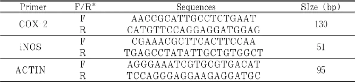
2. 세포 생존율
정상군은 100.00±0.83%, 대조군은 57.82±1.27%, FVL은 1, 5, 50, 100 μg/ml 농도에서 각각 100.17±0.12%, 100.21±0.01%, 100.45±0.74%,
100.56±0.43%로 나타났으며, LJT는 1, 5, 50, 100 μg/ml 농도에서 각각 100.90±0.50%, 100.25±0.33%, 97.50±1.64%, 76.00±4.72%
로 나타났다(Fig. 3).
Fig. 3. Cell viability of FVL and LJT in Raw264.7 cells.
Raw264.7 cells were treated with FVL (1, 5, 50, and 100 μg/ml), LJT (1, 5, 50, and 100 μg/ml), and LPS (1 μg/ml) for 24 h. Cell viability was measured using a WST assay. Each data represents the mean±SD from three independent experiments.
### p
<0.001 compared with the normal group and ***p
<0.001, **p
<0.01 compared with the control group.3. 항산화 효능평가
1) DPPH radical 소거능 측정
FVL은 1, 5, 50 μg/ml 농도에서 각각 3.31±2.33%, 6.59±0.53%, 20.63±0.60%로 나
타났으며, LJT는 1, 5, 50 μg/ml 농도에서 1.17±2.07%, 6.07±2.00%, 62.11±5.99%로 나타나 FVL과 LJT 모두 DPPH radical 소거 능의 농도의존적인 증가가 나타났다(Fig. 4).
Fig. 4. DPPH radical scavenging activity of FVL and LJT.
FVL was incubated at 1, 5 and 50 μg/ml LJT was incubated at 1, 5 and 50 μg/ml with DPPH solution. Activities were determined by measurement of absorbance at 517 nm. Each data represents the mean±SD from three independent experiments.
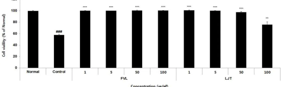
4. 세포 내 ROS 측정
정상군은 9.51±4.17%, 대조군은 100.00±3.94%, FVL는 1, 5, 50 μg/ml 농도에서 각각 91.96±0.39%, 86.27±0.15%, 76.47±1.52%
로 나타났으며, LJT는 1, 5, 50 μg/ml 농
도에서 각각 101.58±1.33%, 85.38±1.99%, 65.72±3.95%로 나타나 LJT 1 μg/ml를 제외한 모든 군에서 감소하는 경향을 보 였다(Fig. 5).
Fig. 5. The effects of FVL and LJT on ROS production in LPS-stimulated Raw264.7 cells.
Raw264.7 cells were pre-treated with FVL (1, 5, and 50 μg/ml), LJT (1, 5, and 50 μg/ml) for 1 h, followed by co-incubation with LPS (1 μg/ml) for 24 h. Level of ROS production was examined by FACS. Each data represents the mean±SD from three independent experiments.
### p
<0.05 compared with the normal group and ***p
<0.001, **p
<0.01, *p
<0.05 compared with the control group.5. Enzyme-linked immunosorbent assay 1) PGE 2
정상군은 0.01±0.00 pg/ml, 대조군은 10603.57±842.09 pg/ml, FVL은 1, 5, 50 μ g/ml 농도에서 각각 9598.67±1224.67 pg/ml, 9221.97±66.59 pg/ml, 6051.15±66.59 pg/ml 로 나타났으며, LJT는 1, 5, 50 μg/ml 농도에 서 각각 7992.46±181.97 pg/ml, 7890.02±420.88 pg/ml, 7603.17±619.05 pg/ml로 나타나 FVL과 LJT 모든 군에서 감소하는 경향 을 보였다(Fig. 6).
2) NO
정상군은 6.85±5.31%, 대조군은 100.00±2.25%, FVL은 1, 5, 50 μg/ml 농도에서 각각 103.94±0.39%, 98.19±0.66%, 88.20±0.79%
으로 나타났으며, LJT는 1, 5, 50 μg/ml
농도에서 각각 109.08±2.02%, 85.65±2.71%, 64.52±4.55%로 나타나 FVL 1, 5 μg/ml 와 LJT 1 μg/ml를 제외한 모든 군에서 유의하게 감소하였다(Fig. 7).
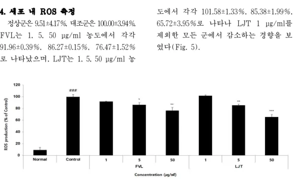
6. mRNA 발현 1) COX-2
정상군은 0.01±0.01, 대조군은 1.00±0.00, FVL은 1, 5, 50 μg/ml 농도에서 각각 0.94±0.05, 0.94±0.06, 0.85±0.07로 나타났 으며, LJT는 1, 5, 50 μg/ml 농도에서 각 각 0.99±0.07, 0.73±0.12, 0.61±0.04로 나타 나 LJT 1 μg/ml를 제외한 모든 군에서 감소하는 경향을 보였다(Fig. 8).
2) iNOS
정상군은 0.33±0.18, 대조군은 1.00±0.00,
FVL은 1, 5, 50 μg/ml 농도에서 각각 0.93±0.17, 0.92±0.09, 0.88±0.07로 나타났 으며, LJT는 1, 5, 50 μg/ml 농도에서 각
각 1.07±0.01, 0.95±0.10, 0.40±0.15로 나타 나 FVL과 LJT 50 μg/ml에서만 감소하 는 경향을 보였다(Fig. 9).
Fig. 6. The effects of FVL and LJT on PGE 2 producion in LPS-stimulated Raw264.7 cells.
Raw264.7 cells were pre-treated with FVL (1, 5, and 50 μg/ml), LJT (1, 5, and 50 μg/ml) for 1 h, followed by co-incubation with LPS (1 μg/ml) for 24 h. Production of PGE
2
was measured using a ELISA. Each data represents the mean±SD from three independent experiments.## p
<0.01 compared with the normal group and **p
<0.01, *p
<0.05 compared with the control group.Fig. 7. The effects of FVL and LJT on NO production in LPS-stimulated Raw264.7 cells.
Raw264.7 cells were pre-treated with FVL (1, 5, and 50 μg/ml), LJT (1, 5, and 50 μg/ml) for 1 h, followed by co-incubation with LPS (1 μg/ml) for 24 h. Production of NO was measured using a griess reagent for nitric oxides. Each data represents the mean±SD from three independent experiments.
### p
<0.001 compared with the normal group and ***p
<0.001 compared with the control group.Fig. 8. The effects of FVL and LJT on COX-2 mRNA expression in LPS-stimulated Raw264.7 cells.
Raw264.7 cells were pre-treated with FVL (1, 5, and 50 μg/ml), LJT (1, 5, and 50 μg/ml) for 1 h, followed by co-incubation with LPS (1 μg/ml) for 24 h. Level of COX-2 mRNA expression was examined by real-time PCR. Each data represents the mean±SD from three independent experiments.
### p
<0.001 compared with the normal group and **p
<0.01 compared with the control group.Fig. 9. The effects of FVL and LJT on iNOS mRNA expression in LPS-stimulated Raw264.7 cells.
Raw264.7 cells were pre-treated with FVL (1, 5, and 50 μg/ml), LJT (1, 5, and 50 μg/ml) for 1 h, followed by co-incubation with LPS (1 μg/ml) for 24 h. Level of iNOS mRNA expression was examined by real-time PCR. Each data represents the mean±SD from three independent experiments.
## p
<0.01 compared with the normal group and **p
<0.01, *p
<0.05 compared with the control group.7. Cytokine level 1) IL-1β
정상군은 3.20±0.00 pg/ml, 대조군은 124.74±7.81 pg/ml, FVL은 1, 5, 50 μg/ml 농 도에서 각각 132.62±14.47 pg/ml, 120.76±0.62 pg/ml, 84.21±4.71 pg/ml로 나타났으며, LJT는 1, 5, 50 μg/ml 농도에서 각각 120.27±6.04 pg/ml, 103.57±3.58 pg/ml, 78.28±
2.40 pg/ml로 나타나 FVL와 LJT 1 μg/ml
를 제외한 모든 군에서 감소하는 경향을 보였다(Fig. 10).
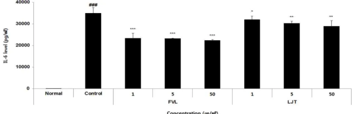
2) IL-6
정상군은 2.68±0.86 pg/ml, 대조군은 35042.93
±2478.71 pg/ml, FVL은 1, 5, 50 μg/ml 농도 에서 각각 23422.05±2309.40 pg/ml, 23238.63
±56.53 pg/ml, 22422.05±349.20 pg/ml로
나타났으며, LJT는 1, 5, 50 μg/ml 농도에
서 각각 32057.20±1578.06 pg/ml, 30271.71
±1023.53 pg/ml, 28910.16±2560.41 pg/ml 로 나타나 FVL과 LJT 모든 군에서 감 소하는 경향을 보였다(Fig. 11).
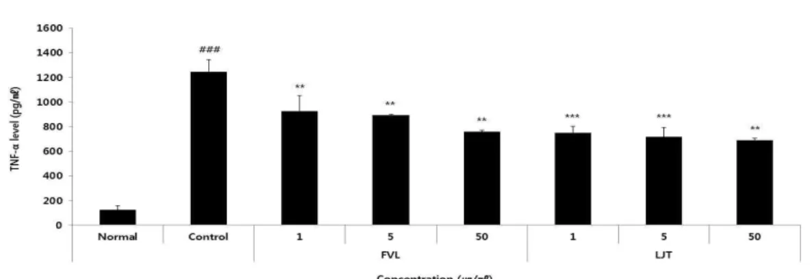
3) TNF-α
정상군은 123.23±33.54 pg/ml, 대조군은 1246.81±96.92 pg/ml, FVL은 1, 5, 50 μg/ml
농도에서 각각 924.66±126.89 pg/ml, 894.93±5.93 pg/ml, 759.21±9.36 pg/ml로 나타났으며, LJT는 1, 5, 50 μg/ml 농도에서 각각 750.62±53.32 pg/ml, 718.07±74.21 pg/ml, 691.01±16.39 pg/ml로 나타나 FVL과 LJT 모든 군에서 감소하는 경향을 보였다(Fig. 12).
Fig. 10. The effects of FVL and LJT on IL-1β level in LPS-stimulated Raw264.7 cells.
Raw264.7 cells were pre-treated with FVL (1, 5, and 50 μg/ml), LJT (1, 5, and 50 μg/ml) for 1 h, followed by co-incubation with LPS (1 μg/ml) for 24 h. Level of IL-1β was measured using a luminex multiplex assays. Each data represents the mean±SD from three independent experiments.
## p
<0.01 compared with the normal group and **p
<0.01, *p
<0.05 compared with the control group.Fig. 11. The effects of FVL and LJT on IL-6 level in LPS-stimulated Raw264.7 cells.
Raw264.7 cells were pre-treated with FVL (1, 5, and 50 μg/ml), LJT (1, 5, and 50 μg/ml) for 1 h, followed by co-incubation with LPS (1 μg/ml) for 24 h. Level of IL-6 was measured using a luminex multiplex assays. Each data represents the mean±SD from three independent experiments.
### p
<0.001 compared with the normal group and ***p
<0.001, **p
<0.01, *p
<0.001 compared with the control group.Fig. 12. The effects of FVL, LJT on TNF-α level in LPS-stimulated Raw264.7 cells.
Raw264.7 cells were pre-treated with FVL (1, 5, and 50 μg/ml), LJT (1, 5, and 50 μg/ml) for 1 h, followed by co-incubation with LPS (1 μg/ml) for 24 h. Level of TNF-α was measured using a luminex multiplex assays. Each data represents the mean±SD from three independent experiments.
### p
<0.001 compared with the normal group and ***p
<0.001, **p
<0.01 compared with the control group.8. Protein expression 1) ERK
정상군은 0.44±0.07, 대조군은 1.00±0.03, FVL은 1, 5, 50 μg/ml 농도에서 각각 0.90±0.03, 0.84±0.01, 0.65±0.01로 나타났 으며, LJT은 1, 5, 50 μg/ml 농도에서 각 각 0.63±0.04, 0.65±0.01, 0.69±0.04로 나타나 FVL과 LJT 모든 군에서 유의하게 감소 하였다(Fig. 13).
2) JNK
정상군은 0.05±0.09, 대조군은 1.00±0.06, FVL은 1, 5, 50 μg/ml 농도에서 각각 1.00±0.04, 0.99±0.01, 0.95±0.00으로 나타 났으며, LJT는 1, 5, 50 μg/ml 농도에서 각각 1.00±0.02, 0.95±0.07, 0.82±0.03으로 나타나 FVL과 LJT 1 μg/ml를 제외한 모 든 군에서 감소하는 경향을 보였다(Fig. 13).
3) p38
정상군은 0.00±0.00, 대조군은, FVL은 1, 5, 50 μg/ml 농도에서 각각 1.00±0.02, 0.87±0.02, 0.84±0.04로 나타났으며, LJT는
1, 5, 50 μg/ml 농도에서 각각 0.81±0.02, 0.80±0.01, 0.72±0.02로 나타나 FVL 1 μg/ml 를 제외한 모든 군에서 감소하는 경향을 보였다(Fig. 13).
4) IκBα
정상군은 6.46±0.14, 대조군은 1.00±0.02, FVL은 1, 5, 50 μg/ml 농도에서 각각 6.56±0.51, 7.56±0.03, 9.05±0.01로 나타났 으며, LJT는 1, 5, 50 μg/ml 농도에서 각 각 9.67±0.18, 9.68±0.07, 10.75±0.14로 나 타나 FVL과 LJT 모든 군에서 유의하게 증가하였다(Fig. 13).
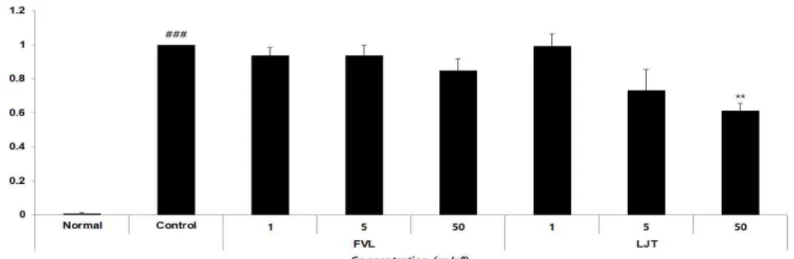
5) NF-κB
정상군은 0.00±0.00, 대조군은 1.00±0.02,
FVL은 1, 5, 50 μg/ml 농도에서 각각
0.84±0.04, 0.75±0.03, 0.63±0.04로 나타났
으며, LJT는 1, 5, 50 μg/ml 농도에서 각
각 0.48±0.02, 0.45±0.01, 0.33±0.03으로 나
타나 FVL과 LJT 모든 군에서 유의하게
감소하였다(Fig. 13).
Fig. 13. The effects of FVL and LJT on ERK, JNK, p38, IκBα and NF-κB in LPS-stimulated Raw264.7 cells.
Raw264.7 cells were pre-treated with FVL (1, 5, and 50 μg/ml), LJT (1, 5, and 50 μg/ml) for 1 h, followed by co-incubation with LPS (1 μg/ml) for 24 h. Level of ERK, JNK, p38, IκBα and NF-κB expression were measured using a western blot. Each data represents the mean±SD from three independent experiments.