http://dx.doi.org/10.14405/kjvr.2015.55.3.175
175
<Original Article>
Characterization of proteases isolated from Kudoa septempunctata
Sang Phil Shin
1,*, Kosuke Zenke
2, Hiroshi Yokoyama
21
Fisheries Laboratory, Kinki University, Wakayama 649-2211, Japan
2
Department of Aquatic Bioscience, Graduate School of Agricultural and Life Sciences, University of Tokyo, Tokyo 113-8657, Japan (Received: July 28, 2015; Revised: August 9, 2015; Accepted: August 17, 2015)
Abstract : Proteases play important roles in parasite development and host parasite interactions. The protease of Kudoa spp. has been recognized as a key factor of severe proteolysis of fish muscle post-mortem; however, there is little information available regarding the protease of Kudoa (K.) septempunctata, which was recently identified as a cause of food poisoning in humans. The present study was conducted to isolate and characterize proteases to elucidate the type of protease contained in the parasite and determine the optimal pH for protease activity. We confirmed the cysteine protease and metalloprotease produced by K. septempunctata. While the cysteine protease showed optimal activity at pH 5 that decreased rapidly with increasing pH, the optimal activity of metalloprotease was pH 7, and it remained stable from pH 6 to pH 8. These results indicate that the pH of cysteine protease is not proper for fish muscle post- mortem, and that metalloprotease can act in human intestines. Overall, the present study provides important information that improves our understanding of the role of protease physiology and the subsequent food poisoning caused by K.
septempunctata.
Keywords : pH, cysteine protease, Kudoa septempunctata, metalloprotease
Introduction
The family Kudoidae (Myxozoa: Multivalvulida) consist of a single genus Kudoa Meglitsch, 1947, having four or more shell valves and polar capsules, and more than ninety spe- cies of Kudoa have been described from marine and estua- rine fish species [10, 27]. Most of Kudoa species are histozoic, inducing macroscopic cysts in various organs including somatic muscles, however some species forming a pseudocyst in the myofibers and cause post-mortem myo- liquefaction [18, 22]. Although myxozoan parasites are gen- erally considered harmless to humans, certain human illnesses have been attributed to Kudoa sp. such as allergic symptoms [14] and food-borne diarrhea. Serial food poison- ings associated with ingestion of raw olive flounder Parali- chthys olivaceus have been recently reported in Japan, and epidemiological analysis demonstrates that Kudoa (K.) sep- tempunctata is associated with these illnesses [11].
Proteases play an integral role in interactions between par- asites and their hosts, are involved in parasite physiology development [3, 19-21], and contribute to parasite virulence [12, 24]. These enzymes have been also described in some of Kudoa species such as K. rosenbuschi, K. paniformis, and K.
thyrsites. They are associated with a degradation of host muscle tissue post-mortem, which was known as myolique-
faction [7, 15]. However, there is currently no information about the proteases of K. septempunctata even the parasite has threatened a public health.
The objective of this study was to isolate and characterize the parasite proteases that may contribute to the physiology and pathogenicity of K. septempunctata. We focused on whether K. septempunctata possesses what kind of pro- teases, and what of pH does work on an optimal for the activity of proteases. The information in the present study will contribute to understand the life cycle or a relationship between the role of protease and food poisoning of K. sep- tempunctata.
Materials and Methods
Sample preparation
Olive flounder samples suspected of being infected with K.
septempunctata were obtained from an olive flounder farm in Japan. The fish samples were examined microscopically to confirm the presence of K. septempunctata spores in squash preparations. The spores were purified using a modified Per- coll (Sigma-Aldrich, USA) density-gradient centrifugation method as previously described [26]. Purified spores were diluted in phosphate buffer solution (PBS) to a final concen- tration of 10
7spores/ml. The lysate of K. septempunctata
*Corresponding author
Tel: +81-739-42-2625, Fax: +81-739-42-2634 E-mail: [email protected]
(LKs) was obtained as described by previous report [25], with some modifications. Briefly, the spores were washed twice by centrifugation at 10,000 × g for 2 min in 0.25 M sucrose. The spores were then disrupted ultrasonically. The homogenate was centrifuged at 15,000 × g for 10 min and the resulting supernatant fraction was stored in aliquots at −80
oC.
Protein concentration in the extract was determined by using the Pierce BCA protein assay kit (Thermo Fisher Scientific, USA) with a microplate reader (MPRA4; Tosoh, Japan) at 570 nm. The bovine serum albumin was used as a standard.
Affinity chromatography
The LKs was dialyzed at 4
oC against four volumes of 25 mM acetate buffer (pH 5.0) and bacitracin was used in affin- ity chromatography as recommended [4] with some modifi- cations. Bacitracin was coupled to cyanogen bromide-activated Sepharose 4B (Sigma-Aldrich) and the dialyzed lysate was applied to a 5 × 1 cm column of bacitracin-Sepharose pre- equilibrated with buffer A (25 mM acetate buffer, pH 5.0), at a flow rate of 0.5 ml/min. The column was washed with 50 ml of buffer A (0.5 mL/min) and eluted with buffer B (buffer A containing 1 M NaCl, 25% of isopropanol) at the same flow rate. The eluted fractions were pooled in conical tubes and concentrated using ultrafiltration through Amicon Ultra-15 filter (30-kDa cutoff; Millipore, USA), and desalted twice using Tris-HCl buffer (TBS; pH 7.0). The partial puri- fied protease of K. septempunctata (PKs) was concentrated again using Amicon Ultra-0.5mL centrifuga filter and the concentration of protein was determined as described above.
SDS-PAGE and gelatin-zymography
The LKs was analyzed by sodium dodecyl sulfate-poly- acrylamide gel electrophoresis (SDS-PAGE). Sample (20 µg) was mixed with SDS-PAGE loading buffer and electrophore- sis was carried out for 100 min in a Tris-glycine buffer sys- tem. The gel was stained with Coomassie blue for the visualization of the proteins, and molecular weights esti- mated by comparison with SDS-PAGE protein standards. In order to visualize the proteases activity of LKs, gelatin was added to the 10% acrylamide running gel for a final concen- tration of 0.2% (w/v) protein. The electrophoresis of two lysate
samples (each 3 µg) was carried out as condition described above. After electrophoresis the gels were incubated for 30 min in 2.5% (v/v) Triton X-100 at room temperature, and then each of gels were incubated for 18 h in 50 mM 2-N- morpholino ethanesulfonic acid (MES) buffer (200 mM NaCl, 0.02% Brij-35 and 0.01% NaN
3, pH 5.0) and TBS (200 mM NaCl, 0.02% Brij-35 and 0.01% NaN
3, pH 7.0) at 37
oC, respectively. The gels were stained with Coomassie blue, and destained until clear gel bands were apparent where proteolytic hydrolysis of the gel embedded substrate had occurred. In addition, we also examined the activity of PKs through gelatin-zymography described above and it was developed in TBS (200 mM NaCl, 0.02% Brij-35 and 0.01%
NaN
3, pH 7.0).
Protease activity and inhibitor assays using azocasein An azocasein assay was used to further characterize the activity of the LKs and PKs. Protease activity was deter- mined by measuring the release of acid-soluble material from azocasein (Sigma-Aldrich). The optimum pH for both the LKs and PKs was determined within a pH range of 4.0 to 8.0, using 50 mM sodium acetate (pH 4.0 −6.0) and 50 mM Tris-HCl (pH 7.0 −8.0) buffers. The reaction was initiated by the addition of 10- µL samples to 90 of µL reaction buffer containing 50 mM sodium acetate/Tris-HCl buffer, pH 4.0 − 8.0, 0.2% azocasein, 200 mM NaCl, 0.02% Brij-35, 0.01%
NaN
3,and the mixtures were incubated for 18 h at 37
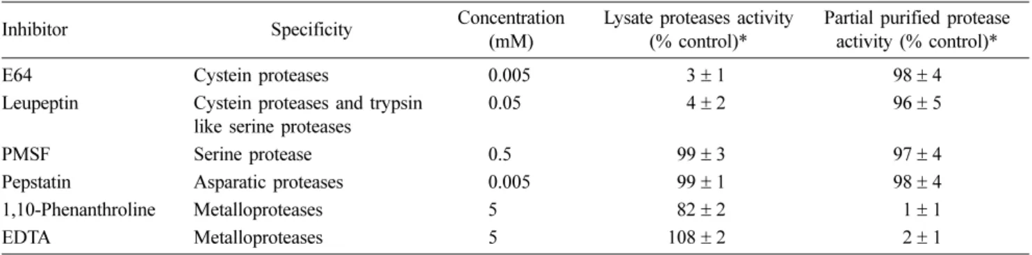
oC. The reaction was stopped by the addition of 100 µL 10% (w/v) trichloroacetic acid (TCA), and the mixtures were incubated at room temperature for 30 min. The mixtures were then cen- trifuged (12,000 × g, 10 min) to pellet the unreacted sub- strate, and 125 µL of the supernatant (containing the TCA- soluble azo-compounds) was withdrawn and mixed with 50 µL 2.0 M NaOH. The absorbance of the mixture was read at 440 nm by MPRA4. The effects of various inhibitors (Pep- statin A, PMSF, EDTA, 1, 10-phenanthroline, E64, and leu- peptin) against the LKs and PKs were evaluated in sodium acetate buffer (pH 5.0) and TBS (pH 7.0), respectively. The final concentrations of inhibitors indicated in Table 1 and the results reported from all assays were carried out in triplicate.
Table 1. Inhibition assay of proteases by various inhibitors
Inhibitor Specificity Concentration
(mM)
Lysate proteases activity (% control)*
Partial purified protease activity (% control)*
E64 Cystein proteases 0.005 3± 1 98± 4
Leupeptin Cystein proteases and trypsin like serine proteases
0.05 4± 2 96± 5
PMSF Serine protease 0.5 99± 3 97± 4
Pepstatin Asparatic proteases 0.005 99± 1 98± 4
1,10-Phenanthroline Metalloproteases 5 82± 2 1± 1
EDTA Metalloproteases 5 108± 2 2± 1
*Enzyme activity was estimated using azocasein as a substrate. The results are mean values from triplicate experiments± SD.
Results
The LKs showed bands of various sizes by SDS-PAGE.
Six of the bands (83, 75, 63, 48, 29, and 26.5 kDa) exhibited gelatinolytic activity at pH 5.0 (lanes 1 and 2 in Fig. 1). Only one of the six LKs bands (83 kDa) had detectable protease activity at pH 7.0 (lane 3 in Fig. 1), which was larger than the only PKs band (45 kDa) that exhibited gelatinolytic activ- ity at pH 7.0 (lane 4 in Fig. 1). The LKs was enzymatically active against the azocasein at pH 5.0, with activity that reduced dramatically at pH 6.0 (Fig. 2A). The PKs showed optimal activity at pH 7.0 and was stable between pH 6.0 − 8.0 (Fig. 2B). While proteolytic activity of LKs was largely inhibited by adding the cysteine protease specific inhibitors E64 and leupeptin, the PKs was inhibited by metalloprotease
inhibitors such as 1,10-phenanthroline and EDTA. The effect of the inhibitors on protease activity of LKs and PKs using azocasein as the substrate are shown in Table 1.
Discussion
Proteases play important roles in interactions between par- asites and their hosts, and underlie the pathogenicity of many organisms [19, 21]. The cysteine proteases have been described in Kudoa spp. and are possibly a key factor of the parasites, which causes post-mortem myoliquefaction [7, 15]. Although K. septempunctata is known to cause food poisoning in humans when accidentally consumed with raw flounder, there are poorly understood about the proteases of this para- site and the K. septempunctata infected fish muscle has showed no myoliquefaction [11, 18]. The present study iden- tified a presence of cysteine proteases through LKs that showed the optimal enzymatic activity at pH 5.0. However, the activity of cysteine protease was decreased rapidly between pH 5.0 and pH 6.0. Following death of the fish, the stored carbohydrate glycogen is anaerobically degraded and lactic acid is accumulated in muscle resulting in a pH drop from 7.4 to 6.0 [2]. In addition, previous studies have reported that the pH of muscle in post-mortem flounder and halibut decreases to pH 6.5 for 3 days [6, 16]. Therefore, we suggest that K. septempunctata does not cause post-mortem myo- liquefaction based on the relationship between the optimal pH of the parasite cysteine protease and the pH of muscle in post-mortem fish.
In previous study, we reported that factors affecting sporo- plasm release in K. septempunctata such as enzymes, culture media, and protease inhibitors. Interestingly, the sporoplasm release was prevented by 1,10-phenanthroline (metallopro- tease) and it suggested that metalloprotease is related the sporoplasm release in K. septempunctata [26]. Metallopro- teases of parasite have been related to pathogenesis and are involved in processes such as immunity evasion, develop- ment, and metabolism [8, 17]. In addition, the activity of metalloproteases are affected by pH of environment [13]. In the present study, we observed one protease (83 kDa) of LKs which showed the proteolytic activity at pH 7.0 and that was purified by affinity chromatography partially. We speculate that the protease was processed as a mature form (45 kDa) for purifying. The PKs have optimal activity at pH 7.0 and showed stable activity from pH 6.0 to pH 8.0. However, the activity was completely prevented by inhibitors of metallo- protease. These results demonstrated that the PKs is a metal- loprotease which has activity from pH 6.0 to pH 8.0. We also observed the prevention of sporoplasm release in acidified culture medium [26], even there are the factors to release it.
It is indicated that the environmental pH for K. septempunc- tata is a key factor to release sporoplasm. Human intralumi- nal pH is rapidly changed from highly acidic in the stomach to about pH 6.0 in the duodenum, and the pH gradually increases in the small intestine from pH 6.0 to about pH 7.4
Fig. 1. Analysis of the lysate proteases (LKs) and partial puri-fied protease (PKs) of Kudoa (K.) septempunctata by 10%
sodium dodecyl sulfate-polyacrylamide and gelatin zymogra- phy. Bands were visualized by Coomassie Blue staining (lane 1) and destaining (lanes 2−4). Lane M, molecular mass protein markers; lane 1, crude supernatant from homogenized purified K. septempunctata; lane 2, LKs was incubated at MES buffer (pH 5.0); lane 3, LKs was incubated at Tris-HCl buffer (TBS;
pH 7.0); lane 4, PKs was incubated at TBS (pH 7.0).
Fig. 2. pH activity profile of LKs and PKs. The activity of the proteases against azocasein substrate was measured at different pHs. The activities are shown relative to that at pH 5.0 and pH 7.0 being 100%, respectively. The results are mean values from triplicate experiments± SD.
in the terminal ileum. The pH drops to 5.7 in the caecum, but again gradually increases, reaching pH 6.7 in the rectum [5].
Therefore, the pH of human intestine is proper to work for the PKs especially that is optimal pH in small intestine. The median latency between eating the K. septempunctata infected raw fish and the presence of the first symptoms is five hours that is corresponding with the time of dietary tran- sit reach to small intestine [1, 9, 28]. Based on these results, we suggest that the PKs contribute the sporoplasm in small intestine, which causes food poisoning by K. septempunc- tata. Two of possible ways to contribute the sporoplasm release were speculated: 1) PKs effect on the mechanism of actin polymerization which discharges the sporoplasm by condensing actin filament [23], 2) the metalloprotease is involved in metabolism of glucose related to the release of sporoplasm in K. setempunctata [26]. In addition, we also observed the proteolytic activity (5 −7%) in LKs at pH 6−8, suggesting that the activity was produced by the metallopro- tease and the protease is not related to post-mortem myo- liquefaction.
In conclusion, the present study demonstrates the presence and kind of proteases in K. septempunctata. The cysteine protease showed the strong activity at pH 5 although it is decreased rapidly at pH 6 over while metalloprotease have stable activity from pH 6 to 8. These findings elucidate the relation between proper pH range of the protease and patho- physiological response such as post-mortem myoliquefaction and sporoplasm release in K. septempunctata. The informa- tion of present study will contribute to reveal the life cycle of K. septempunctata and establish strategies to treat and pre- vent disease caused by this parasite. However, further stud- ies will be required to demonstrate the molecular and immunological mechanism of the proteases effect on physio- logical response in life cycle of K. septempunctata.
Acknowledgments
This research was supported by International Research Fel- low of the Japan Society for the Promotion of Science (P14398; grant no. 26-04398) and Basic Science Research Program through the National Research Foundation of Korea (NRF) funded by the Ministry of Education, Science and Tech- nology (2013R1A6A3A03063215).
References
1. Degen LP, Phillips SF. Variability of gastrointestinal transit in healthy women and men. Gut 1996, 39, 299-305.
2. Delbarre-Ladrat C, Chéret R, Taylor R, Verrez-Bagnis V. Trends in postmortem aging in fish: understanding of proteolysis and disorganization of the myofibrillar structure.
Crit Rev Food Sci Nutr 2006, 46, 409-421.
3. Dörfler C. El-Matbouli M. Isolation of a subtilisin-like serine protease gene (MyxSubtSP) from spores of Myxobolus cerebralis, the causative agent of whirling disease. Dis Aquat Organ 2007, 73, 245-251.
4. Faisal M, Schafhauser DY, Garreis KA, Elsayed E, La Peyre JF. Isolation and characterization of Perkinsus marinus proteases using bacitracin-sepharose affinity chromatography.
Comp Biochem Physiol B Biochem Mol Biol 1999, 123, 417-426.
5. Fallingborg J. Intraluminal pH of the human gastrointestinal tract. Dan Med Bull 1999, 46, 183-196.
6. Foy RJ, Crapo CA, Kramer DE. Investigating the roles of temperature and exercise in the development of chalkiness in Pacific halibut. Report No. 50. 24p. International Pacific Halibut Commission (US), Washington, 2006.
7. Funk VA, Olafson RW, Raap M, Smith D, Aitken L, Haddow JD, Wang D, Dawson-Coates JA, Burke RD, Miller KM. Identification, characterization and deduced amino acid sequence of the dominant protease from Kudoa paniformis and K. thyrsites: a unique cytoplasmic cysteine protease. Comp Biochem Physiol B Biochem Mol Biol 2008, 149, 477-489.
8. Gras S, Byzia A, Gilbert FB, McGowan S, Drag M, Silvestre A, Niepceron A, Lecaille F, Lalmanach G, Brossier F. Aminopeptidase N1 (EtAPN1), an M1 metalloprotease of the apicomplexan parasite Eimeria tenella, participates in parasite development. Eukaryot Cell 2014, 13, 884-895.
9. Iwashita Y, Kamijo Y, Nakahashi S, Shindo A, Yokoyama K, Yamamoto A, Omori Y, Ishikura K, Fujioka M, Hatada T, Takeda T, Maruyama K, Imai H. Food poisoning associated with Kudoa septempunctata. J Emerg Med 2013, 44, 943-945.
10. Jeon CH, Wi S, Song JY, Choi HS, Kim JH.
Development of loop-mediated isothermal amplification method for detection of Kudoa septempunctata (Myxozoa:
Multivalvulida) in olive flounder (Paralichthys olivaceus).
Parasitol Res 2014, 113, 1759-1767.
11. Kawai T, Sekizuka T, Yahata Y, Kuroda M, Kumeda Y, Iijima Y, Kamata Y, Sugita-Konishi Y, Ohnishi T.
Identification of Kudoa septempunctata as the causative agent of novel food poisoning outbreaks in Japan by consumption of Paralichthys olivaceus in raw fish. Clin Infect Dis 2012, 54, 1046-1052.
12. Mackey ZB, O’Brien TC, Greenbaum DC, Blank RB, McKerrow JH. A cathepsin B-like protease is required for host protein degradation in Trypanosoma brucei. J Biol Chem 2004, 279, 48426-48433.
13. Malagón D, Adroher FJ, Díaz-López M, Benítez R.
Collagenolytic activity related to metalloproteases (and serine proteases) in the fish parasite Hysterothylacium aduncum (Nematoda: Anisakidae). Dis Aquat Organ 2010, 90, 129-134.
14. Martínez de Velasco G, Rodero M, Cuéllar C, Chivato T, Mateos JM, Laguna R. Skin prick test of Kudoa sp.
antigens in patients with gastrointestinal and/or allergic symptoms related to fish ingestion. Parasitol Res 2008, 103, 713-715.
15. Martone CB, Spivak E, Busconi L, Folco EJE, Sánchez JJ. A cysteine protease from myxosporean degrades host myofibrils in vitro. Comp Biochem Physiol B Biochem Mol Biol 1999, 123, 267-272.
16. Massa AE, Palacios DL, Paredi ME, Crupkin M.
Postmortem changes in quality indices of ice-stored flounder (Paralichthys patagonicus). J Food Biochem 2005, 29, 570-590.
17. Matheoud D, Moradin N, Bellemare-Pelletier A, Shio MT, Hong WJ, Olivier M, Gagnon E, Desjardins M, Descoteaux A. Leishmania evades host immunity by inhibiting antigen cross-presentation through direct cleavage of the SNARE VAMP8. Cell Host Microbe 2013, 14, 15- 25.
18. Matsukane Y, Sato H, Tanaka S, Kamata Y, Sugita- Konishi Y. Kudoa septempunctata n. sp. (Myxosporea:
Multivalvulida) from an aquacultured olive flounder (Paralichthys olivaceus) imported from Korea. Parasitol Res 2010, 107, 865-872.
19. McKerrow JH. Parasite proteases. Exp Parasitol 1989, 68, 111-115.
20. McKerrow JH. Development of cysteine protease inhibitors as chemotherapy for parasitic diseases: insights on safety, target validation, and mechanism of action. Int J Parasitol 1999, 29, 833-837.
21. McKerrow JH, Sun E, Rosenthal PJ, Bouvier J. The proteases and pathogenicity of parasitic protozoa. Annu Rev Microbiol 1993, 47, 821-853.
22. Moran JDW, Whitaker DJ, Kent ML. A review of the myxosporean genus Kudoa Meglitsch, 1947, and its impact on the international aquaculture industry and commercial fisheries. Aquaculture 1999, 172, 163-196.
23. Ohnishi T, Kikuchi Y, Furusawa H, Kamata Y, Sugita- Konishi Y. Kudoa septempunctata invasion increases the permeability of human intestinal epithelial monolayer.
Foodborne Pathog Dis 2013, 10, 137-142.
24. Que X, Reed SL. Cysteine proteinases and the pathogenesis of amebiasis. Clin Microbiol Rev 2000, 13, 196-206.
25. Shin SP, Han SY, Han JE, Jun JW, Kim JH, Park SC.
Expression and characterization of cathepsin L-like cysteine protease from Philasterides dicentrarchi. Parasitol Int 2014, 63, 359-365.
26. Shin SP, Zenke K, Yokoyama H, Yoshinaga T. Factors affecting sporoplasm release in Kudoa septempunctata.
Parasitol Res 2015, 114, 795-799.
27. Whipps CM, Grossel G, Adlard RD, Yokoyama H, Bryant MS, Munday BL, Kent ML. Phylogeny of the multivalvulidae (Myxozoa: Myxosporea) based on comparative ribosomal DNA sequence analysis. J Parasitol 2004, 90, 618-622.
28. Worsøe J, Fynne L, Gregersen T, Schlageter V, Christensen LA, Dahlerup JF, Rijkhoff NJ, Laurberg S, Krogh K. Gastric transit and small intestinal transit time and motility assessed by a magnet tracking system. BMC Gastroenterol 2011, 11, 145.