大韓獸醫學會誌 (2013) 第 53 卷 第 4 號 Korean J Vet Res(2013) 53(4) : 253~256 http://dx.doi.org/10.14405/kjvr.2013.53.4.253
253
<Short Communication>
Minocycline as a treatment of dog with calcinosis cutis
Hye-Jin Jang
1, Min-Hee Kang
1, Jung-Hyang Sur
2, Hee-Myung Park
1,*
Departments of
1Veterinary Internal Medicine, and
2Veterinary Pathology, College of Veterinary Medicine, Konkuk University, Seoul 143-701, Korea
(Received: May 13, 2013; Accepted: September 6, 2013)
Abstract : An 8-year-old, castrated male, Schnauzer dog was presented for evaluation of gradually worsening erythematous papules. Physical examination revealed multiple erythematous papules having a firm, gritty texture located in bilateral ears, dorsal midline, perianal and inguinal area. Skin biopsy revealed aberrant structure, characterized by atrophic epidermal-dermal layer structure with granular materials which was presumed as calcinosis cutis secondary to iatrognic hyperadrenocotricism. By initiating minocycline for 14 days, there was reduction in size, number of calcium deposit with remarkably decreased erythema. This case report presents the clinical trial of minocycline as a potential agent in treating dogs with calcinosis cutis.
Keywords : calcinosis cutis, dog, iatrogenic hyperadrenocorticism, minocycline
Calcinosis cutis is a term to describe abnormal calcium deposits forming in the dermis, epidermis, or subcutis [2, 3].
Calcification of the skin may occur in a wide variety of dis- orders. It is most commonly developed as a result of natu- rally occurring or iatrogenic hyperadrenocorticism (HAC) in dogs [4, 12]. The mechanisms of calcification are divided into four categories: dystrophic, metastatic, iatrogenic and idiopathic [4]. Dystrophic calcification is the most common type and it is associated with local tissue damage or disor- ders in collagenous, elastic, or subcutaneous tissue [8]. Met- astatic calcification is related to the accumulation of calcium salts in normal tissue, associated with aberrant metabolism of calcium and phosphorus [6]. Iatrogenic calcification appears secondary to penetration of calcium containing products and idiopathic calcification is in the absence of identifiable local or systemic factors such as local tissue damage or metabolic disorders [4]. As dystrophic calcification commonly occurs in association with canine HAC that leads to calcinosis cutis [1], diagnoses on hematologic and biochemical profile present hypercortisolism with normal calcium and phospho- rus levels typically found [4, 5].
Cutaneous lesions consist of erythematous papule, plaques, and nodules frequently ulcerated and secondarily infected.
The lesions often affect especially along the dorsum, axillae and the inguinal region [12]. Radiographs reveal calcified mass in skin and by performing skin biopsy, multifocal deposits of granular materials can be observed in dermal, subcutaneous tissue [3, 8].
As calcinosis cutis is a rare disorder, no standard therapy
has been generally accepted. However, several treatments have been reported beneficial in human medicine, including warfarin, bisphosphonates, probenecid, ceftriaxone, diltiazem and minocycline [9].
This case describes beneficial effects of skin lesions in iatrogenic HAC fallowing minocycline treatment.
An 8-year-old, castrated male, Schnauzer dog was initially presented for evaluation of numerous erythematous papules which appeared 6 weeks ago and gradually increased in num- ber and size. The dog had a history of long term orally administered corticosteroids for 8 years continuously to treat allergic dermatitis. On physical examination, the skin was thin on palpation and there were lesions consisting of multi- ple erythematous, crusted papules located in the dermis of both sides of the ears, dorsal midline, perianal and inguinal area, having a firm, gritty texture (Figs. 1A-C). Also hepatome- galy was present on abdominal palpation. A complete blood count showed thrombocytosis (666 × 10
3/µL; reference range:
200~500 × 10
3/µL) with mild stress leukogram and serum chemistry profile revealed marked elevation in alanine ami- notransferase (278 U/L; reference range: 19~70 U/L), alka- line phosphatase (3817 U/L; reference range: 15~127 U/L), gamma-glutamyl transpeptidase (118 mg/dL; reference range:
0~6 mg/dL) with increased triglycerides (302 mg/dL; refer- ence range: 19~133 mg/dL) and hypercalcemia (12.2 mg/dL;
reference range: 8.8~11 mg/dL) (Table 1).
Dermatological examinations for the skin lesions were conducted. Skin taping, scraping, trichogram and wood’s lamp tests were unremarkable. Bacterial and fungal cultures
*Corresponding author
Tel: +82-2-450-4140, Fax: +82-2-444-4396 E-mail: [email protected]
254
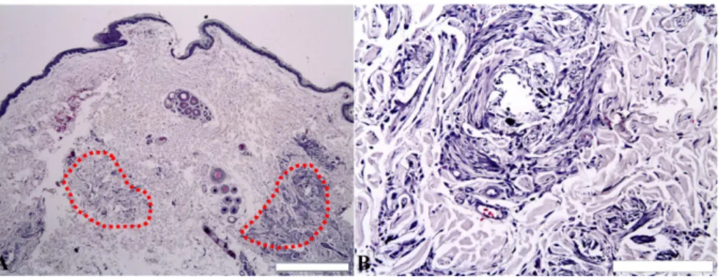
Hye-Jin Jang, Min-Hee Kang, Jung-Hyang Sur, Hee-Myung Parkwere all negative. Representative skin biopsy samples were taken from skin in the inguinal area and were evaluated to discover the underlying cause. Histopathologically, thin epi- dermal-dermal layer characterized by atrophic hair follicles, adnexal glands and multifocal accumulations of coarsely granular amorphous basophilic materials were identified in deep dermal tissue (Figs. 2A and B).
Radiography revealed hepatomegaly and displacement of stomach caudally and dorsally. Multiple rounded, dense, amorphous calcified materials were detected on the dorsal subcutis. On ultrasonography, hyperechoic foci smaller than 1mm were scattered in soft tissues including the spleen and bilateral renal cortex.
Echocardiography was performed because the dog had hypertension (151 mmHg, Cardell model 9401; Dan Scott &
Associates, USA) and slight cardiomegaly (Vertebral Heart Score, VHS = 10.8, normal range; 9.7 ± 0.5) and revealed
thickened cardiac wall with hyperechoic spots of cardiac muscle.
Based on the results described previously, the association of calcinosis cutis with iatrogenic HAC was considered, additional tests including serum parathyroid hormone (PTH) level and adrenocorticotropic hormone (ACTH) stimulation test were performed for confirmation, revealing normal PTH level with normal pre (1.47 µg/dL; reference range: 1.0~6.0 µg/dL) and low post (1.75 µg/dL; reference range: 5.50~
20.00 µg/dL) cortisol level.
Based on history, laboratory tests and dermatologic exami- nations including histopathologic findings, dystrophic calci- nosis cutis following iatrogenic HAC was diagnosed and corticosteroid treatment was discontinued for a month.
At 4 week follow-up, the serum biochemical findings revealed lowered levels of hepatobiliary enzymes including ALT, ALP, GGT. However, there was no improvement in
Fig. 1. Photographs of progress of calcinosis cutis lesion in bilateral ears, dorsal midline and perianal region. Appearance of multiple calcium deposit lesions with marked erythema and alopecia which is predisposed to secondary infection (A-C). Fourteen days after treatment with minocycline. Remarkable reduction of size and number of papules were noted and erythematous cutaneous lesions dis- appeared (D-F).Table 1. Profiles of relevant serum biochemical findings
Parameters Day 1 Day 2 Day 30 Reference range
Alanine aminotransferase (U/L) 278 250 139 19~70
Alkaline phosphatase (U/L) 3817 3766 2617 15~127
Gamma-glutamyl transpeptidase (U/L) 118 102.4 28.1 0~6
Triglyceride (mg/dL) 302 160 399 19~133
Calcium (mg/dL) 12.2 11.1 10 8.8~11
Ionized Calcium (mmol/L) 1.38 1.33 – 1.1~1.3
Inorganic phosphorus (mg/dL) 2.9 3.3 2.4 3.0~6.2
Minocycline as a treatment of dog with calcinosis cutis
255
skin lesions. Thus, we initiated prescription of minocycline (15 mg/kg, PO, q12hr, Minocin; SK chemical, Korea) for 2 weeks and noticed both reduction in size, number of papules and alleviation of erythema with no spreading of calcium deposits to other areas (Figs. 1D-F). After 4 week of treat- ment with minocycline, skin lesions were resolved and no other adverse reaction was noticed.
Calcinosis cutis is characterized by inappropriate deposi- tion of insoluble calcium within the skin. It is the specific type of dystrophic calcium deposits and occurs most com- monly secondary to iatrogenic HAC [2, 3]. It is probably caused by protein catabolic functions of cortisol which results in damage to the structure within collagen and elas- tin, predisposing calcium precipitation in the skin [4]. In addition, local tissue damage and necrosis increases cell mem- brane permeability, allowing cytosolic influx of sufficient cal- cium that exceeds the capacity of mitochondria. As a result, abnormally high mitochondrial calcium and phosphate may lead to crystal deposition and cell necrosis which predis- poses the problems causing local skin irritation, inflamma- tion, and ulceration and encouraging secondary infection [8].
Matrix metalloproteinases (MMPs) are zinc-dependent endopeptidases that play a crucial role in remodeling connec- tive tissue and promoting wound healing but when exces- sive, they may exacerbate the skin lesion by degrading the extracellular matrix which is important to maintain normal skin structure. Thus the inhibition of these enzymes seems to contribute to reduce inflammation and ulceration [9, 11].
Minocycline is a broad spectrum antibiotic that is associ- ated with the tetracycline family, and aside from their antimi- crobial effects as described previously [11], it chelates calcium and directly inhibits collagenolytic enzymes includ- ing matrix MMPs, elastase and cathepsins. As overproduc- tion of these enzymes is possible in calcinosis cutis, the inhibition of these enzymes is important in reducing inflam- mation and ulceration [10, 11]. In addition, the calcium chelating ability of tetracyclines is well known in light of decreased size of the calcium salt deposits [10]. Thus, it is likely that the action of minocycline is also relevant. In
human medicine, minocycline was proved useful in treating cutaneous calcinosis in limited cutaneous systemic sclerosis [10]. Also in rodent models, minocycline was proved to be helpful in prevention of calcium accumulation by inhibiting aortic calcification which was associated with MMP medi- ated elastin degradation. The study results imply that the role of minocycline may be beneficial for dogs with calcium accumulation disorders [7].
In this case, dystrophic calcinosis cutis is thought to be related with iatrogenic HAC, a form of dystrophic calcifica- tion resulting in deposition of calcium salts on dermal col- lagen and elastin fibers, with no systemic disturbance in calcium or phosphorus metabolism being detectable. Discon- tinuation of the steroid did not improve the skin lesions, so we applied minocycline for the purpose of alleviating the lesions of calcinosis cutis and found that there were improve- ment in reduction in size of calcinosis deposit and resolution of erythema located in the area of calcinosis cutis.
Although the effectiveness of minocycline for calcinosis cutis would remain unknown until many studies are con- ducted with large scales, this case suggests that there might be a beneficial effect of minocycline in the treatment of cal- cinosis cutis induced by iatrogenic HAC.
References
1. Blois SL, Caron I, Mitchell C. Diagnosis and outcome of a dog with iatrogenic hyperadrenocorticism and secondary pulmonary mineralization. Can Vet J 2009, 50, 397-400.
2. Danny WS, William HM, Craig EG. Muller and Kirk’s Small Animal Dermatology. 6th ed. pp. 798-815, 1398- 1399, WB Saunders, Philadelphia, 2001.
3. Gross TL, Ihrke PJ, Walder EJ, Affolter VK. Skin Diseases of the Dog and Cat: Clinical and Histopathologic Diagnosis. 2nd ed. pp. 373-378, 484-487, Blackwell Science, Ames, 2005.
4. Hsu K, Snead E, Davies J, Carr A. Iatrogenic hyperadrenocorticism, calcinosis cutis, and myocardial infarction in a dog treated for IMT. J Am Anim Hosp Assoc 2012, 48, 209-215.
Fig. 2. Histopathologic appearance of the biopsy in inguinal region. (A) Atrophy of epidermis and skin adnexa with marked deposition of calcium at the sites of crystal formation and along degenerating collagen fibers (dotted line). (B) The region of calcification is observed as dark purple. H&E stain, Scale bars = 500µm (A), 100 µm (B).
256
Hye-Jin Jang, Min-Hee Kang, Jung-Hyang Sur, Hee-Myung Park5. Kooistra HS, Galac S. Recent advances in the diagnosis of Cushing's syndrome in dogs. Vet Clin North Am Small Anim Pract 2010, 40, 259-267.
6. Pugashetti R, Shinkai K, Ruben BS, Grossman ME, Maldonado J, Fox LP. Calcium may preferentially deposit in areas of elastic tissue damage. J Am Acad Dermatol 2011, 64, 296-301.
7. Qin X, Corriere MA, Matrisian LM, Guzman RJ. Matrix metalloproteinase inhibition attenuates aortic calcification.
Arterioscler Thromb Vasc Biol 2006, 26, 1510-1516.
8. Reiter N, El-Shabrawi L, Leinweber B, Berghold A, Aberer E. Calcinosis cutis: part I. Diagnostic pathway. J Am Acad Dermatol 2011, 65, 1-12.
9. Reiter N, El-Shabrawi L, Leinweber B, Berghold A, Aberer E. Calcinosis cutis: part II. Treatment options. J Am Acad Dermatol 2011, 65, 15-22.
10. Robertson LP, Marshall RW, Hickling P. Treatment of cutaneous calcinosis in limited systemic sclerosis with minocycline. Ann Rheum Dis 2003, 62, 267-269.
11. Sapadin AN, Fleischmajer R. Tetracyclines: nonantibiotic properties and their clinical implications. J Am Acad Dermatol 2006, 54, 258-265.
12. Scott DW, Miller WH, Griffin GE. Muller and Kirk's Small Animal Dermatology. 6th ed. pp. 1379-1381, WB Saunders, Philadelphia, 2001.