https://doi.org/10.12750/JET.2017.32.3.221
제주흑우 정자 생존성 평가를 위해 flow cytometry를 사용한 두가지 형광 염색법의 비교
신상민†, 박설화, 손준규, 조인철, 성필남, 김남영, 우제훈, 신문철, 박남건 농촌진흥청 국립축산과학원 난지축산연구소
Comparison of Two Fluorescent Stain Methods for Jeju Black Cattle Spermatozoa Viability Assessment by Using Flow Cytometry
Sang-Min Shin
†, Seol-Hwa Park, Jun-Gyu Son, In-Cheol Cho, Pil-Nam Seong, Nam-Young Kim, Jai-Hoon Woo, Moon-Cheol Shin and Nam-Geon Park
Subtropical Livestock Research Institute, National Institute of Animal Science, RDA, Jeju 63242, Republic of Korea.
ABSTRACT
Spermatozoa viability can be assessed by microscopy, flow cytometry, and other methods using fluorescent stain. Flow cytometry can be used to examine the morphological and functional characteristics of spermatozoa in a short time. The purpose of this study was to compare the viability of cryopreserved spermatozoa in Jeju black cattle by two dual fluorescent stain methods. Semen of Jeju black cattle raised in Subtropical Livestock Research Institute, National Institute of Animal Science, RDA were collected with artificial vaginal technique. Sperm was diluted with Triladyl®-egg yolk diluent and then was performed cryopreservation. There was no significant difference in viability of spermatozoa according to the two dual fluorescent stain methods. However, when the distribution of spermatozoa according to the staining method was compared, the spermatozoa group stained with 6-CFDA/PI was more clearly distinguished than the spermatozoa group stained with calcein AM/PI.
(Key words: Jeju black cattle, Cryopreserved spermatozoa, Flow cytometry, Viability assessment)
†Correspondence: Sang-Min Shin Phone: +82-64-754-5725 E-mail: adamrib@korea.kr
서 론
한우의 품종 중 제주 흑우는 멸실 위험에 직면한 희소 한우 로서 국가에서 천연기념물로 지정하여 관리되고 있으며, 제주 흑우의 유전자원 보존과 번식 기술 개발을 통한 개체 수 증가 의 노력이 계속되고 있는 실정이다.
인공수정은 유전자의 보급과 가축 유전자의 품질을 향상시키 기 위한 가장 주요한 방법으로 주로 신선 정액과 동결정액의 두 가지 형태의 정액을 이용하며, 특히 동결정액의 경우 액체질소 의 –196℃에서 보관시 시간과 거리의 제약이 없이 인공수정을 실시할 수 있다(Bolten M 등, 2005; Vishwanth R와 Shanonn P, 2000; 최 등, 2011). 동결정액은 냉각 속도, 동결 속도, 동결 방법 등에 따라 품질에 영향을 끼치며 정자 생체막의 손상으로 인한 생존율 감소가 발생된다(Mahadevan M와 Trouson AO.
1984; Rastegarnia A 등, 2013). 정자의 생존율은 정자 기능 평가에 서 중요한 지표이며 평가를 위한 방법으로는 eosin-nigrosin 염색
법, hypo osmotic swelling test(HOST)의 현미경하 검사법과 6-carboxyfluorescein diacetate(6-CFDA), SYBR-14, Hoechst-33342, calcein acetomethyl ester(Calcein AM), propidium iodide(PI), ethidium homodimer-1(EthD-1), cyanine Yo-Pro 등의 형광물질 을 사용하여 형광현미경 및 flow cytometry 검사법이 사용되고 있다(Bratosin D 등, 2005; Chan LL 등, 2012; Gordon KM 등, 2003; Jarnagin JL 등, 1980; Jones KH와 Senft JA, 1985).
Flow cytometry는 수천에서 수만 개의 정자의 형태적, 기능 적 특징 등 여러 가지 항목을 짧은 시간 내에 검사할 수 있는 방법으로 기존의 eosin-nigrosin 염색법, HOST의 실험실적 검 사나 형광현미경 검사에 비하여 시간 경과에 따른 정액 성상 의 변화를 최소화할 수 있으며 보다 객관적인 결과를 얻을 수 있다(Gledhill 등, 1976; Gillan L 등, 2005; Graham, 2001; 홍 등, 2004).
6-CFDA는 세포막 투과성이 있는 비형광물질로 살아있는 세포 내의 esterase에 의하여 carboxifluorescein이라는 세포막
비투과성 형광물질로 변환되어 녹색 형광을 발현하며(Ex:
492nm, EM: 517nm)(Breeuwer P 등, 1995; Decker EM, 2001;
Leeder JS 등, 1989), calcein AM은 세포 내 esterase에 의해 acetomethyl ester 부위가 분해되어 calcein이 세포막 비투과성 형광물질로 변환되어 녹색 형광을 발현한다(EX :496nm, EM:
516nm)(Liminga G 등, 1999). propidium iodide는 세포막 비 투과성 형광물질이나 죽은 세포 또는 손상된 세포막에 통과 하여 핵의 DNA와 결합하는 형광물질로 붉은색 형광을 발현 한다(EX: 538nm, EM: 617nm)(Jones KH와 Senft JA, 1985;
Tawakoli PN 등, 2013).
본 연구는 flow cytometry 방법으로 6-CFDA/PI와 calcein AM/PI의 이중형광염색법을 이용하여 동결 융해 후 정자의 생 존율을 비교, 분석한 연구가 수행되지 않아 두 형광염색법의 flow cytometry를 이용한 생존율 결과를 비교하고자 하였다.
재료 및 방법
1. 제주흑우의 정액 채취
정액 채취에 이용된 흑우는 국립축산과학원 난지축산연구 소의 8~9세의 수컷 2두로서 별도의 공간에서 자유급식하여 사육하였으며, 정액 채취는 인공질을 이용하여 암컷을 보정시 키고 수컷을 승가시켜 채정하였다. 인공질의 온도는 38℃를 유지하였으며, 수컷 penis의 삽입을 원활하게 하기 위해 인공 질의 삽입부에 윤활제를 도포한 후 반대편에 vinyl 콘을 설치 하 정액을 수집, 회수하여 신속히 실험실로 이동하였다.
2. 정액의 동결
원정액의 운동성을 현미경으로 평가하여 90% 이상의 정액 을 본 실험에 이용하였다. 정액 동결을 위한 동결 희석제는 Triladyl®(Minitub©, Germany)을 사용하였으며 200ml의 Triladyl 에 항생제가 포함된 600ml 초순수 물을 첨가한 후 200ml의 난황을 첨가, 교반하여 Triladyl-egg yolk 희석제를 제조하였 다. 채취한 정액을 정자수 2.0×10⁷개/ml이 되도록 희석제로 희석하여 4℃의 냉장실에서 1시간 30분 동안 정치한 후 정액 스트로 포장기를 이용하여 0.5ml 스트로에 충전하였다. 액체 질소가 든 스티로폼 박스 내에서 액체질소 상단 5cm 높이에 서 15분간 정치한 후 액체질소에 침지하여 동결을 실시하였 으며, 액체질소 탱크에 30일 이상 보존하였다.
3. 정액의 융해
액체질소 탱크에 보관된 스트로를 꺼내어 공기 중에 5초간 노출 후 37℃ 온수에 침지하여 1분간 융해하였다.
4. 정자의 형광염색 및 flow cytometry
동결보존된 스트로를 융해, 원심분리하여 상층액을 제거하 고 PBS로 5.0×10⁵개/ml 농도로 희석 후 2개로 분획하였으며, 분획에 각각 5㎕ 6-CFDA(1㎍/ml, Sigma®, USA)와 5㎕ PI(
0.1mg/ml) 그리고 0.25㎕ calcein AM(4uM, InvitrogenTM, USA)와 5㎕ PI(0.1mg/ml, Sigma®, USA)로 염색 후 37.0℃, 인큐베이터에서 15분간 배양 후 flow cytometry를 실시하여 생존율을 분석하였다.
Flow cytometry는 FACS calibur flow cytometer(Becton Dickinson©, USA)를 사용하였으며, FL1(6-CFDA, calcein AM) signal은 500-530nm의 band pass filter를, FL3(PI) signal 은 >630nm band pass filter를 거쳐 검출하였으며 샘플 당 총 10,000개의 정자를 검사하였다.
6-CFDA/PI와 calcein AM/PI의 이중염색법을 통한 정자의 비율은 Cell Quest pro software(Becton Dickinson©, USA)와 FCS Express 6 plus(De novo software™, USA)을 이용하여 조 사하였다.
5. 통계처리
실험 결과의 통계처리는 SPSS 프로그램(ver 18.0)을 사용 하였으며 paired T test를 실시하여 분석하였다.
결과 및 고찰
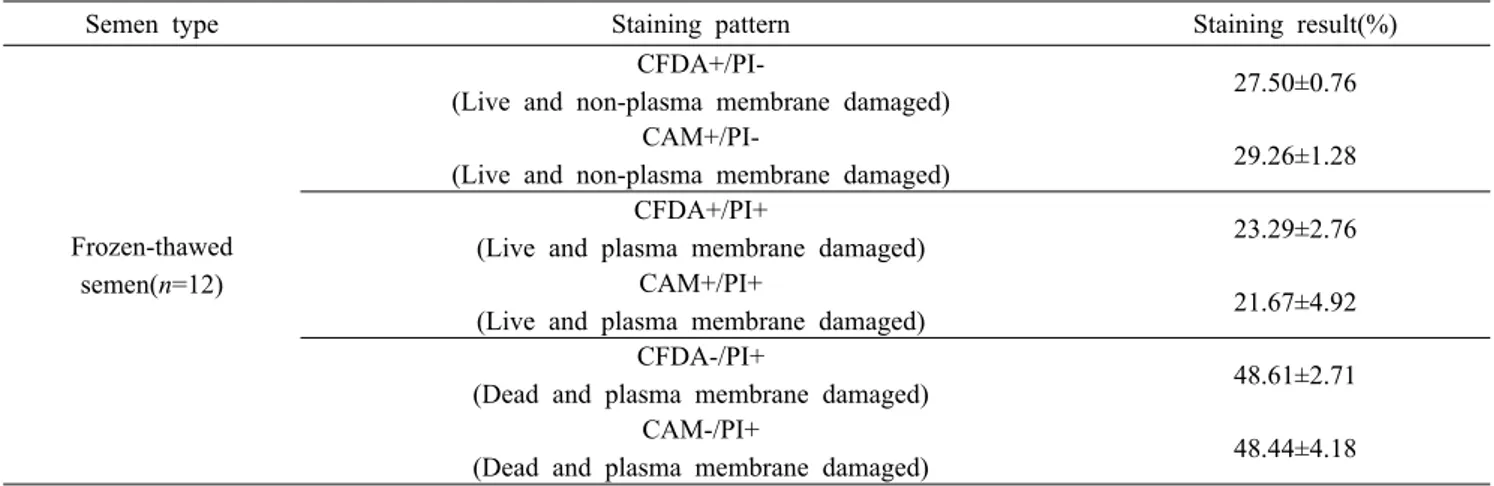
1. Flow cytometry를 이용한 동결 융해 정자의 생존율 검사결과 동결보존된 제주흑우 2마리의 정액은 융해, 희석, 분획하여 6-CFDA(CFDA)/PI와 Cacein AM(CAM)/PI로 각각 이중염색 하였으며, flow cytometry 방법으로 정자의 생존율을 평가하 였다. 결과는 Table 1과 같다.
형광염색 양상에서 CFDA+/PI-와 CAM+/PI-는 정자는 살아 있으며 세포막 손상이 없는 상태, CFDA+/PI+와 CAM+/PI+는 정자는 살아있으나 세포막 손상이 있는 상태, CFDA-/PI+와 CAM-/PI+는 세포막 손상으로 인해 정자가 죽은 상태로 해석하 였다. flow cytometry 분석 결과 CFDA+/PI-와 CAM+/PI- 분포 는 각각 27.50±0.76%, 29.26±1.28%이었으며, 두 형광염색군간 유의적인 차이는 없었다(p>0.05). CFDA+/PI+와 CAM+/PI+ 분 포는 각각 23.29±2.76%, 21.67±4.92%로서 두 군간 유의적인 차이는 없었다(p>0.05). 또한 CFDA-/PI+와 CAM-/PI+ 분포는 각각 48.61±2.71%, 48.44±4.18%로서 역시 두 군간 차이가 없었 다(p>0.05).
2. Flow cytometry를 이용한 정자세포 분포 및 형광염색된 집단의 분포 양상
Table 1. The results of stain percentage of 6-CFDA(CFDA)/PI and calcein AM(CAM)/PI by flow cytometry
Semen type Staining pattern Staining result(%)
Frozen-thawed semen(n=12)
CFDA+/PI-
(Live and non-plasma membrane damaged) 27.50±0.76 CAM+/PI-
(Live and non-plasma membrane damaged) 29.26±1.28 CFDA+/PI+
(Live and plasma membrane damaged) 23.29±2.76 CAM+/PI+
(Live and plasma membrane damaged) 21.67±4.92 CFDA-/PI+
(Dead and plasma membrane damaged) 48.61±2.71 CAM-/PI+
(Dead and plasma membrane damaged) 48.44±4.18 Data are presented as the Mean±SEM. The values isn’t differ between CFDA/PI and CAM/PI
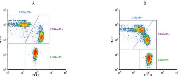
A B
Fig. 1. Flow cytometry scatter profile of frozen-thawed spermatozoa(A: 6-CFDA/PI staining group, B: calcein AM/PI staining group).
Gate 1; spermatozoa population set on forward scatter-height(FSC-H) versus side scatter-height(SSC-H).
제주흑우의 동결정액을 융해, 희석한 후 flow cytometry를 이용하여 forward scatter(FSC)로 세포의 크기와 side scatter (SSC)로 세포 내 과립도를 측정하여 density plot으로 정자세 포의 분포를 나타낸 결과는 Fig. 1과 같다. Fig. 1의 세포 분포 에서 gate를 설정하여 정자세포의 크기와 과립도가 균일한 세 포의 집단을 정자세포로 판단하였으며, 히스토그램으로 표현 한 FSC와 SSC에 따른 세포의 수는 Fig. 2, Fig. 3과 같다. Fig.
1~3에서 6-CFDA/PI와 calcein AM/PI 염색 후 정자 세포의 크 기와 세포 내 과립도에 따른 정자세포의 분포는 서로 유사하 였으며, 세포의 크기와 과립도에 따른 세포의 수를 나타낸 히 스토그램 역시 서로 비슷한 양상을 보였다.
6-CFDA/PI와 Calcein AM/PI의 이중염색 후 flow cytometry 법을 이용하여 형광 발현에 따른 density plot에서 정자의 분포 를 비교하였을 때, Fig. 4와 같이 6-CFDA/PI 염색법과 calcein
AM/PI 염색법은 정자세포의 집단의 염색상에 따른 구분에는 어려움이 없었으나 6-CFDA/PI 염색법이 calcein AM/PI 염색보 다 정자세포 집단의 구분이 뚜렷함을 알 수 있었으며, Fig. 5와 같이 6-CFDA와 Calcein AM의 형광강도에 따른 정자세포 수와 집단에 따른 분포를 표현한 히스토그램 비교에서도 6-CFDA의 정자세포의 형광 발현에 따른 구분이 calcein AM보다 명확함을 알 수 있었다.
6-CFDA와 calcein AM의 염색 후 flow cytometry를 이용하여 정자 세포의 생존율을 비교한 연구는 보고되지 않았으나, 효모의 생존율 검사에서 세포 내 esterase 효소의 작용으로 형광을 발현하 는 CFDA-AM(5-Carboxyflourescein diacetate, acetoxymethly ester)와 DiBAC4(5)(Bis-(1,3-dibutylbarbituric acid)pentamethine oxonol)의 이중 염색법과 calcein AM와 DiBAC4(5)의 이중 염색 법에서 CFDA-AM/DiBAC4(5)가 Calcein AM/DiBAC4(5)보다
A B
Fig. 2. Histogram of forward scatter-height(FSC-H) on Gate 1 and all cells(A: 6-CFDA/PI staining group, B: calcein AM/PI staining group).
A B
Fig. 3. Histogram of side scatter-height(SSC-H) on Gate 1 and all cells(A: 6-CFDA/PI staining group, B: calcein AM/PI staining group).
A B
Fig. 4. Cytogram of spermatozoa stained with dual fluoresceins(A: 6-CFDA/PI staining group, B: calcein AM/PI staining group, FL1:
6-CFDA(A), calcein AM(B), FL3: PI on spermatozoa population(gate 1). CFDA-/PI+, CAM-/PI+(only red-stained) represents dead sperm and damaged plasma membrane. CFDA+/PI+, CAM+/PI+(green- and red-stained) represents live sperm and damaged plasma membrane. CFDA+/PI-, CAM+/PI-(only green- stained) represents live sperm and non-damaged plasma membrane.
A B
Fig. 5. Histogram of 6-CFDA and calcein AM staining on FL1-H(A: 6-CFDA/PI staining group, B: Calcein AM/PI dual stain group)
A B
Fig. 6. Histogram of PI staining on FL3-H(A: 6-CFDA/PI dual stain group, B: Calcein AM/PI dual stain group)
flow cytometry에서 보다 적합하다고 보고하였다(Hernlem B와 Hua SS, 2010). 다른 연구에 따르면 일부 효모와 세균에서 calcein AM의 염색이 생존율 검사에 적합하지 않다는 보고가 있었으나 (Kaneshiro 등, 1993; Diaper JP와 Edwards c. 1994), 특정 세균 (Clavibacter michiganesis subsp. Michiganesis)의 생존성 검사에 서 calcein AM/PI 염색법이 CFDA/PI 염색법보다 적합하다는 보고 (Luiz GC 등, 2006)도 있었다. 이를 토대로 유추해 볼 때, 세포의 종류에 따라 형광염색의 발현도가 차이가 있다고 생각된다.
결 론
본 연구에서 세포 내 estrase 작용에 의해서 형광 발현되는 6-CFDA와 calcein AM를 PI와 함께 정자세포를 염색하여 생 존율을 flow cytometry로 비교 분석한 결과 염색상 각각의 정 자세포의 비율에는 유의적인 차이가 없었으며, 정자세포의 크
기(FSC)와 정자세포의 과립도(SSC)에 따른 정자세포의 분포 를 비교하였을 때 분포 양상이 비슷함을 알 수 있었다. 형광 염색에 따른 정자세포의 집단을 density plot으로 나타내어 비 교하였을 때 6-CFDA/PI 염색법과 calcein AM/PI 염색법은 염 색상에 따른 정자세포 집단의 구분은 둘 다 가능하였으며, 특 히 6-CFDA/PI 염색법이 calcein AM/PI 염색법보다 정자세포 집단의 구분이 명확하였다.
본 연구 결과에서는 flow cytometry를 이용하여 정자의 생존 율을 측정할 경우 6-CFDA/PI 염색법과 Calcein AM/PI 염색법 은 정자세포의 생존율 검사 결과에서 서로 유의적인 차이가 없어 둘 다 이용 가능하지만 6-CFDA/PI 염색법이 calcein AM/PI 염색법보다 염색상에 따른 정자세포의 집단을 구별할 때 더욱 명확하므로 정자세포 분석에는 6-CFDA/PI 염색법이 사용이 유용할 것으로 판단된다.
ACKNOWLEDGMENTS
이 논문은 농촌진흥청 연구사업(세부과제명: 제주흑우 품 종 보존 및 산업화 이용 축군 개발 연구, 세부과제번호 : PJ01010501)의 지원에 의해 이루어진 것임.
REFERENCES
Bolten M, Weissbach L, and Kaden R. 2005. Cryopreserved human sperm deposits: Usability after decades of storage.
Urologe A. 44:904-908.
Bratosin D, Mitrofan L, Palii C, Estaquier J, and Montreuil J.
2005. Novel fluorescence assay using calcein-AM for the determination of human erythrocyte viability and aging.
Cytometry A. 66A:78-84.
Breeuwer P, Drocourt JL, Bunschoten N, Zweitering MH, Rombouts FM, and Abee T. 1995. Charaterization of uptake and hydrolysis of fluorescein diacetate and carboxyfluorescein diacetate by intracellular esterase in Saccharomyces cerevisiae, which result in accumulation of fluorescent product. Appl.
Environ. Microbiol. 61:1614-1619.
Chan LL. Wilkinson AR, Paradis BD, and Lai N. 2012. Rapid Image-based Cytometry for Comparison of Fluorescent Viability staining Methods. J. Fluoresc. 22:1301-1311.
Chitarra LG, Breeuwer P, Abee T, and Bulk RW. 2006. The Use of Fluorescent Probes to Assess Viability of the Plant Pathogenic Bacterium Clavigbacter michiganensis subsp.
Michiganensis by Flow Cytometry. Fitopatol. Bras. 31:349-356.
Decker EM. 2001. The ability of direct fluorescence-based, two colour assays to detect different physiological states of oral streptococci. Lett. Appl. Microbiol. 33:188-192.
Diaper JR and Edwards C. 1994. The use of fluorogenic esters to detect viable bacteria by flow cytometry. J. Appl.
Bacteriol. 77:221-228.
Gillan L, Evans G, and Maxwell WMC. 2005. Flow cytometric evaluation of sperm parameters in relation to fertility potential. Theriogenology 63:445-457.
Gledhill BL, Lake S, Steinmetz LL, Gray JW, Crwaford JR, Dean PN, and Van Dilla MA, 1976, Flow micro fluorometric analysis of sperm DNA content: Effect of cell shape on the fluorescence distribution. J. Cell Physiol. 87:367-375.
Graham JK. 2001. Assessment of sperm quality: A flow cytometric approach. Anim. Reprod. Sci. 68:239-247.
Gordon KM, Duckett L, Daul B, and Petrie HT. 2003. A simple
method for detecting up to five immunofluorescent parameters together with DNA staining for cell cycle or viability on a benchtop flow cytometer. J. Immunol. Methods. 275:113-121.
Hernlem B and Hua SS. 2010. Dual fluorochrome Flow Cytometric Assessment of Yeast Viability. Curr. Microbiol. 61:57-63.
Jarnagin JL and Luchsinger DW. 1980. The use of fluorescein dacetate and ethidium bromide as a stain for evaluating viability of mycobacteria. Biotech. Histochem. 55:253-258.
Jones KH and Sanft JA. 1985. An improved method to determine cell viability by simultaneous staining with flulorescein diacetate-propidium iodide. J. Histochem. Cytochem. 33:77-79.
Kaneshiro ES, Wyder MA, Wu YP, and Cushion MT. 1993.
Reliability of calcein acetoxy methyl ester and ethidium homodimer or propidium iodide for viability assessment of microbes. Journal of Microbiological Methods 17:1-16.
Leeder JS, Dosch HM, Harper PA, Lam P, and Spielberg SP.
1989. Fluorescence-based viability assay for studies of reactive drug intermediates. Anal. Biochem. 177:364-372.
Liminga G, Jonsson B, Nygren P, and Larsson R. 1999. On the mechanism underlying calcein-induced cytotoxicity.
Eur. J. Pharmacol. 383:321-329.
Mahadevan M and Trounson AO. 1984. Effect of cooling, freezing and thawing rates and storage conditions on preservation of human spermatozoa. Andrologia 16:52-60.
Rastegarnia A, Shahverdi A, Topraggaleh TR, Ebrahimi B, and Shafipour V. 2013. Effect of Different Thawing Rates on Post-Thaw Viability, Kinematic Parameters and Chromatin Structure of Buffalo (Bubalus bubalis) Spermatozoa. Cell J.
14:306-313.
Tawakoli PN, Al-Ahmad A, Hoth-Hannig W, Hannig M, and Hannig C. 2013. Comparison of different live/dead stainings for detection and quantification of adherent microorganisms in the initial oral biofilm. Clin. Oral Invest. 17:841-850.
Vishwanath R and Shannon P. 2000. Storage of bovine semen in liquid and frozen state. Anim. Reprod. Sci. 62:23-53.
최선호, 고민희, 강태영, 조상래, 박용상, 오신애. 2011. 제주 흑우 동결 정액 제조에 있어 Glycerol 농도에 따른 생존율 및 정자 첨체 양상의 변화. 한국수정란이식학회지 26:187-193.
홍유미, 김용준, 유일정, 지동범, 김명순. 2004. Flow Cytometry 에 의한 개 신선정액과 동결정액의 생존성 분석. 한국동물 번식학회지 28:167-172.
Received July 11 2017, Revised September 13, 2017, Accepted September 15, 2017