http://dx.doi.org/10.12750/JET.2013.28.4.361
Gonadotropins Improve Porcine Oocyte Maturation and Embryo Development through Regulation of Maternal Gene Expression
Qing-Ling Wang, Ming-Hui Zhao, Yong-Xun Jin, Nam-Hyung Kim and Xiang-Shun Cui
†Department of Animal Sciences, Chungbuk National University, Chungbuk 361-763, South Korea.
ABSTRACT
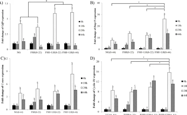
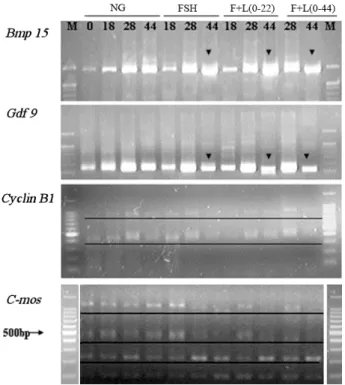
The present study assessed the effect of FSH and LH on oocyte meiotic, cytoplasmic maturation and on the expression level and polyadenylation status of several maternal genes. Cumulus-oocyte complexes were cultured in the presence of FSH, LH, or the combination of FSH and LH. Significant cumulus expansion and nuclear matura- tion was observed upon exposure to FSH alone and to the combination of FSH and LH. The combination of FSH and LH during entire IVM increased the mRNA level of four maternal genes, C-mos, Cyclin B1, Gdf9 and Bmp15, at 28 h. Supplemented with FSH or LH significantly enhanced the polyadenylation of Gdf9 and Bmp15;
and altered the expression level of Gdf9 and Bmp15. Following parthenogenesis, the exposure of oocytes to com- bination of FSH and LH during IVM significantly increased cleavage rate, blastocyst formation rate and total cell number, and decreased apoptosis. In addition, FSH and LH down-regulated the autophagy gene Atg6 and upre- gulated the apoptosis gene Bcl-xL at the mRNA level in blastocysts. These data suggest that the FSH and LH enhance meiotic and cytoplasmic maturation, possibly through the regulation of maternal gene expression and poly- adenylation. Overall, we show here that FSH and LH inhibit apoptosis and autophagy and improve parthenogene- tic embryo competence and development.
(Key words : apoptosis, autophagy, embryo development, maternal gene, oocyte maturation)
Qing-Ling Wang and Ming-Hui Zhao equally contribute to this work.
This work was supported by the research grant of the Chungbuk National University in 2012.
†
Correspondence : E-mail : [email protected]
INTRODUCTION
In mammals, meiotic resumption and subsequent ovulation are controlled by two gonadotropins, follicle stimulating hor- mone (FSH) and luteinizing hormone (LH) (Richards, 1980).
Previous studies reported that gonadotropins influence the resumption of meiotic maturation and cumulus expansion in porcine cumulus-oocyte complexes (COCs) in vitro. Further addition of gonadotropins to the culture medium increased developmental competence, suggesting that gonadotropins could improve cytoplasmic maturation (Sun et al., 2003). Exposure to FSH alone also affected the nuclear and cytoplasmic matu- ration of sow oocytes. Cleavage and blastocyst development rates showed that FSH was most effective in the first 20 hr of maturation culture (Schoevers et al., 2003). Although most in vitro maturation (IVM) protocols currently utilize FSH, LH, or a combination of both, the effect of gonadotropins on IVM and subsequent early embryo development is still controversial and little is known about their molecular mechanism.
During the growth period, the oocyte synthesizes and accu- mulates transcripts and proteins that are vital to growth, matura- tion and embryo development (Lonergan et al., 2003, Lonergan et al., 2003). Translation of these transcripts is generally asso- ciated with changes in polyadenylation (Bettegowda et al., 2007). Several oocyte genes, such as growth differentiation factor 9 (Gdf9) and bone morphogenetic protein 15 (Bmp15), play crucial roles in oocyte maturation; these two members of the Transforming growth factor (TGF) superfamily regulate granulosa cell functions including proliferation, differentiation and cumulus expansion (Otsuka et al., 2011, Sugiura et al., 2010). C-mos is a proto-oncogene first identified as a regulator of oocyte maturation in pigs (Newman et al., 1996). Mos is a serine/threonine kinase that activates the cascade through direct phosphorylation of the MAPK activator MAPK kinase (Prasad et al., 2008). Mos acts almost exclusively in the second meiotic metaphase arrest and is an essential component of the cytostatic factor in mouse oocytes (Hashimoto et al., 1994).
Another essential regulator of meiosis resumption is formed by
Cyclin B1, which gene product complexes with p34 (cdc2) to form the maturation-promoting factor (MPF) (Sartor et al., 1992). It has been reported that two key cell cycle regulator mediated the activity MPF through MAPK pathway by their cytoplasmic polyadenylation in porcine oocyte (Zhang et al., 2009).
Apoptosis and autophagy are common features of mam- malian development. They are tightly regulated processes that play a fundamental role in cell growth, development and ho- meostasis. Recent reports have shown that apoptotic genes are expressed during the preimplantation stage of embryo develop- ment in human, and bovine embryos (Cui et al., 2004, Fear et al., 2011, Metcalfe et al., 2004). Bcl-2 gene products act as essential regulators of the apoptotic signal and may either suppress (Bcl-2, Bcl-xL) or promote (Bak, Bad) the induction of apoptosis (Hetz, 2010). Caspases are cysteine proteases involved in programmed cell death that have recently been found to have vital functions in mouse preimplantation develop- ment (Busso et al., 2010). Autophagy is initiated by class III phosphoinositide 3-kinase and theautophagy gene Atg6. In addition, other systems are involved, including the ubiquitin- like protein Atg8 that is known as microtubule-associated protein 1 light chain 3 (Lc3) in mammalian cells (Schmid et al., 2007). A recent study also suggests that apoptosis and autophagy are involved in early embryo development (Bou- mela et al., 2011).
Hence, to identify the molecular mechanism (s) triggered by FSH and LH, the effects of FSH and LH on the expression and polyadenylation of maternal genes and on the developmen- tal competence of matured oocytes following parthenogenesis were assessed. Our results show that FSH and LH affect por- cine oocyte maturation, regulate maternal gene expression and improve embryo developmental competence by inhibiting auto- phagy and apoptosis.
MATERIALS AND METHODS
All chemicals were obtained from Sigma-Aldrich Co. unless otherwise indicated. Each experiment was repeated at least three times.
1. Collection of Porcine Oocytes, In Vitro Maturation (IVM) and Embryo Culture
COCs aspirated from 3 to 6 mm follicles were matured in IVM medium based with tissue culture medium (TCM) 199
under paraffin oil at 38.5 ℃ in a humidified atmosphere of 5%
CO
2inair. FSH (F2293, Sigma,) (5 μg/ml) and LH (L5269, Sigma) (5 μg/ml) were used in experiment. All experimental groups are showed in Table 1. Following maturation, oocytes were activated for parthenogenesis with 5 μM Ca
2+ionophore and cultured in North Carolina State University (NCSU) 37 medium with 0.4% (w/v) Bovine Serum Albumin (BSA) at 38.5 ℃ in an atmosphere of 5% CO
2. During culture, the percentages of 2cell, 4cell and blastocysts were measured.
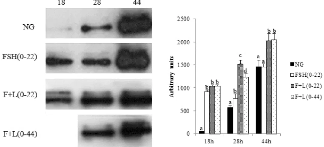
2. Western Blot Analysis
Pig oocytes were denuded in 0.1% Hyaluronidase and wash three times in PVA/PBS and stored in —80℃ until use. Before western blot, oocytes were thawed at room temperature, and lysised by 25 μl Laemmli Sample Buffer. Total protein content was subsequently separated by electrophoresis through a Mini- PROTEAN TGXTM Precast Gel for 2 hr at 100 V. Proteins were then transferred onto a polyvinylidene difluoride (PVDF) membrane. After blocking with 5% BSA in Tris Buffered Sa- line (TBS) for 1 hr, the membrane was incubated with primary phospho-p44/42 MAPK antibody (1 : 500) for 1 hr at room tem- perature. After washing three times in TBS with 1.0% Tween- 20 (TBST), the membrane was incubated for 1 hr with horse- radish peroxidase (HRP)-linked anti-rabbit IgG diluted 1 : 2000 in blocking solution at room temperature. Finally, after three 10 min washes in TBST, bands were visualized using Enhanced Chemiluminescece Luminol Reagent.
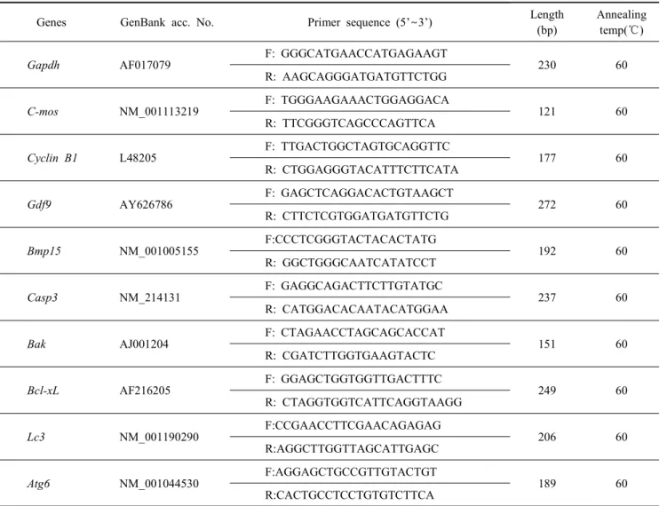
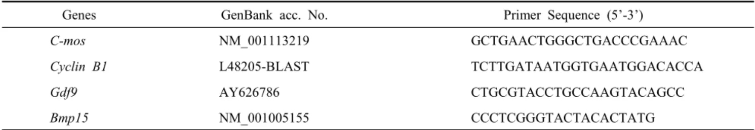
3. Real-Time RT-PCR with SYBR Green
To detect gene expression, mRNA was extracted from por- cine oocytes or blastocyst using Dynabeads mRNA Direct Kit based the instructions. Primer sequences of maternal genes,
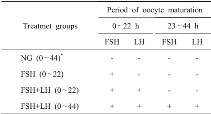
Table 1. List of treatment groups supplemented with FHS and/or LH
Treatmet groups
Period of oocyte maturation 0 ∼22 h 23 ∼44 h
FSH LH FSH LH
NG (0 ∼44)
*- - - -
FSH (0 ∼22) + - - -
FSH+LH (0 ∼22) + + - -
FSH+LH (0 ∼44) + + + +
*