Ⅰ. 서 론
수면무호흡증은 수면 중에 최소 10초 이상의 무호흡 상태 가 1시간 중 5회 또는 7시간 중 30회 이상 발생하는 경우로 정의되며, 중추성(central), 폐쇄성(obstructive) 그리고 혼합형(mixed)으로 크게 분류 된다
1,2). 중추성은 호흡중추 에서 호흡자극이 없거나 호흡에 관여하는 근육기능의 중지 로 인하여 무호흡이 유발되는 경우이며, 폐쇄성은 정상적인 호흡자극에도 불구하고 상부 호흡기도의 간헐적 폐쇄로 인 하여 수면무호흡증이 발생되는 경우를 말한다
3). 폐쇄성 수 면무호흡증후군 (Obstructive Sleep Apnea Syndrome,
OSAS)은 호흡계, 심혈관계, 신경 근육계의 복합적인 질환 으로서 생리학적으로 또는 사회적으로 심각한 문제를 발생 시킬 수 있는 질병으로 특히 악안면 기형과 밀접한 연관을 갖는 것으로 알려져 있다
1). 임상적으로는 수면 중에 야기되 는 반복적인 저산소증으로 인해 심폐기능의 이상을 초래하 며 인지기능장애, 주간의 과도한 졸림에 따른 정상생활 장 애 및 교통사고의 가능성을 증가시킬 수 있다고 보고되고 있다
4).
폐색의 위치는 매우 다양하여 상기도인 비인두강에서 후 두까지 어디나 위치할 수 있으며, 진단을 위하여 두부규격 방사선계측(cephalometry), 전산화단층촬영(CT), 자기공 박광호ㆍ허종기ㆍ안제영ㆍ김지용ㆍ임재형
연세대학교 치과대학 구강악안면외과학교실 영동세브란스병원
폐쇄성 수면무호흡증 진단을 위한 한국인 성인 부정교합자의 두부방사선 사진 계측 분석에 의한 연구
MEAN VALUES OF CEPHALOMETRIC ANALYSIS FROM KOREAN ADULTS WITH ABNORMAL OCCLUSION IN RELATION TO THE DIAGNOSIS
OF OBSTRUCTIVE SLEEP APNEA SYNDROME
Kwang-Ho Park, Jong-Ki Huh, Je-Young Ahn, Ji-Yong Kim, Jae-Hyung Lim Department of Oral and Maxillofacial Surgery, College of Dentistry, Yonsei University,
Yongdong Severance Hospital
Obstructive sleep apnea syndrome (OSAS) is characterized by sleep-induced obstruction of the upper air- way that results in cessation of airflow. Obstruction can occur at a number of points in the airway, but fre- quently in the oropharynx.
A diagnostic evaluation includes cephalometry, computed tomography, magnetic resonance imaging, acoustic reflection technique, polysomnography and fibroptic endoscopy. Cephalometric measurements of the patients with obstructive sleep apnea have revealed that posterior airway anatomy has strong relations with the symptoms of them. A lateral cephalogram is routinely obtained in the radiologic evaluation of sleep apnea patients.
The purpose of this study is to provide a the lateral cephalometric korean norms for the diagnosis and treatment of the patients with obstructive sleep apnea by analyzing the abnormal occlusion of Korean adults.
Key words : Obstructive sleep apnea, Cephalometric analysis, Korean norms Abstract
※ 본 논문은 2000학년도 연세대학교 학술연구비에 의하여 연구되었음.
명영상(MRI), acoustic reflection technique, 다원수면검 사(polysomnography), 섬유광학내시경 검사 등이 주로 이 용된다
5,6). 이 중 두부방사선계측법은 오래 전부터 두개 악 안면 성장과 발달의 평가 및 치아, 골격, 연조직의 구조 및 안면 형태 등을 분석하는 데 사용되어져 왔으며, 진단 범위 를 넓혀 코골이 환자를 포함한 폐쇄성 수면무호흡증 환자들 이 악안면의 구조와 연구개, 혀, 하인두(hypopharynx) 등 의 연조직 구조가 밀접한 상관관계가 있다는 점에 착안하여 진단의 한 방법으로 이용되고 있다
5,7,8,9). 이 질환으로 초래 될 수 있는 심각성을 고려할 때 질환의 진단을 위한 기본적 인 연구가 필요하리라 사료되나 현재 국내에서는 이에 대한 진단 및 치료방법의 개발이 부족한 실정이다.
본 교실에서는 일차적으로 정상 교합자들을 대상으로 시 행한 자료들과 이차적으로 골격성 제 2급과 3급 부정교합
자에서 얻은 결과를 비교 분석하여 폐쇄성 수면 무호흡증에 대한 진단과 치료분석을 위한 기초 작업으로 삼고자 한다.
Ⅱ. 연구대상 및 방법
1. 연구대상
연세대학교 신입생 및 재학생을 대상으로 안모와 교합관계 및 치열궁 형태에 관한 일차적인 임상검사를 시행한 후, 정 상적인 안모 및 I 급 교합관계를 가진 남자 52명, 여자 50명 및 II 급 부정교합을 가진 남자 29명, 여자 24명과 영동세브 란스병원 구강악안면외과에 내원하여 3급 부정교합으로 진 단 받은 남자 29명, 여자 30명을 연구대상으로 하였다.
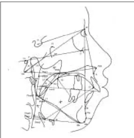
Fig. 1. Cephalometric tracing for Class Ⅱ Fig. 2. Skeletal parameters
Fig. 3. Position of the hyoid Fig. 4. Dimension of the tongue & the soft palate
2. 연구방법
측모두부규격 방사선사진을 촬영하여 정상군은 2명의 구 강악안면외과 의사가 묘사지 위에 각각 2회씩 그린 투사도 상에서 계측점과 기준선을 설정하고 거리 및 각도를 계측하 였고, 실험군에서는 1명의 구강악안면외과 의사에 의해 동
일 조건하에 1회의 투사도를 완성하였다(Fig. 1). 모든 방 사선 사진은 경조직과 연조직 구조를 0.1mm 두께의 투사 지상에 중첩시켜 계측점을 컴퓨터 디지타이저를 이용하여 입력시킨 후 계측값을 컴퓨터상에서 계산하였다. 계측에 사 용된 계측점과 계측항목은 Table 1-4와 Fig. 1-4에 정리하 였다.
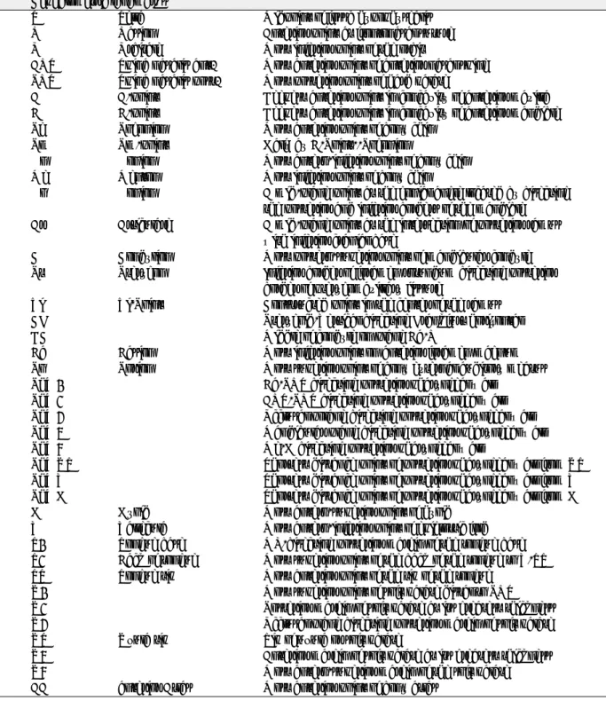
Table 1. Cephalometric Landmarks Cephalometric landmarks
S Sella Midpoint of fossa hypophysealis
N Nasion Anterior point at frontonasal suture
O Orbitale Most inferior point of the orbit
ANS Spina nasalis ant. Most anterior point of anterior nasal spine PNS Spina nasalis post. Most posterior point of hard palate
A A-point Deepest anterior point in concavity of anterior maxilla B B-point Deepest anterior point in concavity of anterior mandible
Pg Pogonion Most anterior point of bony chin
Pm Pm-point Halfway B-Point--Pogonion
Gn Gonion Most antero-inferior point of bony chin
Me Menton Most inferior point of bony chin
Go Gonion A mid-plane point at the gonial angle located by bisecting the posterior and inferior borders of the mandible
Ar Articulare A mid-plane point at the intersection of posterior ramus with inferior cranial base
C Condylion Most postero-superior point of mandibular condyle Pt Pterygon Inferior border of foramen rotundum bisecting posterior
border of pterygomaxillary fissure
Xi Xi-Point Constructed point in the center of the ramus
CF Pterygoid-Vertical bisecting Frankfurt horizontal
DC Middle of condyle on plane Ba-N
Ba Basion Most inferior point on anterior foramen magnum
Po Porion Most superior point of bony external auditory meatus
PhW1 Ba-PNS bisecting posterior pharyngeal wall
PhW2 ANS-PNS bisecting posterior pharyngeal wall
PhW3 Occlusal polane bisecting posterior pharyngeal wall
PhW4 Mandibular plane bisecting posterior pharyngeal wall
PhW5 Me-H bisecting posterior pharyngeal wall
PhWUT Shortest distance point of posterior pharyngeal wall from UT
PhWV Shortest distance point of posterior pharyngeal wall from V
PhWH Shortest distance point of posterior pharyngeal wall from H
H Hyoid Most antero-superior point of hyoid
V Vallecula Most antero-inferior point of epiglottic fold T1 Tongue base ML-bisecting posterior margin of the tongue base T2 Back of tongue Most superior point of the back of the tongue to V-TT TT Tongue tip Most anterior point of the tip of the tongue
U1 Most superior point of soft palate distal to PNS
U2 Posterior margin of soft palate at its greatest thickness
U3 Occlusal plane bisecting posterior margin of soft palate
UT Uvula tip Tip of uvula or soft palate
U4 Anterior margin of soft palate at its greatest thickness
U5 Most antero-superior margin of the soft palate
AA anterior Atlas Most anterior point of bony atlas
Table 2. Cephalometric Distance
Cephalometric distances (mm).
S-Go Posterior facial height
N-Gn Anterior facial height
A/N-Pog Convexity
PAS (ML) Distance posterior pharyngeal wall-tongue base on ML
PAS (Occl.) Distance posterior pharyngeal wall-tongue base on occlusal plane PAS (NL) Distance posterior pharyngeal wall-tongue base on NL
PAS (UT) Distance posterior pharyngeal wall-uvula tip AA-PNS Distance ant. atlas-post. nasal spine Ba-PNS Distance basion-post. nasal spine
Ba-PhW1 Distance basion-posterior pharyneal wall on Ba-PNS PhW1-PNS (PAS) Distance posterior pharyngeal wall-PNS on Ba-PNS Go-PNS Posterior lower facial height
Ba-A Distance basion-point A
PNS-UT Length of the soft palate (uvula-length) U2-U4 Thickness of the soft palate (uvula-thickness)
V-Me Distance vallecula-menton
V-ANS Distance vallecula-ant. nasal spine
V-S Distance vallecula-sella
T1-ANS Distance tongue base-ant. nasal spine
T1-B Distance tongue base-point B
T1-PNS Distance tongue base-post. nasal spine
T1-TT Distance tongue base-tongue tip
V-PhW (PAS) Shortest distance V-posterior pharyngeal wall
V-TT Axis of the tongue tip
T2/V-TT Tongue height
H-ML Shortest distance hyoid to mandibular plane
H-Me Distance hyoid-menton
H-B Distance hyoid-point B
H-PhW (Me-H) Disstance hyoid-posterior pharyngeal wall on Me-H H-PhW Shortest distance hyoid to posterior pharyngeal wall
AA-H Distance hyoid-ant. atlas
H-S Distance hyoid-Sella
Table 3. Cephalometric Reference-lines Cephalometric reference-lines NSL Nasion-sella-line NL Nasal-line (ANS-PNS) ML Mandibular-line (Me-Go) FH Frankfurt horizontal line (O-Po)
Table 4. Cephalometric Angles Cephalometric angles ( 。).
SNA Angle between S-N and N-A
NL-NSL Angle between NSL and NL
N-S-Ba Angle between N-S and S-Ba
ML-NSL Angle between NSL and ML
SNB Angle between S-N and N-B
ML-NL Angle between NL and ML
Saddle-Angle Angle between S-N and S-Ar
Articular Angle Angle between S-Ar and Ar-Go
Gonion Angle Angle between Ar-Go and Go-Me
Sum Angle Sum of saddle-, articular-, gonion angle
Lower Gonion Angle Angle between N-Go and Go-Me
Facial Axis Angle between Pt-Gn and Ba-N
Facial Depth Angle between FH and N-Pog
Mandibular Plane Angle Angle between FH and ML
LFH-Angle Angle between ANS-Xi and Xi-Pm
Mandibular Arc Angle Angle between DC-Xi and Xi-Pm
Maxillary Depth Angle between FH and N-A
zMaxillary Height Angle between N-CF and CF-A
Palatal Plane Angle between FH and NL
Ramus Position Angle between FH and CF-Xi
Uvula-Angulation Angle between NL and PNS-UT
V-TT/ML Angle between V-TT and ML
V-TT/FH Angle between V-TT and FH
N-S-H Angle between N-S and S-H
NSL/Ar-H Angle between NSL and Ar-H
ML/H Angle between Go-Me-H
Table 5. Analysis of Pharyngeal Dimensions (Male)
Male ClⅠ Male ClⅡ Male ClⅢ Multiple
Measurements (N = 52) (N = 29) (N = 29) p-value comparision
Mean (SD) Mean (SD) Mean (SD) ⅠⅡ ⅠⅢ ⅡⅢ
SGO 93.27 ± 5.07 90.18 ± 7.99 91.25 ± 6.28 0.0878
NGN 131.35 ± 5.26 134.26 ± 6.48 137.93 ± 7.41 0.0001 *
PAS (ML) 13.47 ± 3.11 13.60 ± 3.72 16.12 ± 5.32 0.0116 *
PAS (OL) 19.08 ± 2.78 20.70 ± 3.17 20.55 ± 4.85 0.0750
PAS (NL) 28.79 ± 2.54 29.51 ± 4.46 28.00 ± 4.38 0.2910
PAS (UT) 11.60 ± 2.25 11.91 ± 3.45 15.72 ± 4.63 0.0001 * *
AA-PNS 36.16 ± 3.14 37.68 ± 4.52 34.99 ± 3.54 0.0215 *
Ba-PNS 50.29 ± 3.26 50.10 ± 4.89 50.04 ± 3.56 0.9648
Ba-PhW1 22.01 ± 2.93 21.22 ± 3.37 22.70 ± 3.28 0.2028
PhW1-PNS (PAS) 28.25 ± 2.43 28.88 ± 4.01 27.35 ± 4.63 0.2571
Go-PNS 56.10 ± 3.92 56.02 ± 6.02 50.40 ± 3.84 0.0001 * *
Ba-A 100.02 ± 5.03 101.76 ± 7.20 99.63 ± 5.01 0.3025
PNS-UT 37.02 ± 3.10 38.67 ± 3.88 32.17 ± 4.28 0.0001 * *
U2-U4 11.18 ± 1.31 9.87 ± 1.70 9.80 ± 1.66 0.0001 * *
V-Me 60.44 ± 5.34 56.54 ± 7.58 65.86 ± 8.56 0.0001 * *
V-ANS 98.43 ± 4.64 105.22 ± 5.46 98.26 ± 5.66 0.0001 * *
V-S 114.74 ± 5.66 117.45 ± 7.86 117.70 ± 7.36 0.0909
T1-ANS 90.77 ± 4.58 93.06 ± 5.83 86.02 ± 5.70 0.0001 * *
T1-B 69.90 ± 3.94 68.06 ± 6.72 75.39 ± 5.56 0.0001 * *
T1-PNS 53.89 ± 3.41 53.55 ± 4.78 49.39 ± 3.14 0.0001 * *
T1-TT 74.51 ± 4.45 76.03 ± 5.39 70.85 ± 6.33 0.0008 * *
V-PhW (PAS) 18.66 ± 3.93 18.91 ± 4.54 19.59 ± 4.95 0.6562
V-TT 77.90 ± 4.66 83.23 ± 5.04 76.80 ± 6.54 0.0001 * *
T2/V-TT 37.77 ± 3.04 38.46 ± 3.52 36.11 ± 4.30 0.0349 *
H-ML 10.20 ± 4.71 13.55 ± 5.40 10.71 ± 6.22 0.0239 *
H-Me 43.52 ± 4.62 40.22 ± 7.26 47.55 ± 7.52 0.0001 * *
H-B 51.84 ± 4.75 52.62 ± 6.64 55.53 ± 7.68 0.0356 *
H-PhW(MeH) 36.00 ± 3.74 35.67 ± 3.32 39.55 ± 5.22 0.0003 * *
H-PhW (PAS) 35.00 ± 3.01 35.47 ± 3.55 38.23 ± 4.46 0.0015 * *
AA-H 69.68 ± 5.65 69.03 ± 7.39 75.32 ± 8.46 0.0007 * *
H-S 118.24 ± 5.68 118.81 ± 8.06 120.30 ± 8.07 0.4509
SNA 83.12 ± 3.20 82.49 ± 4.10 81.86 ± 3.91 0.3228
NLNSL 8.80 ± 3.29 9.32 ± 4.21 9.33 ± 3.43 0.7418
NSBA 130.51 ± 3.95 130.82 ± 6.19 131.83 ± 4.57 0.4894
MLNSL 30.99 ± 3.96 34.50 ± 8.51 34.98 ± 6.05 0.0058 * *
SNB 80.51 ± 3.01 76.31 ± 4.41 84.80 ± 5.23 0.0001 * * *
MLNL 22.19 ± 3.47 25.18 ± 7.09 25.65 ± 5.35 0.0056 * *
SADDLEA 124.58 ± 4.12 125.00 ± 5.80 123.84 ± 4.77 0.6419
ARTICA 148.78 ± 5.00 152.09 ± 8.07 143.59 ± 7.09 0.0001 * *
GONIONA 117.63 ± 4.72 117.41 ± 7.81 127.55 ± 6.99 0.0001 * *
SUMA 390.99 ± 3.96 394.50 ± 8.51 394.98 ± 6.05 0.0058 * *
LOWGNA 73.67 ± 3.08 74.28 ± 6.33 79.90 ± 4.99 0.0001 * *
FACIALAX 94.36 ± 3.36 92.01 ± 11.17 90.43 ± 3.90 0.0288 *
FACIALD 89.19 ± 2.32 84.08 ± 3.89 91.76 ± 3.91 0.0001 * * *
MNPLA 23.15 ± 3.35 27.38 ± 8.17 28.72 ± 5.05 0.0001 * *
LFHA 46.44 ± 3.12 51.18 ± 7.05 48.55 ± 4.18 0.0002 *
MNARCA 142.77 ± 4.27 150.61 ± 8.95 147.26 ± 5.37 0.0001 * *
MXD 90.96 ± 2.37 89.61 ± 3.48 88.12 ± 3.24 0.0003 *
MXH 66.59 ± 3.46 66.13 ± 3.73 66.49 ± 3.12 0.8417
PALATALP 2.56 ± 1.54 3.32 ± 2.16 3.45 ± 2.39 0.0891
RAMUSPO 71.82 ± 3.05 73.36 ± 3.59 76.75 ± 4.10 0.0001 * *
UVULAA 127.81 ± 4.19 130.25 ± 6.47 121.13 ± 10.07 0.0001 * *
VTTML 51.70 ± 5.10 55.28 ± 8.72 56.86 ± 7.62 0.0038 *
VTTFH 28.55 ± 4.22 27.90 ± 6.15 28.14 ± 6.21 0.8603
NSH 89.10 ± 3.63 92.42 ± 4.85 86.71 ± 4.26 0.0001 * * *
NSLARH 74.00 ± 4.08 78.39 ± 5.72 72.72 ± 4.63 0.0001 * *
MLH 13.69 ± 6.71 21.00 ± 9.90 13.38 ± 8.29 0.0002 * *
*: Comparisons significant at the 0.05 level (unit : mm)
S.D. = standard deviation
Table 6. Analysis of Pharyngeal Dimension (Female)
Male ClⅠ Male ClⅡ Male ClⅢ Multiple
Measurements (N = 50) (N = 24) (N = 30) p-value comparision
Mean (SD) Mean (SD) Mean (SD) ⅠⅡ ⅠⅢ ⅡⅢ
SGO 84.66 ± 5.09 83.13 ± 7.32 80.43 ± 4.87 0.0065 *
NGN 124.23 ± 5.62 128.09 ± 6.18 129.02 ± 5.94 0.0010 * *
PAS (ML) 11.55 ± 2.95 11.22 ± 2.32 14.01 ± 4.43 0.0024 * *
PAS (OL) 18.47 ± 3.25 19.96 ± 3.47 17.81 ± 2.32 0.0376 *
PAS (NL) 27.54 ± 2.51 28.35 ± 3.51 27.17 ± 3.65 0.3761
PAS (UT) 11.05 ± 3.22 11.00 ± 2.25 11.89 ± 2.62 0.3831
AA-PNS 33.90 ± 2.74 35.53 ± 3.35 32.54 ± 3.58 0.0033 *
Ba-PNS 46.36 ± 2.71 47.65 ± 2.03 47.25 ± 3.84 0.1684
Ba-PhW1 19.46 ± 2.10 20.29 ± 2.26 20.94 ± 2.79 0.0245 *
PhW1-PNS (PAS) 26.91 ± 2.40 27.36 ± 3.44 26.32 ± 3.97 0.4754
Go-PNS 51.53 ± 4.26 52.23 ± 5.56 44.27 ± 5.38 0.0001 * *
Ba-A 94.77 ± 4.24 97.49 ± 3.08 94.68 ± 4.08 0.0138 * *
PNS-UT 34.35 ± 3.44 35.63 ± 4.55 31.78 ± 3.12 0.0005 * *
U2-U4 9.18 ± 1.33 9.18 ± 0.99 9.27 ± 1.26 0.9458
V-Me 57.42 ± 5.21 57.03 ± 5.50 61.41 ± 5.00 0.0018 * *
V-ANS 91.51 ± 4.00 97.52 ± 5.76 90.34 ± 5.06 0.0001 * *
V-S 103.18 ± 5.53 103.23 ± 6.39 104.17 ± 5.86 0.7431
T1-ANS 85.28 ± 4.22 89.34 ± 4.64 80.47 ± 4.95 0.0001 * * *
T1-B 66.60 ± 4.16 67.49 ± 3.80 70.85 ± 3.35 0.0001 * *
T1-PNS 49.36 ± 3.73 50.25 ± 5.17 43.71 ± 4.87 0.0001 * *
T1-TT 69.93 ± 4.33 74.75 ± 4.44 67.27 ± 4.45 0.0001 * * *
V-PhW (PAS) 15.25 ± 2.54 15.18 ± 2.69 16.05 ± 3.17 0.3892
V-TT 72.11 ± 4.38 78.78 ± 5.09 71.11 ± 4.83 0.0001 * *
T2/V-TT 35.73 ± 2.72 35.07 ± 2.70 33.84 ± 3.65 0.0283 *
H-ML 7.72 ± 3.89 9.42 ± 5.14 8.61 ± 4.73 0.2937
H-Me 43.01 ± 5.03 42.24 ± 3.68 47.75 ± 4.15 0.0001 * *
H-B 49.82 ± 4.52 51.49 ± 3.51 54.06 ± 3.48 0.0001 *
H-PhW (Me-H) 30.30 ± 3.01 30.31 ± 3.16 32.28 ± 3.56 0.0211 *
H-PhW(PAS) 29.76 ± 2.95 29.85 ± 3.13 30.58 ± 3.40 0.5067
AA-H 60.87 ± 5.38 59.46 ± 5.24 63.62 ± 6.27 0.0217 *
H-S 105.55 ± 5.58 104.37 ± 5.70 104.89 ± 7.02 0.7215
SNA 81.09 ± 2.66 80.24 ± 2.95 80.94 ± 3.70 0.5251
NLNSL 10.62 ± 3.01 11.29 ± 3.69 9.88 ± 2.71 0.2528
NSBA 132.35 ± 4.87 134.53 ± 5.80 133.96 ± 4.76 0.1640
MLNSL 33.80 ± 4.40 36.56 ± 6.78 38.85 ± 6.04 0.0005 *
SNB 78.02 ± 2.67 74.23 ± 3.55 82.98 ± 4.04 0.0001 * * *
MLNL 23.17 ± 3.95 25.26 ± 6.41 28.96 ± 6.64 0.0001 * *
SADDLEA 126.11 ± 5.10 127.69 ± 5.60 124.72 ± 5.51 0.1261
ARTICA 150.30 ± 6.15 151.77 ± 6.06 144.73 ± 6.11 0.0001 * *
GONIONA 117.39 ± 6.16 117.09 ± 6.04 129.40 ± 7.10 0.0001 * *
SUMA 393.80 ± 4.40 396.56 ± 6.78 398.85 ± 6.04 0.0005 *
LOWGNA 73.75 ± 4.10 74.10 ± 4.82 80.97 ± 5.45 0.0001 * *
FACIALAX 90.07 ± 4.34 84.67 ± 9.23 91.05 ± 3.98 0.0002 * *
FACIALD 87.64 ± 2.45 84.05 ± 3.03 91.50 ± 3.24 0.0001 * * *
MNPLA 24.94 ± 4.74 27.16 ± 6.18 30.56 ± 6.19 0.0001 *
LFHA 46.82 ± 3.09 50.76 ± 5.99 48.33 ± 3.82 0.0010 *
MNARCA 141.07 ± 4.47 148.97 ± 6.82 146.60 ± 5.77 0.0001 * *
MXD 89.94 ± 2.42 89.63 ± 2.47 89.23 ± 3.45 0.5391
MXH 67.03 ± 2.87 66.77 ± 3.07 67.12 ± 3.35 0.9105
PALATALP 2.98 ± 1.81 2.47 ± 2.05 3.00 ± 2.06 0.5254
RAMUSPO 70.41 ± 2.73 72.67 ± 4.17 75.64 ± 3.21 0.0001 * * *
UVULAA 127.75 ± 5.04 132.75 ± 5.80 125.82 ± 6.42 0.0001 * *
VTTML 49.01 ± 5.55 48.83 ± 7.64 54.57 ± 6.29 0.0004 * *
VTTFH 24.06 ± 3.99 21.67 ± 5.07 24.01 ± 3.93 0.0606
NSH 91.80 ± 3.66 95.71 ± 5.52 89.01 ± 4.08 0.0001 * * *
NSLARH 76.57 ± 4.31 81.38 ± 6.53 75.30 ± 5.03 0.0001 * *
MLH 10.60 ± 5.77 13.17 ± 7.84 10.54 ± 6.06 0.2209
*: Comparisons significant at the 0.05 level (unit : mm)
S.D. = standard deviation
이렇게 얻어진 자료는 ANOVA(Scheffe’s test)분석을 통해 통계 처리하였다.
Ⅲ. 연구 결과
측모두부규격 방사선사진상에서 골격구조분석, 인후기도 공간구조분석 및 설골의 위치분석을 위한 계측항목에 대하 여 남녀의 평균치와 표준편차를 구하였다(Table 5, 6).
골격구조분석 항목 중에서 SNA는 남자에서 정상교합자 (1급), 골격성 제2급 부정교합자(2급), 골격성 제3급 부정 교합자(3급)가 각각 83.12, 82.49, 81.86, 여자의 경우는 각각 81.09, 80.24, 80.94로 차이가 거의 없었으며, 이는 본 실험군들에서 상악은 정상발육을 보인다고 할 수 있다.
상악의 수직성장을 나타내는 상악고경 또한 남녀 모두 세 집단간에서 유의성 있는 차이를 보이지 않았다. SNB는 1 급, 2급, 3급이 남녀 각각 80.51, 76.31, 84.80과 78.02, 74.23, 82.98로 세 집단간에 모두 유의성 있는 차이를 보 였으며(p=0.0001, 0.0001), 그 외에도 하악과 관련된 수 치는 1급에 대해 2, 3급의 대부분이 차이를 나타내었고, 특 히 3급에서 그 정도가 더욱 빈번하게 나타났다.
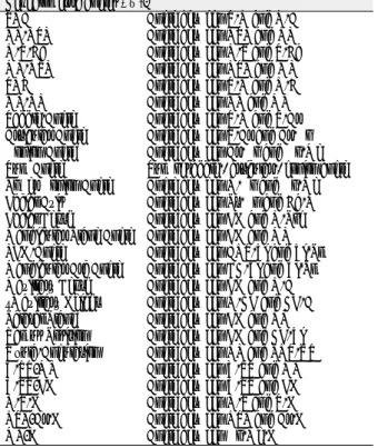
인후기도공간분석 항목 중에서 PAS(ML)는 남녀 모두에 서 1급, 2급, 3급의 측정값(mm)이 각각 13.47, 13.60, 16.12(p=0.0116)와 11.55, 11.22, 14.01 로 유의성 있 는 차이를 보였다(p=0.0024). PAS(UT)는 남자의 경우 1급과 3급에서 11.60, 15.72로 차이를 보였으나 남자의 1, 2급간이나 여자의 경우에는 비슷한 측정값을 얻었다. 하인 두 부위에서의 기도공간은 집단간의 차이가 두드러졌으며, 특히 1, 3급 간에서 유의성 있는 차이가 있었다 (Fig. 5, 6).
설골의 위치분석 항목에서는 H-ML 측정값이 제시하듯이
설골은 남자의 경우 1급(10.20mm)에 비해 2급 (13.55 mm)에서 더욱 하방에 위치해 있으나, 여자는 측정값의 분 포도가 넓어서 그 유의적인 차이를 검증할 수 없었으며, 3 급은 남녀 모두에서 1급에서의 값과 큰 차이를 보이지 않 았다. 설골의 전후방 위치를 나타내는 N-S-H, NSL/Ar- H, ML/H는 2급, 1급, 3급의 순으로 감소되었으며, N- S-H 의 경우 세 집단 간에서 모두 유의적인 차이를 나타 내었다.
Ⅳ. 총괄 및 고찰
폐쇄성 수면무호흡증은 코나 입을 통한 공기의 출입은 없 어도 가슴이나 복부의 호흡운동은 있는 것으로, 수면 중 상 기도의 폐쇄에 기인한 간헐적 무호흡을 특징으로 하는 복잡 한 수면장애이다
5,6). 중추성 수면 무호흡증은 코나 입을 통 한 공기의 유입이 없을 뿐만 아니라 가슴이나 복부의 호흡 운동도 없는 것으로, 중추 신경계 중 호흡중추의 자율조절 이 안됨으로써 호흡근의 활동성 감소로 인해 생기는 무호흡 이다. 혼합형 수면 무호흡증은 폐쇄성과 중추성 수면 무호 흡증이 함께 나타나는 경우이다
3). 폐쇄성 수면무호흡증의 수면 중에 일어나는 증상은 코골음, 호흡중단, 수면단절, 호 흡곤란, 야뇨증, 발한 등을 들 수 있고 수면 중의 호흡장애 로 인해 부정맥, 빈맥, 서맥, 고혈압 등 심혈관계의 기능장 애를 초래할 수 있다. 수면 시 나타나는 이러한 증상들로 인 해 충분한 수면을 취하지 못한 결과, 아침 기상 시 두통, 판별 능력 감소, 기억력 감퇴, 우울증 등이 나타날 수 있다
10,11,12,21).
Fujita는 폐쇄 위치를 구인두-구개(연구개), 구인두-하인 두, 하인두(설기저부)로 분류하였다
13). 폐쇄성 수면 무호흡 증에 연구개만 단독으로 관련된 경우가 18%, 설기저부와
Fig. 6. Pharyngeal dimension (Female)
Fig. 5. Pharyngeal dimension (Male)
연구개가 관련된 경우가 80%, 설기저부와 인후측벽이 관 련된 경우는 거의 없다고 보고 된 바가 있다
14). 폐쇄성 수면 무호흡증의 원인과 병인에 대해서 명확하게 밝혀지지는 않 았으나 수면 중 인후 근육이 병적 혹은 생리적으로 긴장도 가 감소된 상태에서 음주, 마취제, 근육이완제, 진정제 등으 로 인해 근육의 긴장도가 더 떨어지는 경우, 또는 해부학적 으로 상기도의 비정상적 협착이 있는 경우 발생된다고 알려
져 있다
5,15,16,17). 따라서 비정상적으로 좁아진 상기도 부위나
폐쇄부위를 찾아내는 것이 폐쇄성 수면 무호흡증의 진단과 치료에 중요하다.
폐쇄성 수면무호흡증의 진단을 위해서는 병력 청취와 임 상검사를 비롯하여 두부규격 방사선 사진, 전산화단층촬영 (CT), 자기공명영상(MRI), acoustic reflection tech- nique, 다원수면검사(polysomnography), 섬유광학내시경 검사 등이 주로 이용된다. 두부규격 방사선 사진은 다른 검 사에 비해 손쉽게 많은 환자들에게 적용가능하고, 치료 전 후에 비교가 용이하며 경제적이라는 장점이 있어 진단에 유 용하게 사용될 수 있다
5). 측모두부규격 방사선 사진은 일반 적으로 골조직은 잘 평가할 수 있지만, 구강인후의 연조직 을 평가하는 데에는 많은 어려움이 있다. 따라서 이것만으 로 단독으로 진단하는 것보다는 수면 중 생리적 변화까지 검사할 수 있는 수면다원검사와 상기도의 전후좌우 공간과 역동적인 움직임까지 관찰 가능한 섬유광학내시경 검사 등 의 진단방법이 상호보완적으로 이루어져야 할 것이다.
1983년 Riley 등이 측면 두부규격 방사선 사진을 이용하여 처음으로 폐쇄성 수면무호흡증 환자를 연구하였고, 폐쇄성 수면 무호흡증 환자군이 하악의 열성장과 함께 연구개의 길 이, 설골 위치, 상기도 수평 길이 등에서 유의성 있는 차이 가 있음을 보고하였다
18).
연구방법은 Hochban 등
19)이 1994년 제안한 분석법을 토 대로 하여, 상기도의 수평 거리, 연구개, 혀, 설골 등과 관련 된 연조직구조분석 및 안면 골격구조분석을 시행하였다. 정 상교합자와 골격성 2급, 3급 부정교합자를 대상으로 시행 한 결과에 따르면, 골격구조 항목과 인후기도공간구조 항목 에서 남녀간의 뚜렷한 차이를 보였으며 1, 3급 부정교합자 간의 하인두 기도공간에서 유의할 만한 차이가 나타났다 (p<0.05).
본 연구는 코골이를 포함한 폐쇄성 수면무호흡증 환자의 진단법의 하나인 두개방사선 계측법을 한국인에게 적용하 기 위해 이미 일차적인 기초 작업의 일환으로 한국인 정상 성인 교합자를 대상으로 연구한 결과
20)와 연계하여, 이번 연 구에서는 부정교합자를 대상으로 계측점과 계측값의 남녀 기준치를 구함으로서 실제 임상에서 진단 및 치료를 위한 기초 자료수립을 위해 시행되었다.
참고문헌