with Argon Plasma Coagulation in a Patient with Resectable Primary Lung Cancer
Departments of
1Internal Medicine,
2Chest Surgery,
3Pathology,
4Diagnostic Radiology, Konyang University College of Medicine, Daejon, Korea
Mi-Hye Kwon, M.D.
1, Mi-Il Kang, M.D.
1, Ji-Hyun Jeong, M.D.
1, Hee-Kwan Won, M.D.
1, Hyun-Woong Park, M.D.
1, Jung-Ho Park, M.D.
1, Sung-Tae Kim, M.D.
1, Sun-Jung Kwon, M.D.
1, Eugene Choi, M.D.
1, Moon-Jun Na, M.D.
1, Hyun-Min Cho, M.D.
2, Young-Jin Kim, M.D.
2, Yoon-Mee Kim, M.D.
3, Young-Jun Cho, M.D.
4, Ji-Woong Son, M.D.
1수술적 절제가 가능한 원발성 폐암 환자에서 병발된 방사선학적으로 발견되지 않은 동시성 원발성 폐암을 아르곤 플라스마 응고소작술로 치료한 1예
권미혜1, 강미일1, 정지현1, 원희관1, 박현웅1, 박정호1, 김성태1, 권선중1, 최유진1, 나문준1, 조현민2, 김영진2, 김윤미3, 조영준4, 손지웅1
건양대학교 의과대학
1내과학교실,
2흉부외과학교실,
3조직병리학교실,
4영상의학교실
1990년대 초부터 형광 기관지 내시경이 임상 진료에 사용되면서 방사선검사에서 나타나지 않는 상피내 폐암이나 미세하게 진행된 조기 폐암의 진단 빈도가 늘어났으며, 이러한 상피내 폐암의 과반수 이상에서 진행성 폐암으로 진행하므로, 근치적 목적의 치료가 더욱 조기에 도입될 수 있어 폐암 치료에서의 중요한 진단 도구가 되었다.
치료적 내시경술의 발달로 기존의 진행된 폐암에서 기도 폐쇄 감소 혹은 출혈 부위의 지혈 등의 완화적 목적뿐 아니라, 조기 폐암이지만 심폐 기능 등 전신 상태의 문제로 수술이 불가능한 환자에서 근치적 치료로도 이용되고 있으며, 특히, 수술의 절제 범위를 축소시키는 효과를 가져올 수 있다. 아르곤 플라스마 응고소작술(argon plasma coagulation, APC)은 레이저와 광역학 치료법(photodynamic therapy, PDT) 등에 비하여 조기 폐암 병변의 치료에 서 근치적 치료 및 진행성 폐암에서의 완화요법으로서 효과면에서 동등하고, 특히 침투 범위가 얕으므로 표재성 의 병변에서 탁월하며, 경제적 접근성이 용이하다. 저자들은 우측 상엽의 절제 가능한 폐암에 우측 하엽에 상피내 암이 동반된 동시성 원발성 폐암 환자를 우측상엽절제와 APC로써 치료한 증례 1예를 경험하여 보고한다. (Tuberc Respir Dis 2008;65:137-141)
Key Words: Synchronous Roentgenographically Occult Lung Carcinoma (ROLC), Carcinoma in situ, Argon Plasma Coagulation (APC)
Address for correspondence: Ji-Woong Son, M.D.
Department of Internal Medicine, Konyang University Hospital, 685, Kasuwon-dong, Seo-gu, Daejeon 302-718, Korea
Phone: 82-42-600-8817, Fax: 82-42-600-9090 E-mail: [email protected]
Received: Jun. 9, 2008 Accepted: Jul. 14, 2008
Introduction
The development of the autofluorescence broncho- scopy (AFB) technique in the early 1990s in addition to traditional sputum cytology methods enabled early detection of both bronchial intraepithelial neoplasia and
lung cancer in central airways. Even though this ad- vancement allowed for earlier intervention, it un- fortunately did not achieve significantly increased sur- vival rates1. Recently the prevalence of synchronous roentgenographically occult lung carcinoma (ROLC) in patients with resectable primary lung cancer was de- termined to be relatively high, 9.3%, by AFB and biop- sy2. Carcinoma
in situ
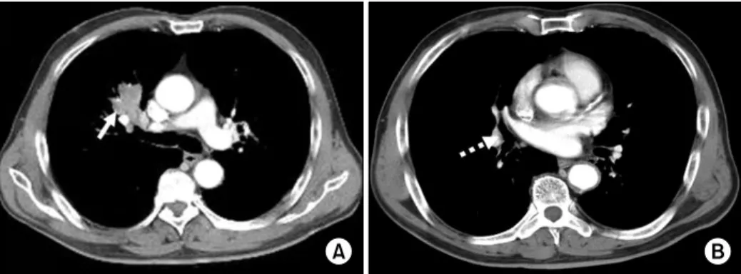
(CIS) diseases from the bronchus are known to progress to invasive carcinomas in more than half of the cases. Surgical resection remains the primary curative treatment of lung cancer. However, several endobronchial treatment modalities are available for curative or palliative purpose in inoperable patientsFigure 1. Preoperative images of CT showed (A) about 3.7×2.7 cm sized heterogenous enhancing low density mass with total obstruction of anterior segmental bronchus of right upper lung (solid arrow), (B) proximal portion of right middle and lower bronchus is unremarkable (dashed arrow).
with limited cardiopulmonary reserve3. Synchronous second primary lung cancer in non small cell lung can- cer (NSCLC), is not a rare phenomenon and current guidelines recommend considering curative surgical re- section for both of types of lesions, invasive mediastinal staging and extrathoracic imaging. We report a case of synchronous double primary lung cancers that were treated with lobectomy for lung mass and argon plasma coagulation (APC) for another lesion of CIS.
Case Report
A 68 year-old male patient visited our hospital for eval- uation of an incidental right upper lung mass that was found in a chest x-ray performed at a routine check- up.
The patient was a current smoker with 15 pack-years of smoking history. In addition, he had been diagnosed with hypertension and diabetes mellitus and was on regu- lar medications. He was free from any respiratory symp- toms, and had no systemic complaints such as weight loss, general weakness and decreased appetite.
Furthermore, the patient’s physical examination revealed no abnormal findings and his vital sign was stable upon admission. Laboratory findings revealed a white cell count of 9.21×10⁹/L, 17.9 g/dL hemoglobin, 50.7% hem- atocrit, and platelet count 227×109/L. Routine chemistry and ABGA were also unremarkable. However in the chest x-ray the right hilum was prominent, and the chest dy- namic CT showed 3.7×2.7 cm sized heterogenously en-
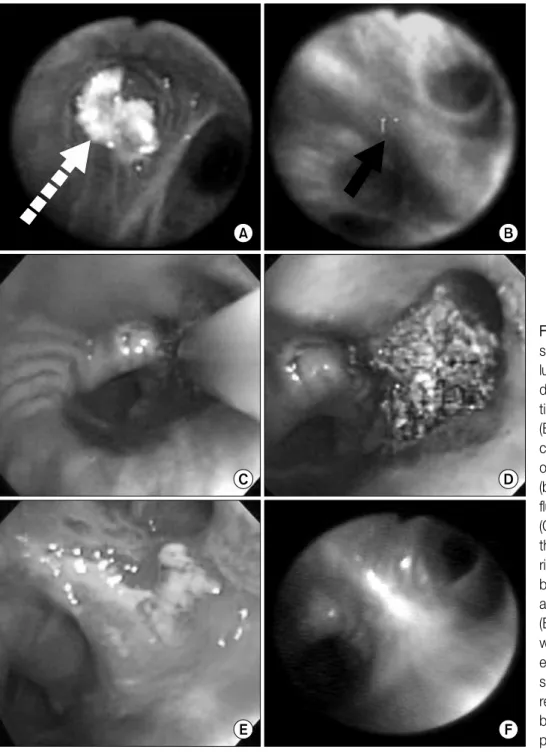
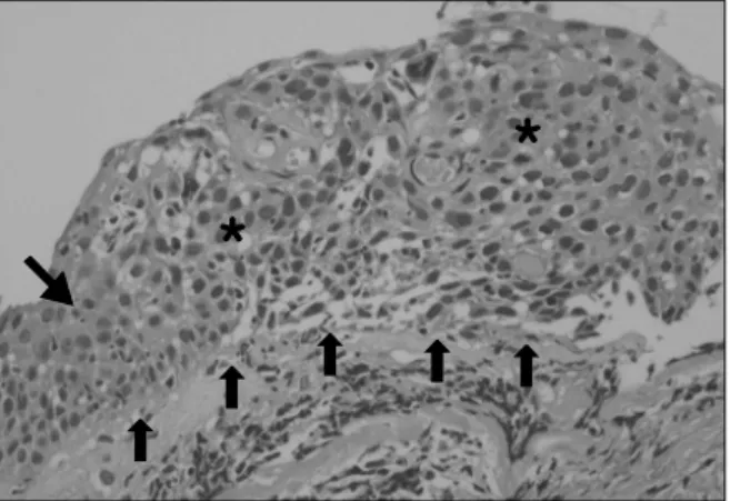
hanced low density mass that completely obstructed the anterior segmental bronchus of the right upper lobe (Figure 1). We performed autofluorescence broncho- scopy (OncoLIFEⓇ, Xillix, British Columbia) and obtained a tissue sample, using forcep biopsy, from the obstructing intraluminal mass in the right upper lobe (Figure 2A) and the superior segment of the right lower lobe where a loss of autofluorescence was observed without mucosal abnormalities (Figure 2B). The pathology reports in- dicated that the mass in the right upper lung was squ- amous cell carcinoma and the lesion in the superior seg- ment consisted of squamous cell carcinoma
in situ
(Figure 3). No distant metastasis was observed in the brain MRI, whole body bone scan, and whole body PET/CT. In the preoperative physiologic evaluation, the patient’s per- formance was good and the postbronchodilator FEV1 and MVV were 2.39 L (92.2% of pred), and 61.8 L (60.4%of pred), respectively, therefore the right pneumo- nectomy was planned. During the operation, the right lobectomy and mediastinal lymph node dissection were done because he could not tolerate one- lung ventilation.
After 3 weeks, the superior segment of the right lower lung lesion was treated with one session of APC (ERBEⓇ, Elektromedizin Tübingen, Germany) and no immediate complication was encountered (Figure 2C, D). On the 18th day post-APC, an ulcerative lesion with mild edema was observed by bronchoscopy (Figure 2E), and the biop- sy report revealed acute and chronic inflammation. He was discharged uneventfully and there was no evidence
Figure 2. Initial broncho- scopy showed (A) intra- luminal mass in RB3 (white dashed arrow) by conven- tional bronchoscopy, and (B) ‘loss of autofluores- cence’ in superior segment of right lower bronchus (black solid arrow) by auto- fluorescence bronchoscopy.
(C) APC was performed on the superior segment of right lower bronchus, (D) bronchoscopy immediate after APC showed debris, (E) and after two and half weeks ulcerative and mild edematous changes were shown. (F) No evidence of recurrence was shown on bronchoscopy on 3 months post-APC.
of recurrence in a follow-up bronchoscopy at 3 month post-APC (Figure 2F), and in CT at 1 year post-APC.
Discussion
Previously, bronchoscopic intervention was used for the removal of foreign bodies or toileting bronchial se- cretions, and the applications have been widened
nowadays. Tumors that are located in the central air- ways can be treated with several bronchoscopic techni- ques, such as lasers (Nd:YAG), photodynamic therapy (PDT), brachytherapy, cryotherapy and electrocautery (argon, CO2), for palliation of airway obstruction in ad- vanced cancers and for curative treatment of ROLCs.
APC delivers a high-frequency alternating current to the tissue through an ionized argon gas in non-contact
Figure 3. The report from pathologist revealed ‘carcinoma in situ’ for superior segment of right lower bronchus. The five small upward arrows shows intact basement mem- brane, and the region marked as ‘*’ is filled with cancer cells with mitosis, pleomorphism. The downward arrow (left middle) revealed normal stratified squamous epi- thelium (H&E stain, ×200).
mode and has been extensively evaluated in open sur- gery of liver, spleen, and kidneys. From these evalua- tions it was shown that APC is an effective tool for su- perficial bleeding and controlling bleeding during gas- trointestinal endoscopies4. In addition, it is attractive for the treatment of hemorrhagic superficial spreading tu- mor and also suitable for treating tumors ‘around the corner’ at a sharp angle. APC causes more acute super- ficial tissue destruction, thus, is more effective than any other method for the management of hemostasis5. However, it is less efficient for the in-depth tissue de- struction of bulky tumors because it has a limited pene- tration depth of 2∼3 mm. The cost of APC is less ex- pensive and more readily available in many centers than lasers and photodynamic therapy (PDT). The possible complications are imminent respiratory failure, hemor- rhage and life-threatening airway obstruction.
The results of previous studies showed that when electrocautery of rigid or flexible bronchoscopes for pal- liative treatment of lung cancer was used it was equally effective in achieving tumor coagulation and debulking compared to an Nd:YAG laser but with less excessive complication rates6-10. For microinvasive lung cancer, PDT is preferentially used because of its strength of
‘selective’ damage where only relatively minor destruc- tion occurs in normal tissue. However, any kinds of bronchoscopic technique can be effectively used when less than 3 millimeters of invasion of the bronchial mu- cosa and visible distal margin is required if the patient is not a candidate for surgery because of poor car- diopulmonary function11. The cases treated with electro- cautery for occult cancer and typical intraluminal carci- noid were reported12.
Schuurman et al.13 reported a case of microinvasive and premalignant lesion detected by AFB and treated with APC and surgery. In addition, Kato et al.14 reported several cases where early lung cancers treated with PDT resulted in a reduction in the extent of required surgery.
These reports clearly demonstrate the benefits of inter- ventional bronchoscopy in that it provided an additional option of curative treatment to patient unable to tolerate surgery. In the present report, the patient was a candi- date for pneumonectomy, with good preoperative phys- iologic function, but when on a mechanical ventilator in the operation room the patient was not able to toler- ate one-lung ventilation and oxygen saturation dropped while the right upper lobectomy was performed. The pathology report of the
in situ
lesion revealed cancer cells of 1∼2 millimeters’ thickness and the basement membrane was intact. Therefore, we treated thein situ
lesion with one session of APC, since the therapeutic depth of the diseased region was within the limits of APC. After APC treatment, the patient was free from im- mediate complications and from long-term sequelae, such as airway stenosis. Furthermore, there was no evi- dence of recurrence after 1 year. Consequently the pa- tient was treated successfully with reduced-extent sur- gery by using APC as a curative treatment. We think interventional bronchoscopy, especially APC, is an ef- fective, safe and economical tool for the curative treat- ment of ROLCs with superficial diseases, and it can be an alternative for patients who can’t tolerate surgery.Summary
We treated synchronous double primary lung can-
cers, where one site resulted from CIS disease, with lo- bectomy and argon plasma coagulation (APC) in a pa- tient who couldn’t tolerate pneumonectomy, which re- sulted in a reduction of the extent of surgery. APC could be a reasonable alternative for CIS disease of lung in inoperable patients.
References
1. Lam S, MacAulay C, Hung J, LeRiche J, Profio AE, Palcic B. Detection of dysplasia and carcinoma in situ with a lung imaging fluorescence endoscope device.
J Thorac Cardiovasc Surg 1993;105:1035-40.
2. Pierard P, Vermylen P, Bosschaerts T, Roufosse C, Berghmans T, Sculier JP, et al. Synchronous roentgeno- graphically occult lung carcinoma in patients with re- sectable primary lung cancer. Chest 2000;117:779-85.
3. Venmans BJ, van Boxem TJ, Smit EF, Postmus PE, Sutedja TG. Outcome of bronchial carcinoma in situ.
Chest 2000;117:1572-6.
4. Johanns W, Luis W, Janssen J, Kahl S, Greiner L. Argon plasma coagulation (APC) in gastroenterology: experi- mental and clinical experiences. Eur J Gastroenterol Hepatol 1997;9:581-7.
5. Freitag L. Interventional endoscopic treatment. Lung Cancer 2004;45:S235-8.
6. Morice RC, Ece T, Ece F, Keus L. Endobronchial argon plasma coagulation for treatment of hemoptysis and neoplastic airway obstruction. Chest 2001;119:781-7.
7. Okada S, Yamauchi H, Ishimori S, Satoh S, Sugawara H, Tanaba Y. Endoscopic surgery with a flexible bron- choscope and argon plasma coagulation for tracheo- bronchial tumors. J Thorac Cardiovasc Surg 2001;121:
180-2.
8. Stephens KE Jr, Wood DE. Bronchoscopic management of central airway obstruction. J Thorac Cardiovasc Surg 2000;119:289-96.
9. Hooper RG. Electrocautery in endobronchial therapy.
Chest 2000;117:1820-1.
10. Ledingham SJ, Goldstraw P. Diathermy resection and radioactive gold grains for palliation of obstruction due to recurrence of bronchial carcinoma after external irradiation. Thorax 1989;44:48-51.
11. Sutedja G, Schramel F, Postmus PE. Bronchoscopic treatment modalities in lung cancer, indications and limitations. Ann Oncol 1995;6:951-2.
12. Shah H, Garbe L, Nussbaum E, Dumon JF, Chiodera PL, Cavaliere S. Benign tumors of the tracheobronchial tree. Endoscopic characteristics and role of laser resec- tion. Chest 1995;107:1744-51.
13. Schuurman B, Postmus PE, van Mourik JC, Risse EK, Sutedja TG. Combined use of autofluorescence bron- choscopy and argon plasma coagulation enables less extensive resection of radiographically occult lung cancer. Respiration 2004;71:410-1.
14. Kato H, Konaka C, Ono J, Kawate N, Nishimiya K, Shinohara H, et al. Preoperative laser photodynamic therapy in combination with operation in lung cancer.
J Thorac Cardiovasc Surg 1985;90:420-9.